Introduction
Poly(3-alkylthiophenes) (P3ATs) are commonly studied conjugated polymers for organic electronics due to their relatively high hole mobilities, low optical band gaps, and well-controlled synthesis.[Reference Osaka and McCullough1] Although straight chain aliphatic side chains were originally introduced to improve the thermal and solvent processability of the unsubstituted polythiophene analog,[Reference Elsenbaumer, Jen and Oboodi2] it is well known that the structural as well as optoelectronic properties are affected by the length, architecture, and chemical composition of the side chain.[Reference Causin, Marega, Marigo, Valentini and Kenny3–Reference Hong and Collard7] More recently, it has been shown that substituting straight-chain alkyl substituents for branched compounds results in a significant depression in thermal transitions.[Reference Ho, Boudouris and Segalman8] Specifically, it is well known that the melting temperature of poly(3-hexylthiophene) (P3HT) is over 200 °C, whereas the melting transitions of poly(3-(2′-ethyl)hexylthiophene) (P3EHT) are approximately 80 °C. The lower melting transition has proven ideal for controlling the nanoscale morphology in block copolymers and provides a novel handle for studying structure–property relationships in homopolymers.[Reference Boudouris, Ho, Jimison, Toney, Salleo and Segalman9–Reference Iovu, Jeffries-El, Zhang, Kowalewski and McCullough12]
In addition to morphological control afforded by manipulation of the thermal transition, consideration of the optical properties is important for potential application to devices. The random copolymerization of P3AT monomers has been presented as a convenient synthetic approach for controlling both thermal and optoelectronic properties simultaneously. In copolymers of poly(3-butylthiophene) and poly(3-octylthiophene), Jenekhe and co-workers[Reference Wu, Ren and Jenekhe13] have shown that both the melting transitions and (h00) lattice spacings are linear functions of composition and span the range between the two endpoints. Furthermore, the lattice spacings shift is not correlated with a shift in the optical absorption edge.[Reference Wu, Ren and Jenekhe13] More recently, random copolymers of 3EHT and 3HT have been synthesized by several groups. Zhang et al.[Reference Zhang, Tajima and Hashimoto14] showed that copolymers of P3EHT and P3HT resulted in melting transitions, which were significantly depressed from that of P3HT and could be controlled through the relative composition. In that work, Hashimoto and co-workers suggest that increasing the 3EHT content introduces crystalline defects; however, we show in this work that both monomers must necessarily co-crystallize in order to observe universally accepted signatures of crystallinity in UV–vis, x-ray diffraction (XRD), and differential scanning calorimetry (DSC) over the entire molar composition range (and in particular the symmetric case). Additionally, Thompson and co-workers[Reference Burkhart, Khlyabich and Thompson15] observed that the open-circuit voltage of bulk heterojunctions with P3AT copolymers and phenyl-C61-butyric acid methyl ester (PCBM) extended the range of the two components, though the complicated relationship between bulk heterojunction solar cell morphology and performance precludes definitive fundamental explanation for the optimal composition. Although these papers present application and optimization of random copolymers for optoelectronic devices, the relationship between the copolymer composition and the adopted crystal structures was beyond the scope of these papers. Furthermore, trends with monomer composition were presented, but the fundamental role of the crystal structure on the resulting thermal and optical properties has not been explored.
Herein we show that changes in the crystal structure alone can have a significant effect on the absorption profile of copolymers although the conjugated unit is identical. The lattice of P3EHT-co-P3HT is shown to depend on the copolymer composition and the differences in the lattice can be correlated with the energy of the 0–0 transition in UV–vis absorbance. However, while optical absorbance is tied to the symmetry of the unit cell, the melting transitions are not solely dependent on the crystal lattice but are more strongly related to the overall chemical composition. This work presents a model system in which the thermal properties can be tuned independently from the optoelectronic properties and provides design rules for the synthesis of conjugated copolymers. In particular, in this work we not only demonstrate tuning of the melting temperature through composition, but we also show how the optical band gap can be attributed to the unit cell adopted by each random copolymer.
Results and discussion
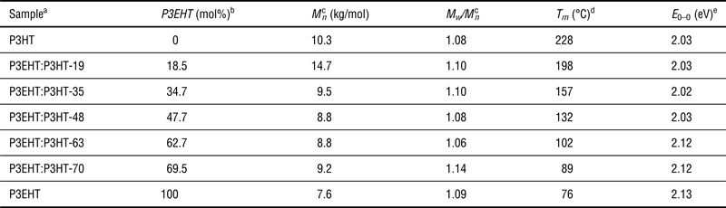
Copolymers of 3EHT and 3HT were synthesized via the Grignard Methathesis (GRIM) polymerization route developed by McCullough and co-workers[Reference Loewe, Ewbank, Liu, Zhai and McCullough16] with a fixed monomer ratio; molecular characteristics presented in Table I confirm low polydispersities and similar molecular weights. Copolymer compositions were determined by relative areas of peaks—via the thiophene ring's aromatic proton—in the Reference Osaka and McCullough1H NMR spectra and range from 19 to 70 mol% 3EHT.
Polymer molecular characterization.

aCopolymers named according to 3EHT mol% composition.
bAs determined by 1H NMR spectroscopy.
cAs determined by gel permeation chromatography. Values given in polystyrene equivalents.
dMelting temperatures determined by DSC.
eValues determined by fitting the Spano model (details in Supplemental Information).
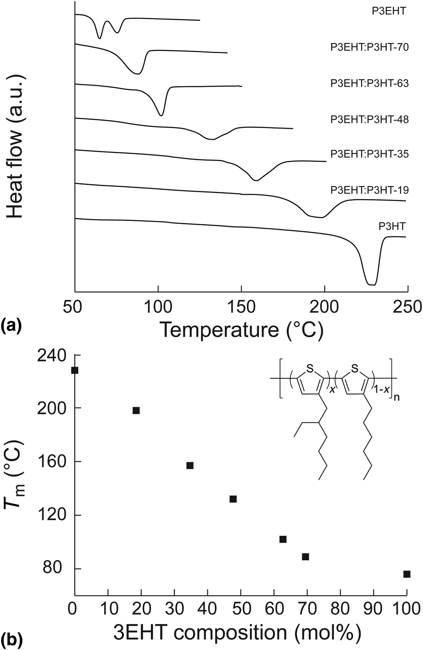
As expected, the melting transitions of random copolymers are highly dependent on the relative composition. Figure 1(b) shows the melting transition temperatures for all polymers as a function of 3EHT composition obtained from the second heating scan in DSC traces [Fig. 1(a)]. Importantly, although DSC endotherms of the random copolymers are broad, no samples exhibit a second melting transition as would be expected for phase separation of the two components. P3EHT:P3HT-19 displays a melting transition at 198 °C, 30 °C lower than homopolymer P3HT. While further increases in the 3EHT content to 35 and 48 mol% 3EHT results in a similarly large decrease in the melting temperatures (157 and 132 °C, respectively) these large variations in melting temperature cease at high 3EHT compositions. In contrast, both transitions of P3EHT:P3HT-61 and P3EHT:P3HT-70 are within 25 °C of P3EHT even though the minority repeat unit contents of copolymers are comparable with that of P3EHT:P3HT-35.
(a) Differential scanning calorimetry heating traces for copolymers showing a single melting transition for all copolymers. Traces are offset for clarity. (b) Melting temperatures as a function of 3EHT composition. Chemical structure for P3EHT:P3HT-x is presented as an inset.
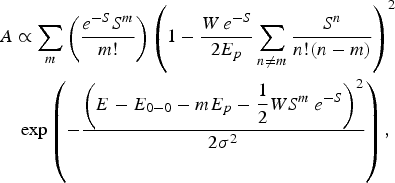
The melting transitions are inherently tied to the relative composition of the random copolymer, a simple synthetic handle for controlling the melting transition. However, it is not known a priori what the effect of copolymer content will be on the absorbance profile of conjugated systems. Figure 2 shows normalized UV–vis absorption profiles of random copolymer thin films. Although the difference in the absorption onset is clear from these data, fits to the model of the absorption from aggregates in the weak excitonic coupling regime developed by Spano provide a quantitative comparison between the absorption profiles.[Reference Turner, Pingel, Steyrleuthner, Crossland, Ludwigs and Neher17–Reference Spano19] The absorption data were fit to Eq. (1),
 $$\eqalign{&A \propto \sum\limits_m {\left({\displaystyle{{e^{ - S} S^m } \over {m!}}} \right)\left({1 - \displaystyle{{W\, e^{ - S} } \over {2E_p }}\sum\limits_{n \ne m} {\displaystyle{{S^n } \over {n!\lpar n - m\rpar }}} } \right)} ^2 \cr & \quad \exp \left({ - \displaystyle{{\left({E - E_{0 - 0} - mE_p - \displaystyle{1 \over 2}WS^m \, e^{ - S} } \right)^2 } \over {2\sigma ^2 }}} \right)\comma \; }$$
where S is the Huang–Rhys factor (which has been set to 1 for this study), m and n are vibrational states, W is the excitonic bandwidth, E p is the energy of the C = C stretching mode, E 0–0 is the energy of the 0–0 transition, and σ is the Gaussian line width. In particular, a comparison of the 0–0 transition energy provides a spectroscopic quantity to compare the “gas-to-crystal shifts” induced by differences in copolymer crystal structures. Owing to the complex origin of the excitonic bandwidth, W, a parameter which has been shown to be dependent on morphological features on multiple length scales, the value is only of interest in the context of this work as a validation of the weak excitonic coupling assumption underlying the model. Further details of the model and fitting parameters (Table S1) can be found in the Supplemental Information.
Optical properties for random copolymers and homopolymers. (a) In the normalized UV–vis absorption profiles, the absorption edge for 3HT-rich copolymers align exactly, showing no difference in the band gap with increasing comonomer content. The band gap of the 3EHT-rich copolymers is blue-shifted significantly from that of the other copolymers. (b) The energy of the 0–0 transition as a function of 3EHT composition. A large increase in the transition energy is observed as the 3EHT content increases beyond the symmetric composition.
Values for the P3HT homopolymer agree well with literature values for thin films cast from chlorobenzene.[Reference Duong, Toney and Salleo20–Reference Niles, Roehling, Yamagata, Wise, Spano, Moule and Grey23] The P3EHT homopolymer exhibits an absorbance profile which is blue-shifted by 100 meV from that of P3HT, as noted in prior work.[Reference Ho, Boudouris and Segalman8] Application of the Spano model to the P3EHT homopolymer results in E 0–0, E p, and W values slightly larger than that of P3HT, and although the energy of the vibrational transition and the excitonic bandwidth are larger than that presented for P3HT in this work, such values have been observed in thin films of P3HT in the past.[Reference Duong, Toney and Salleo20] Importantly, the value of the excitonic bandwidth is still less than the energy of the vibrational transition, which indicates that the P3EHT homopolymer lies in the weak excitonic coupling regime and the Spano model is generally applicable.
Unlike the continual decrease in melting transition temperature with increasing 3EHT content (up to high 3EHT contents), parameters from the fit to the Spano model do not follow a simple monotonic trend. Specifically, the random copolymers can be divided into two groups: those with 0–0 transition energies similar to P3HT (P3EHT:P3HT-19, P3EHT:P3HT-35, and P3EHT:P3HT-48) and those nearer that of P3EHT (P3EHT:P3HT-63 and P3EHT:P3HT-70). We will first discuss the copolymers with high P3HT content. P3EHT:P3HT-19, P3EHT:P3HT-35, and P3EHT:P3HT-48 have values of E 0–0 and Gaussian linewidths that are almost identical to that of pure P3HT. That the energy of the 0–0 transition is so closely matched suggests that 3EHT repeat units included within the copolymer aggregate are indistinguishable electronically from 3HT. This is not surprising, as the only difference between monomers is in the optically inactive alkyl component. Indeed, similar observations with copolymer composition can be made for copolymers with majority 3EHT. P3EHT:P3HT-63 and P3EHT:P3HT-70 having similar transitions, which are nearly 100 meV removed from that of P3HT.
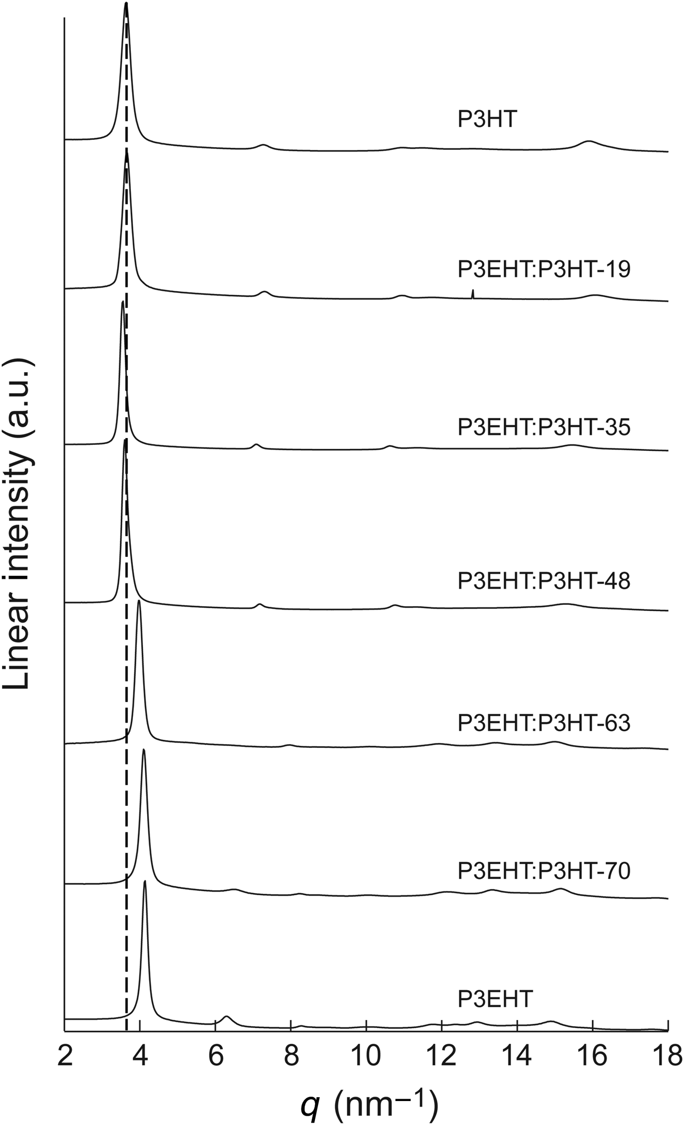
The origin of these distinct regimes in the UV–vis absorption profiles is not immediately obvious, but it is well known that optoelectronic properties are inherently tied to the crystal structure of a material.[Reference Fox24, Reference Kittel25] It is therefore imperative to investigate fully the atomic structure in these polymers using wide-angle x-ray scattering (WAXS), as presented in Fig. 3. Peak positions are tabulated in the Supplementary Information (Table S3). While P3HT is known to adopt an orthorhombic unit cell[Reference Prosa, Winokur, Moulton, Smith and Heeger26] (or a monoclinic unit cell with γ very near 90°),[Reference Kayunkid, Uttiya and Brinkmann27] additional diffraction peaks in P3EHT have been assigned in thin film to a triclinic lattice.[Reference Boudouris, Ho, Jimison, Toney, Salleo and Segalman9] Differences between these two symmetries are readily observable in the WAXS profiles of Fig. 3. The pure P3HT profile has peaks at 3.6, 7.3, and 10.9 nm−1, corresponding to the (h00) family of planes or the alkyl side-chain spacing in P3ATs, and at a higher q value of 15.9 nm−1, the (020) peak, corresponding to the π–π lattice dimension, is observed. In contrast, the pure P3EHT diffraction pattern exhibits reflections at 4.1 and 8.3 nm−1, which are assigned to the alkyl chain dimension, but a non-integer peak at 6.3 nm−1 can be observed as well. Although it is tempting to assign this reflection to the same family of planes with one-half magnitude of the currently proposed reciprocal lattice vector, a peak corresponding to a (100) reflection for such an assignment is not observed at lower angles and thin-film grazing-incidence x-ray diffraction (GI-XD) indicates that these non-integer peaks do not lie on the same reciprocal line as the (h00) reflections.[Reference Boudouris, Ho, Jimison, Toney, Salleo and Segalman9] As such, this peak has been identified as the (010) reflection, which is no longer disallowed by symmetry.
WAXS profiles for homopolymers and copolymers. Profiles are offset for clarity. Below 50 mol% P3EHT, the conventional orthorhombic P3HT unit cell is observed with no changes to the (h00) family of planes (the 100 is highlighted with the dashed line). A small shift to smaller q is observed in the (020) reflection of P3EHT:P3HT-35 and P3EHT:P3HT-48 suggesting a small expansion of the unit cell is necessary to accommodate the bulkier EHT side chain. Alternatively, above 63 mol% P3EHT, a shift in the (100) to higher q and higher-order reflections are observed, indicative of the triclinic unit cell observed in homopolymer P3EHT.
In random copolymers, unit cells tend to trend with the majority monomer present in the polymer sample. For copolymers with sufficiently high P3HT content, the copolymers adopt the P3HT orthorhombic unit cell, as evidenced by nearly identical lattice spacings to that of pure P3HT and the lack of peaks between the (100) and (200) reflections observed in P3EHT. For compositions above 52 mol% P3HT, the (100) peaks for all polymers have q-values of approximately 4.1 nm−1, indicating that the incorporation of P3EHT repeat units does not increase the alkyl chain spacing of the adopted P3HT unit cell. This is not surprising as the side-chain interdigitation in P3AT homopolymers is poor,[Reference Kline, DeLongchamp, Fischer, Lin, Richter, Chabinyc, Toney, Heeney and McCulloch5] and therefore the ethyl chain does not provide the steric bulk required to disturb the spacing in this lattice dimension during growth of orthorhombic crystallites. However, the (020) reflection is shifted to lower q-values for the P3EHT:P3HT-35 and P3EHT:P3HT-48 copolymers from 15.9 nm−1 for pure P3HT to 15.5 and 15.3 nm−1, respectively. This corresponds to an increase in this lattice dimension from 3.95 Å for P3HT to 4.11 Å at 48 mol% 3EHT. Furthermore, the breadth of the (020) peak increases with increased 3EHT content suggesting a decrease in the coherence length, likely dominated by paracrystallinity similar to observations in other semicrystalline polymers.[Reference Rivnay, Noriega, Kline, Salleo and Toney28, Reference Noriega, Rivnay, Vandewal, Koch, Stingelin, Smith, Toney and Salleo29]
Unlike P3EHT:P3HT-35 and P3EHT:P3HT-48, the crystal structures of the copolymers with higher compositions of 3EHT are not orthorhombic. At 63 and 70 mol% 3EHT the (h00) planes lie at the same positions as P3EHT, and reflections at non-integer multiples of the (100) peak are observed. It should be noted that a small shift to lower angle may be observed in the 63 mol% case but the large peak breadth makes definitive assignment difficult. Although it is tempting to suggest that the observation of identical diffraction patterns for the P3EHT:P3HT-70 and P3EHT homopolymer may indicate that the 3HT units are not incorporated into the crystalline P3EHT domains (such that the copolymer diffraction corresponds solely to pure P3EHT crystallites), the melting transition for the copolymers is not depressed as would be expected for an exclusion model of crystallization in random copolymers and the percent aggregates is not significantly decreased (Table S1).[Reference Flory30]
In a manner analogous to the UV–vis spectroscopy, the analysis of the diffraction data groups the unit cells of the random copolymers into one of two groups. The first group (P3EHT:P3HT-19, P3EHT:P3HT-48, and P3EHT:P3HT-35) maintain the P3HT orthorhombic unit cell although the d-spacing of the (020) increases with increasing 3EHT content. This agrees with the observed trend that these polymers have approximately the same E 0–0 and linewidths in UV–vis spectra. Additionally, it can be noted that the trend of increasing paracrystalline disorder follows the increase in the excitonic bandwidth, but further studies are warranted before any direct correlation can be proposed. In the P3EHT-rich copolymers, the diffraction and UV–vis spectra are significantly different from that of the P3HT. The energy of the 0–0 transition abruptly increases by nearly 100 meV once the unit cell transitions away from the orthorhombic symmetry. As the content is increased from 63 to 70 mol% 3EHT, slight changes in the unit cell result in a blue-shift in the absorption profile as well, further supporting that the absorption profile is intimately tied to the unit cell adopted by the P3AT. In contrast with the work of Jenekhe and co-workers in which only the (100) dimension was altered, the observed change in the unit cell results in increases of the π–π stacking dimension, which affects the optical band gap of the material.
Conclusions
3HT and 3EHT units co-crystallize when randomly copolymerized over all compositions and adopt a crystalline structure similar or identical to that of one of the homopolymers. In these cases, the optical band gap for the copolymers and the corresponding homopolymer indicate that both thiophene repeat units are conjugated along the backbone and electronically indistinguishable in the crystalline domains. However, while P3EHT:P3HT-63 exhibits a melting transition within 25 °C of P3EHT, P3EHT:P3HT-35 melts 71 °C below that of P3HT even though it contains a comparable minority repeat unit content. This gradual change in melting temperature with a simultaneous abrupt change in crystal symmetry is commonly observed for isodimorphism[Reference Allegra and Bassi31, Reference Kamiya, Sakurai, Inoue and Chujo32] in copolymers and has been observed in multiple systems in the past including polyolefins,[Reference Matsumoto, Nakamae, Ogoshi, Kawasoe and Oka33–Reference Voigt-Martin and Mandelkern35] isotactic polystyrene,[Reference Natta36] polyimides,[Reference Kreuz, Hsiao, Renner and Goff37] and polyamide[Reference Edgar and Hill38] copolymers. Our observations plant P3AT copolymers within the context of well-established literature, while providing a platform for modifying the melting temperature of these conjugated polymers and maintaining the optical band gap. From the data, it can be determined that the aliphatic side chain in P3ATs, although optoelectronically inactive, plays an important role in dictating the crystalline packing in the solid state. While changes in the alkyl chain dimension have been observed in the past and that this does not affect the optical absorbance in such systems, this work highlights that drastic changes to the crystalline structure can be affected which result in subsequent shifts in the absorbance spectra. Overall, the P3EHT–P3HT random copolymer system rather elegantly illustrates the correlation between the optical properties and crystalline structure of P3ATs and provides a simple synthetic handle for modifying the melting transition temperature over a range of 96 °C without affecting the absorbance edge.
Acknowledgments
This material is based upon work supported by the National Science Foundation under Grant no. 1206296. WAXS was performed at beam lines 1–4 at the Stanford Synchrotron Radiation Lightsource, a Directorate of SLAC National Accelerator Laboratory and an Office of Science User Facility operated for the US Department of Energy Office of Science by Stanford University. We thank Professor Bryan W. Boudouris for helpful discussion.
Supplementary materials
For supplementary material for this article, please visit http://dx.doi.org/10.1557/mrc.2014.9
Supplemental Information Available: Sample preparation, detailed synthetic procedure, 1H NMR spectroscopy, details of the Spano model, and fits to absorption profiles.