Introduction
Congenital choanal atresia is a rare abnormality, affecting approximately 1 in 7000–8000 newborns, with a female predominance.Reference Ravindhra, Elluru, Stucker, Kenyon, Lian, Draf and Schick1 In congenital choanal atresia, the widened vomer fuses with the atretic plate, resulting in an hourglass configuration of the nasopharynx and the choanal region.Reference Ravindhra, Elluru, Stucker, Kenyon, Lian, Draf and Schick1
Currently, nasal endoscopy and multi-slice, high resolution, axial computed tomography (CT) are the procedures of choice for the diagnosis of congenital choanal atresia, whether bony or mixed, and for facilitating surgical planning.Reference Brown, Pownell and Manning2
Earlier literature suggested that 90 per cent of choanal atresia cases are of the bony type and 10 per cent are membranous.Reference Brown, Pownell and Manning2 However, other studies have stated that the mixed type (bony-membranous) is the most common (70 per cent), with a 30 per cent incidence of pure bony atresia.Reference Park, Brockenbrough and Stankiewicz3, Reference Josephson, Vickery, Giles and Gross4
Neonates with bilateral congenital choanal atresia and respiratory distress are obligate nasal breathers. Affected children have cyclical cyanosis, which is relieved by crying. Approximately 20–50 per cent of patients with congenital choanal atresia have other associated congenital anomalies or syndromes. The most frequent association, in about 20 per cent of cases, is coloboma, heart defects, atresia of nasal choanae, retarded growth, genital abnormalities, ear defects and deafness (‘CHARGE’) syndrome. Other isolated anomalies associated with congenital choanal atresia include meningocele, hypertelorism and cleft palate.Reference Harrison, Calvert and Longhurst5
Multiple surgical approaches have been proposed to repair the congenital choanal atresia, evolving from blind transnasal puncture,Reference Carpenter and Neel6 transpalatal,Reference Freng7 sublabialReference Krespi, Husain, Levine and Reede8 and transseptal approaches,Reference Hall, Watanabe, Kenan and Baylin9 to the currently favoured endoscopic transnasal approach. However, there remains no general consensus about the optimal surgical technique. The ideal surgical procedure should be safe, with restoration of a normal nasal passage, without damage to any surrounding craniofacial structures, and which results in a low rate of re-stenosis post-operatively.
This study aimed to evaluate the outcomes of endoscopic repair of bilateral congenital choanal atresia utilising a mirrored L-shaped septonasal flap without stenting versus endoscopic repair using stenting and powered instrumentation (drilling) without a flap.
Materials and methods
A prospective randomised controlled study, comprising 72 newborns with congenital bilateral choanal atresia, was conducted at Tanta University Hospital (Egypt), a tertiary referral hospital, between March 2009 and December 2016. Our study was approved by the Tanta University institutional review board.
The exclusion criteria included: associated pyriform aperture stenosis; coloboma, heart defects, atresia of nasal choanae, retarded growth, genital abnormalities, ear defects and deafness syndrome association; revision cases; unilateral cases; and patients with severe neurological deficits. Physical examination and complete investigations were carried out for all patients to detect other associated congenital anomalies.
All patients underwent pre-operative axial, non-contrast CT and nasal fibre-optic endoscopy, to confirm the diagnosis and assess the type of atretic plate.
Randomisation
After obtaining consent from parents of the newborns with congenital bilateral choanal atresia, the patients were randomly allocated to one of two groups using a simple randomisation method. Randomisation was performed by statistics personnel using a computer-generated random list. Group A consisted of 42 patients who underwent endoscopic transnasal repair using a mirrored L-shaped septonasal flap, without stenting or powered instrumentation. Group B, a control group, consisted of 30 patients who underwent endoscopic transnasal repair using powered instrumentation (drilling) and stenting without a flap.
Operative procedure
A 0-degree, 2.7-mm endoscope was utilised under general anaesthesia. In group A, we used the classical otological instruments, including a round ear knife, straight and curved ear scissors, a House curette, and ear forceps for elevation of the septonasal flap. We used 1-mm and 2-mm Kerrison forceps and straight circular-cutting punch forceps instead of powered drills, to avoid excessive trauma to the surrounding structures and to achieve maximal preservation of mucosa and the harvested flaps.
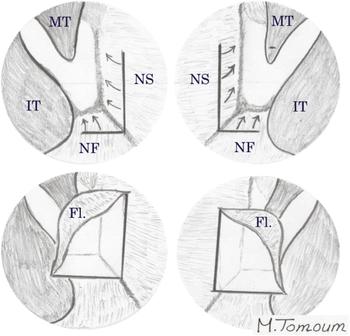
After application of adrenaline-soaked pledgets to the nasal cavity, laterally based septonasal flaps were elevated. This was done by creating a vertical incision at the cartilaginous/bony junction through septal mucosa about 1 cm anterior to the atretic plate, and then elevating the mucoperiosteal flap over the vomer and atretic plate using a round ear knife and ear scissors. Flap elevation was continued over the nasal floor (hence the name ‘L-shaped’). The same was done on the other side, creating a mirrored L-shaped septonasal flap, as shown in Figure 1.
Schematic diagram showing elevation of the mirrored L-shaped septonasal flap. MT = middle turbinate; IT = inferior turbinate; NS = nasal septum; NF = nasal floor; Fl. = flap

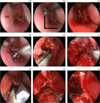
The atretic plate was then penetrated through its inferomedial portion, which is the safest part, using either an olive-tipped suction or a small curette (e.g. House curette). This penetration was then widened with 1-mm and 2-mm Kerrison forceps and straight circular-cutting punch forceps. The posterior-most part of the septum was also resected, using paediatric back biting forceps to widen the opening to the nasopharynx. This allowed for an endoscopic view bilaterally, and enabled the endoscope to be placed on one side and the instrument on the other, to facilitate bone removal. The newly formed choana was then widened laterally to the perpendicular plate of palatine bone and medial pterygoid plate, inferiorly to the palatine bone, and superiorly to the level of the nasopharynx roof. Finally, the elevated flap was laid down to cover most of the raw surfaces of the newly formed choana (Figure 2). Non-absorbable nasal packing and an oral airway were used for 1 day to keep the flap in place. No stenting was used.
(a) Endoscopic view of the choanal atresia. (b) L-shaped incision of the nasal septum and nasal floor made using a circular ear knife. (c) Elevation of the L-shaped septonasal flap. (d) Penetration of the septo-atretic plate through its inferomedial part using a House curette. (e) Removal of the most posterior part of the nasal septum using paediatric back biting forceps. (f) Straight circular-cutting punch forceps used to widen the newly formed choana. (g) Release of the septo-atretic flap using ear scissors. (h) & (i) The newly formed choana with the flap covering the superior and lateral raw surface, with visualisation of the soft palate. NS = nasal septum; MT = middle turbinate; AP = atretic plate; V = Vomer; FL = flap; SP = soft palate

In group B, the choana was enlarged by drilling the medial pterygoid plate and vomer instead of using cold instrumentation, with no attempts to preserve mucosal flaps. At the end of surgery, a nasal stent fashioned from a size 3.5 Portex® endotracheal tube was inserted and left for two to three weeks.
Post-operative care
The parents were instructed to irrigate the nose (and the stents in the second group) with saline drops and to carefully aspirate for one week after surgery. Parents were instructed to irrigate with saline twice daily, and then apply topical nasal steroid drops, for one month.
Post-operative assessment was carried out at monthly intervals for 6 months, and then at 12, 18 and 24 months post-operatively. This involved both subjective and objective assessment, via evaluation of nasal obstruction symptoms and nasal endoscopy, respectively.
Sample size, power and statistical analysis
Using a 2-sided Z test and alpha level of 0.05, the power analysis showed that recruiting 36 patients in each group would give 80 per cent power to detect a difference in the outcomes between both groups. We used simple randomisation to eliminate selection bias.
The data were analysed using SPSS version 20 statistical software (IBM, Armonk, New York, USA). Quantitative data were expressed as means ± standard deviations. Qualitative data were expressed as frequencies and percentages. Independent-samples t-test of significance was used for comparisons of two means. A chi-square test of significance was used to compare proportions between two qualitative parameters. P values of less than 0.05 were considered statistically significant.
Results
A total of 72 patients were included in our study, ranging from 3 to 10 days old; 25 patients were male and 47 were female. Thirteen patients (18.05 per cent) had associated malformations; of these, seven had ear anomalies, four had a genital defect and two had facial asymmetry.
Forty-two patients were randomly allocated to group A, and underwent endoscopic transnasal repair of choanal atresia using a mirrored L-shaped septonasal flap, without stenting or powered instrumentations. Thirty patients were randomly allocated to group B and underwent endoscopic transnasal repair using powered instrumentation (drilling) and stenting, without flaps. The difference in sample size between both groups resulted from using a simple randomisation method.
Based on radiological and intra-operative findings, 26 patients (36.11 per cent; 14 in group A and 12 in group B) had a pure bony plate, while 46 patients (63.89 per cent; 28 in group A and 18 in group B) had mixed bony and membranous components. There were no cases of isolated membranous atresia. All of our patients had bilateral choanal atresia.
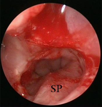
Surgery was considered successful if the patient showed no symptoms of airway obstruction. Objectively, the operation was considered successful if the new choana remained patent post-operatively during the follow-up period, as shown in Figure 3.
Six months post-operative endoscopic view of the choana after stentless endoscopic repair of bilateral congenital choanal atresia using the mirrored L-shaped flap technique. SP = soft palate

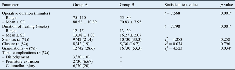
The average operative duration in group A was 88.52 ± 10.89 minutes, ranging from 75 to 110 minutes. The average operative duration in group B was 70.83 ± 7.95 minutes, ranging from 55 to 80 minutes. There was a significant difference between the two groups (p = 0.001).
The average follow-up period for all patients was 18.2 months, ranging from 8 to 24 months after surgery. Endoscopic assessment revealed a patent posterior choana in 34 out of 42 patients (81 per cent) in group A, and in 25 out of 30 patients (83.33 per cent) in group B, with an overall patency of 81.94 per cent. In both groups, failure was commonly evident in the first three to six months. Revision surgery was conducted for all failed cases (13 cases). This involved removal of granulation tissue and adhesions using cold instrumentation, with a patency rate of 76.93 per cent (10 out of 13 cases). The remaining three cases required a second revision surgery procedure, which was finally successful.
Choanal narrowing and stenosis occurred in 9 out of 42 patients (21.40 per cent) in group A, and in 10 out of 30 patients (33.33 per cent) in group B. There were no significant differences between the two treatment groups when comparing the incidence of closure and narrowing of the new choana between group A and group B (p > 0.05).
There were no major intra-operative complications, including spinal or skull base injuries. The average duration of healing in group A was 13.38 ± 1.03 weeks, ranging from 12 to 15 weeks. The average duration of healing in group B was 16.27 ± 2.07 weeks, ranging from 13 to 20 weeks. There was a significant difference between the two groups (p = 0.001).
Granulation tissue was observed in 12 out of 42 patients (28.6 per cent) in group A, compared to 16 out of 30 patients (53.3 per cent) in group B, with a significant difference between two groups (p = 0.034). Complications related to the stents – dislodgement, premature extrusion and columellar injury – were observed in 10 per cent, 6.67 per cent and 20 per cent of patients, respectively (Table I).
Comparison between endoscopic transnasal repair groups in bilateral congenital choanal atresia patients

Group A (42 patients) underwent endoscopic repair using a mirrored L-shaped flap without stenting. Group B (30 patients) underwent endoscopic repair using stenting without a flap. *Indicates statistical significance between groups. SD = standard deviation; χ2 = Chi-square test; t = t-test
Discussion
A variety of surgical approaches and techniques have been suggested for the repair of bilateral congenital choanal atresia, including traditional and endoscopic procedures. Traditional approaches include transnasal puncture, transpalatal repair and the sublabial transseptal approach.Reference Carpenter and Neel6, Reference Owens10 Stankiewicz was the first to describe endoscopic techniques for choanal atresia repair.Reference Stankiewicz11 The use of endoscopes has mostly replaced blind approaches and other techniques. It is preferred today because of the excellent visualisation and magnification it provides, which results in increased safety and reduced surgical time.
The two most common controversies regarding surgical repair of congenital bilateral choanal atresia are the utilisation of stenting after surgery and the creation of nasal flaps to cover the raw areas. The benefits versus the risks of stenting after repair have been debated in the otolaryngology literature for all airway procedures.Reference Schoem12 The risks related to stenting in congenital choanal atresia repair include circumferential pressure, leading to ischaemia, osteoblastic and fibroblastic reactions, and a lack of re-epithelisation in the choanal aperture, as well as pressure-related lesions of the columella and alar cartilages.Reference Pasquini, Sciarretta, Saggese, Cantaroni, Macri and Farneti13, Reference Khafagy14 Granulation tissue has been associated with a 30 per cent failure rate with the use of stents, regardless of the surgical approach.Reference Sharma, Lee, Gunasekaran, Knight and Bielby15
Regardless of the risks, some authors believe that stenting is necessary for all cases,Reference Osguthorpe, Singleton and Adkins16 while others believe that stenting is necessary only in bilateral cases.Reference Holzmann and Ruckstuhl17 Some authors recommend using long-term stents for at least three to four weeks,Reference Richardson and Osguthorpe18, Reference Samadi, Shah and Handler19 while others recommend using short-term stents for less than one week.Reference Pasquini, Sciarretta, Saggese, Cantaroni, Macri and Farneti13, Reference Van Den Abbeele, Francois and Narcy20
However, several publicationsReference Sharma, Lee, Gunasekaran, Knight and Bielby15, Reference Van Den Abbeele, Francois and Narcy20–Reference Durmaz, Tosun, Yldrm, Sahan, Kvrakdal and Gerek22 and a Cochrane reviewReference Cedin, Atallah, Andriolo, Cruz and Pignatari23 reported no difference in patency rates for patients who received or did not receive stenting after surgery. For example, Schoem argued that the use of a stent in choanal atresia repair seems unnecessary with the utilisation of endoscopes, high-speed protected drill bits and microdebriders.Reference Schoem12 He performed transnasal endoscopic repair of congenital choanal atresia in 13 patients without stents. Zuckerman et al. suggest that the placement of stents for bilateral choanal atresia repair may result in a higher rate of re-stenosis and the need for multiple subsequent procedures during infancy.Reference Zuckerman, Zapata and Sobol24 Cedin et al. reported that the avoidance of stenting allowed for fast recovery in one-stage surgery, with better long-term results, as it stimulates granulation tissue formation that frequently leads to stenosis.Reference Cedin, Fujita and Cruz25 Van Den Abbeele et al. treated their patients with very short-term stenting 2 days after surgery; they concluded that post-operative stenting is not necessary and may lead to the formation of granulation tissue and nasal synechia.Reference Van Den Abbeele, Francois and Narcy20 Other investigators have also reported the successful repair of congenital choanal atresia without stenting.Reference Gujrathi, Daniel, James and Forte26, Reference Shivakumar, Naik, Prashanth, Vishwanath and Praveen27
To the authors’ best knowledge, this is the largest prospective study comparing the outcomes of endoscopic repair of congenital bilateral choanal atresia using a flap technique without stenting versus endoscopic repair using stenting and powered instrumentation without a flap. In addition, we are the first authors to coin the term ‘mirrored L-shaped septonasal flap’ for this type of flap. Seventy-two patients with congenital bilateral choanal atresia were included in our study. We reported pure bony plate atresia in 8 cases (36.11 per cent), and mixed bony and membranous atresia in 14 cases (63.89 per cent), as compared to respective rates of 37 per cent and 63 per cent in Gosepath and colleagues’ study.Reference Gosepath, Santamaria, Lippert and Mann21
In our series, we performed revision surgery on 13 patients (18.05 per cent). This figure compares favourably with rates in the literature; various patency rates of 70–85 per cent after choanal atresia surgery have been reported.Reference Pasquini, Sciarretta, Saggese, Cantaroni, Macri and Farneti13, Reference Holzmann and Ruckstuhl17, Reference McLeod, Brooks and Mair28–Reference Gulsen, Baysal, Celenk, Aytac, Durucu and Kanlikama30
We believe that re-stenosis after endoscopic repair of congenital bilateral choanal atresia is largely due to granulation tissue formation from the raw areas, especially associated with excessive drilling. In group A, our technique depended mainly on mucosal preservation by utilising otological instruments, which is unique in the literature. Instrumentation included: otological scissors, a House curette, a circular knife, as well as 1-mm and 2-mm Kerrison forceps and straight circular-cutting punch forceps, to avoid the stripping of mucosa, with preservation of the harvested flap. We utilised a mirrored L-shaped septonasal flap to cover the raw areas, avoiding the formation of exuberant granulation tissue and scarring, in an effort to decrease the incidence of re-stenosis.
Our study supports the notion that stenting may be unnecessary in the endoscopic transnasal repair of congenital choanal atresia, especially when using septo-atretic flaps to cover denuded bone, in an effort to decrease the incidence of post-operative granulation tissue formation and synechiae. Our results compare favourably with prior reports using stents, with avoidance of associated complications, including discomfort, columellar necrosis and stimulation of granulation tissue formation.
• Congenital choanal atresia is a rare abnormality affecting 1 in 7000–8000 newborns, with a female predominance
• The transnasal endoscopic approach is the most popular for managing congenital choanal atresia
• Posterior nasal septum (‘vomer’) resection and mucoperiosteal flap creation prevent post-operative granulation tissue and stenosis
• Stent use is not mandatory for long-term patency of the newly formed choana
We recognise that a limitation of our study is the comparison of two different surgical techniques with the additional variable of stenting; however, in our hospital, we usually utilise stents if the flap technique is not employed. In this paper, we have tried to show that the outcomes of endoscopic repair of bilateral congenital choanal atresia using a mucoperiosteal flap without stenting are comparable to endoscopic repair with stenting, taking into consideration the avoidance of stent-related complications. The second limitation in our study is the use of the simple randomisation method, which explains the difference in sample size between the two groups. We recommend using the block randomisation method in the future research, which is designed to randomise participants into groups, and ensures a balance in sample size across groups over time.
Conclusion
The advent of less traumatic endoscopic techniques, miniaturised endoscopes, careful removal of the posterior septal/vomer segment, meticulous creation and preservation of intranasal flaps, and avoidance of stenting provide the best chance of success in congenital choanal atresia repair. A novel technique is presented, in which drilling and post-operative stenting are avoided. Specifically, conventional and otological instrumentation are favoured, with utilisation of a mirrored L-shaped mucoperiosteal flap to cover the raw surface of the newly formed choana, in order to avoid post-operative granulation tissue and stenosis. Outcomes were comparable to those reported in the literature in terms of immediate success, complication rates and longer-term patency.