INTRODUCTION
The actin cytoskeleton controls pivotal cellular processes such as motility, cytokinesis, cell-cell and cell-substrate interactions, vesicular and organelle transport, and the establishment and maintenance of cell morphology (Puius et al. Reference Puius, Mahoney and Almo1998; Paavilainen et al. Reference Paavilainen, Bertling, Falck and Lappalainen2004). Assembly and spatial organization of actin filaments are dynamic events regulated by a large repertoire of actin-binding proteins (Carlier, Reference Carlier1998; Disanza et al. Reference Disanza, Steffen, Hertzog, Frittoli, Rottner and Scita2005). A limited subset of these proteins is conserved from protozoa to higher eukaryotes. Importantly, the fundamental modular structure can be modified, shuffled, and combined to form multidomain proteins to carry out specific actin-dependent processes in higher eukaryotes (Puius et al. Reference Puius, Mahoney and Almo1998; Paavilainen et al. Reference Paavilainen, Bertling, Falck and Lappalainen2004).
A great deal of information about the actin cytoskeleton has been obtained mainly from studies involving budding yeasts and metazoan cells (Lanzetti et al. Reference Lanzetti, Di Fiore and Scita2001; Kim et al. Reference Kim, Galletta, Schmidt, Chang, Blumer and Cooper2006). However, very little is known about the actin cytoskeleton of protozoan parasites such as trypanosomatids (Gull et al. Reference Gull1999; De Souza, Reference De Souza2002). Actin and several actin-binding protein sequences are present in trypanosomatid genomes, but were not classified (El-Sayed et al. Reference El-Sayed, Myler, Blandin, Berriman, Crabtree, Aggarwal, Caler, Renauld, Worthey, Hertz-Fowler, Ghedin, Peacock, Bartholomeu, Haas, Tran, Wortman, Alsmark, Angiuoli, Anupama, Badger, Bringaud, Cadag, Carlton, Cerqueira, Creasy, Delcher, Djikeng, Embley, Hauser, Ivens, Kummerfeld, Pereira-Leal, Nilsson, Peterson, Salzberg, Shallom, Silva, Sundaram, Westenberger, White, Melville, Donelson, Andersson, Stuart and Hall2005b). Only recently, actin characterization has been addressed in T. brucei, which seems to play distinct roles along the parasite life-cycle. For instance, in bloodstream forms, actin co-localizes with the endocytic pathway, while in procyclic forms it is distributed throughout the cell (Garcia-Salcedo et al. Reference Garcia-Salcedo, Perez-Morga, Gijon, Dilbeck, Pays and Nolan2004). Depletion of actin expression by RNAi in bloodstream forms blocked endocytosis by interfering with vesicle traffic from the flagellar pocket and led to cell death, revealing that actin is an essential protein (Garcia-Salcedo et al. Reference Garcia-Salcedo, Perez-Morga, Gijon, Dilbeck, Pays and Nolan2004). Nevertheless, vesicle traffic in the opposite direction, delivering newly synthesized surface proteins to the flagellar pocket membrane, was not affected by actin depletion (Nolan and Garcia-Salcedo, Reference Nolan and Garcia-Salcedo2008). Leishmania donovani has an unconventional actin protein assuming a non-filamentous form. It was found not only in the flagellum, flagellar pocket, and nucleus but it was also associated with the kDNA network and subpellicular microtubules (Sahasrabuddhe et al. Reference Sahasrabuddhe, Bajpai and Gupta2004). Leishmania coronin, the only actin-binding protein identified till now in trypanosomatids, co-localizes with actin and, besides, its over-expression promotes enrichment of filament-like structures as well as in patches in the flagellar pocket region (Nayak et al. Reference Nayak, Sahasrabuddhe, Bajpai and Gupta2005). In fact, nothing is known about the dynamics of the actin cytoskeleton in trypanosomatids and no bona fide actin filaments have been observed to date.
In Trypanosoma cruzi, the chromosomal locus of actin, as well as the stability and processing of its cognate transcripts, has been extensively analysed (Cevallos et al. Reference Cevallos, Lopez-Villasenor, Espinosa, Herrera and Hernandez2003). However, the actin protein of T. cruzi has been poorly described, either by localization with antibodies produced against actin from other organisms (De Souza et al. Reference De Souza, Meza, Martinez-Palomo, Sabanero, Souto-Padron and Meirelles1983; Mortara, Reference Mortara1989), or by being the presumed target of cytochalasin in the inhibition of peroxidase (Bogitsh et al. Reference Bogitsh, Ribeiro-Rodrigues and Carter1995) of transferrin (Corrêa et al. Reference Corrêa, Atella, Batista and Soares2008) uptake. The exact subcellular localization remains uncertain. The complex gene repertoire of actin-binding proteins in T. cruzi and the identification of a Rho homologue, TcRho1 (Nepomuceno-Silva et al. Reference Nepomuceno-Silva, Yokoyama, de Mello, Mendonca, Paixao, Baron, Faye, Buckner, Van Voorhis, Gelb and Lopes2001; De Melo et al. Reference De Melo, Nepomuceno-Silva, Sant'Anna, Eisele, Ferraro, Meyer-Fernandes, de Souza, Cunha-e-Silva and Lopes2004, Reference De Melo, Eisele, Nepomuceno-Silva and Lopes2006), suggest the presence of an actin microfilament pathway in this parasite. In this study, the subcellular localization of the actin homologue of T. cruzi (TcActin) is demonstrated, and putative proteins capable of regulating the dynamics of the actin cytoskeleton in T. cruzi are investigated.
MATERIALS AND METHODS
Parasites
T. cruzi Dm28c epimastigotes were maintained at 28°C in LIT medium (liver infusion tryptose medium) (Camargo, Reference Camargo1964) and supplemented with 10% FBS (Cultilab) and 0·025 μg/ml hemin (Sigma-Aldrich). Bloodstream trypomastigote forms, derived from the blood of Swiss mice infected by intraperitoneal injection 7 days before, were used to infect the LLC-MK2 cell type. At 5–7 days after infection, the trypomastigotes released in the supernatant were purified by centrifugation. Intracellular amastigotes were obtained by mechanical disruption of the LLC-MK2 cell type after 4 days of infection with bloodstream trypomastigotes. These evolutive forms were later used for experimental procedures.
Cloning and expression of Trypanosoma cruzi actin
Primers were designed to amplify the actin alleles available from the GenBank (Accession numbers AF494294, and AF494295). The putative ORF of 1131 kb actin was amplified by PCR using the primers forward: 5′-CGGGATCCATGTCTGACGAAGAACAG-3′, and reverse: 5′-GCCTTAAGYTAAAAGCATTTGTTG-3′ and the genomic DNA as template. PCR products flanked by BamHI and EcoRI sites were cloned into a pCRTOPO2.1 vector (Invitrogen) and sequenced by way of the BigDye Terminator v1.1 Cycle Sequencing Kit (Applied Biosystems). In addition, the clones were subsequently subcloned into a pGEX-2TK vector (GE Healthcare) at the BamHI and EcoRI sites and then over-expressed in the Escherichia coli strain BL21 (DE3).
Actin polyclonal antibody
Polyclonal antiserum against the conserved N-terminal region present in all 3 T. cruzi actin alleles (SKKLFVGDEAQAKR) was raised and submitted to affinity purification using the same peptide coupled with CNBr-activated Sepharose 4 Fast Flow (GE Healthcare), as previously described (Nepomuceno-Silva et al. Reference Nepomuceno-Silva, Yokoyama, de Mello, Mendonca, Paixao, Baron, Faye, Buckner, Van Voorhis, Gelb and Lopes2001).
Western blotting
After 2 h of induction with 1 mm IPTG, E. coli strain BL21 (DE3) cultures were diluted and lysed, after which the expressed proteins were resolved on SDS-PAGE (12%) gels in parallel with 50 μg of proteins from Dm28c epimastigotes. Following transfer of the proteins to nitrocellulose, the membrane was incubated at room temperature for 1 h in blocking buffer: 150 mm NaCl, 10 mm Tris-HCl, pH 8·0, 5% (w/v) BSA, and 0·2% (v/v) Tween-20. The membrane was then incubated for 2 h with blocking buffer containing 0·1 μg/ml of anti-actin. For the negative control (peptide competition), anti-actin was pre-incubated overnight at 4°C with 150 μg of actin peptide (SKKLFVGDEAQAKR) or with the unrelated TcArf1 peptide (RIGKARQELEKMFTEDDVRNAVLLVF) (De Sá-Freire et al. Reference De Sá-Freire, Nepomuceno-Silva, da Paixão, de Mendonça, de Melo and Lopes2003). After 3 washings in blocking buffer, the nitrocellulose was incubated for 1 h with (1:2500) HRP-conjugated anti-rabbit IgG (Santa Cruz Biotechnology). The membrane was then washed 3 times with blocking buffer and revealed via the ECL kit (GE Healthcare).
Cytoskeleton preparations were obtained by treating parasites with 0·1%, 0·04%, or 0·02% Nonidet P-40 in PBS (150 mm NaCl, 10 mm sodium phosphate buffer, pH 7·2) at 4°C for 3 min. After centrifugation, soluble fractions were collected and the insoluble pellets were washed with chilled PBS. Soluble and insoluble fractions were electrophoresed, blotted, and immunolabelled either with (1:300) anti-TcActin or with (1:8000) anti-α-tubulin antibody (Clone b-5-1-1) (Sigma-Aldrich) followed by the (1:2500) HRP-conjugated anti-rabbit IgG or the (1:2500) HRP-conjugated anti-mouse IgG (Santa Cruz Biotechnology), respectively, and revealed via the ECL kit.
Immunofluorescence microscopy
Parasites were fixed in 4% (w/v) paraformaldehyde in PBS for 30 min at room temperature. After 2 washings in PBS, the cells were adhered to glass cover-slips pre-coated with 0·1% (w/v) poly-L-lysine in PBS for 30 min. The parasites were permeabilized with PBS containing 0·5% (v/v) Triton X-100 for 5 min, washed with PBS, and quenched for 30 min with 50 mm ammonium chloride in PBS. After blocking with 3% (w/v) BSA in PBS, samples were incubated for 1 h with a 1:1000 dilution of anti-TcActin. After washing, samples were incubated with goat anti-rabbit IgG Alexa Fluor 546 (Molecular Probes) at a 1:800 dilution. After extensive washing, the cover-slips were mounted onto glass microscope slides using 0·2 mn-propyl-gallate as anti-fade. Samples were examined using a Zeiss Axioplan epifluorescence microscope coupled with a CCD C5810 Hamamatsu camera. Images were processed by Adobe Photoshop CS (Adobe Systems, Inc.). To assay the specificity of antibody binding, 0·6 μg anti-actin was pre-incubated with 60 μg TcActin peptide or 60 μg of unrelated TcArf1 peptide in 3% (w/v) BSA in PBS overnight at 4°C before immunofluorescence assay. All images were captured as described above and processed under the same gain and sensitivity conditions.
Alignments and phylogenetic analysis
A large set of actin-binding protein sequences of mammals, yeasts, and protozoa was used as bait on BLAST searches of the T. cruzi genome database (GeneDB release v4.0 [http://www.genedb.org]) to identify putative orthologues. The hits with significant E-values were then obtained with successive PSI-BLAST searches (http://www.ncbi.nlm.nih.gov/blast) to confirm their conservation in T. cruzi. Searches against the PFAM database (http://www.ncbi.nlm.nih.gov/Structure/cdd/wrpsb.cgi) were also carried out to identify conserved domains in these sequences and to evaluate the significant scores. As a result, the hits found in T. cruzi were used as bait on BLAST searches to identify the reciprocal orthologues in L. major and T. brucei. In addition, BLAST searches with E-values <0·001 undertaken in D. discoideum and H. sapiens databases also facilitated estimation of the homologues with the best matches to these organisms. Percentages of identity compared to T. cruzi sequences were determined by ALIGN (http://www.ch.embnet.org/software/LALIGN_form.html) using default settings.
Sequence Accession numbers (GenBank)
The actin sequences utilized in alignments were previously designated: D. discoideum XP_637509, S. cerevisiae AAA34391, H. sapiens AAH15695, as well as the protozoan sequences of T. cruzi AAA62142, T. brucei XP_827205, L. major CAC22667, and P. falciparum NP_703241. Accession numbers of actin-binding proteins identified in T. cruzi: TcForminA: ABF13405 and allele ABF13407; TcForminB: ABF13406 and allele ABF13408; TcForminC: ABF13404; TcArp2: ABF13397; TcArp3: ABF13398; TcARPC1: ABF13399; TcARPC2: ABF13400; TcARPC3: ABF13401; TcARPC4: ABF13402; TcARPC5: ABF13403; TcProfilin: ABF13409; TcCofilin: ABF13410; TcTwinfilin: ABF13411; TcAIP1: ABF13413; TcSrv2: ABF13414; TcCapZ-β: ABF13412; TcCapZ-α isoform 1: ABF13415; TcCapZ-α isoform 2: ABF13416; TcCoronin: ABF58736; TcEB1: ABF58737.
Sequences of T. brucei and L. major identified and annotated by this study or sequences partially annotated by the genome project (El-Sayed et al. Reference El-Sayed, Myler, Blandin, Berriman, Crabtree, Aggarwal, Caler, Renauld, Worthey, Hertz-Fowler, Ghedin, Peacock, Bartholomeu, Haas, Tran, Wortman, Alsmark, Angiuoli, Anupama, Badger, Bringaud, Cadag, Carlton, Cerqueira, Creasy, Delcher, Djikeng, Embley, Hauser, Ivens, Kummerfeld, Pereira-Leal, Nilsson, Peterson, Salzberg, Shallom, Silva, Sundaram, Westenberger, White, Melville, Donelson, Andersson, Stuart and Hall2005b) as a member of a family and/or subunit of a complex were also submitted to GenBank. The Accession numbers of T. brucei sequences are: TbForminA: ABQ43156; TbForminB: ABQ43157; TbArp2: ABF58729; TbArp3: ABF58730; TbARPC1: ABF58731; TbARPC2: ABF58732; TbARPC3: ABF58733; TbARPC4: ABF58734; TbARPC5: ABF58735; TbAIP1: ABQ43158; TbSrv2: ABQ43159; TbEB1: ABQ43160, to L. major are: LmForminA: ABQ43161; LmForminB: ABQ43162; LmArp2: ABF58723; LmArp3: ABF58724; LmARPC1: ABF58725; LmARPC-like: ABF58726; LmARPC4: ABF58727; LmARPC5: ABF58728; LmAIP1: ABQ43163; LmSrv2: ABQ43164.
RESULTS
Structural features and subcellular localization
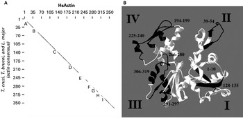
Structural properties of actin may be useful to explain the functional particularities of the T. cruzi actin (TcActin). The homologue of T. cruzi presents similar levels at 95%, 99%, 71%, 69%, and 86% with the eukaryotic actins of L. major, T. brucei, P. falciparum, D. discoideum, and H. sapiens, respectively. Comparison of a consensus sequence based on 3 trypanosomatids with the H. sapiens sequence showed that the most divergent regions corresponded to the following residues: 1–10, 39–54, 128–135, 194–199, 225–240, 266–280, 291–297, 306–319, 324–330 (see Fig. 1A and Supplementary Fig. 1 – in Online version only). Furthermore, the homology modelling method (Arnold et al. Reference Arnold, Bordoli, Kopp and Schwede2006) applied to TcActin showed that the overall structure was similar to eukaryotic actins (Sheterline and Sparrow, Reference Sheterline and Sparrow1994) and that it maintained a highly conserved hydrophobic cleft (Fig. 1B).
Comparison of the actin protein of trypanosomatids with the Homo sapiens protein. (A) Divergent regions in trypanosomatids. Multiple alignment of Trypanosoma cruzi actin with their homologues in Leishmania major, T. brucei, and H. sapiens was performed to construct a pairwise comparison of the trypanosomatid consensus sequence with the H. sapiens actin. Afterwards the divergent level was analysed by dot plot pairwise sequence comparison using a low threshold value of 5·33 with assistance of BioEdit 7.0 software. The X-axis values correspond to HsActin amino acids while the Y-axis values correspond to the trypanosomatid consensus. The most divergent regions are represented by lacunas in dot plot line (corresponding to letters A: 1–10; B: 39–54; C: 128–135; D: 194–199; E: 225–240; F: 266–280; G: 291–297; H: 306–319; I: 324–330). (B) T. cruzi actin monomer model. Homology modelling was carried out by SWISS-MODEL (Arnold et al. Reference Arnold, Bordoli, Kopp and Schwede2006). Structures in white correspond to conserved regions as compared to eukaryotic actins and structures in black with numbers correspond to divergent regions and gaps of residues, respectively. Numerals (I, II, III, IV) correspond to usual actin subdomains.

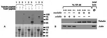
The specificity of the TcActin polyclonal antibody was assayed using Western blotting. The antibody recognized a polypeptide in the whole protein extract of the epimastigote forms, corresponding to the endogenous TcActin, of 42 kDa, and also the fusion protein GST-TcActin of 68 kDa expressed in E. coli (Fig. 2A). Accordingly, TcActin antiserum also recognized TcActin in the trypomastigote and amastigote forms (data not shown). While the competition with an unspecific peptide did not affect the binding of the TcActin antibody, previous competition with a specific peptide for TcActin abolished signal recognition (Fig. 2A).
Specificity of anti-TcActin antibody and cytoskeleton preparations by NP-40 lysis. (A) Antibody recognition is specific for TcActin. Upper panel: Western blotting with anti-TcActin plus competition with the non-specific peptide TcArf or the specific peptide TcActin; lower panel: SDS-PAGE mass control. (Lane 1): protein extract of E. coli pGEX2TK-TcActin not induced with IPTG; (lane 2): protein extract of E. coli pGEX2TK-TcActin induced with IPTG; (lane 3): protein extract of Trypanosoma cruzi Dm28c epimastigotes. Standard molecular weights in kDa are indicated at right. (B) T. cruzi actin is concentrated in a soluble fraction after lysis with different concentrations of NP-40. Anti-tubulin antibody was used as the cytoskeleton marker to identify the subpellicular microtubules enriched in the insoluble fraction. Lower panel: anti-actin Western blotting; upper panel: anti-tubulin Western blotting with a heterologous anti-α-tubulin antibody. Lane with total lysis corresponds to parasites lysed directly in Laemmli buffer [60 mm Tris-HCl, pH 6·8, 10% glycerol, 2% SDS, 1% 2-mercaptoethanol and 0·002% bromophenol blue].

To determine actin distribution in T. cruzi, cytoskeleton preparations were prepared by extraction with increasing detergent concentrations (Schneider et al. Reference Schneider, Plessmann and Weber1997; Scott et al. Reference Scott, Sherwin and Gull1997). As a result, parasite protein extracts prepared with 0·02%, 0·04%, or 0·1% NP-40 in PBS (Fig. 2B) revealed the presence of actin only in the cytoplasmic-soluble fractions and its complete absence in the insoluble fraction, which was enriched with nuclei, kinetoplasts and subpellicular microtubules (as observed by electron microscopy, data not shown).
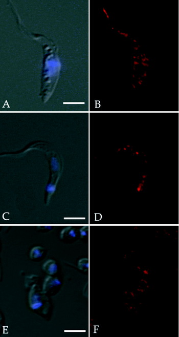
The subcellular localization of TcActin in the different evolutive forms of T. cruzi analysed by immunofluorescence revealed many rounded and punctuated structures similar to patches distributed throughout the cytoplasm. Pointedly, no pattern differences could be found within epimastigotes, trypomastigotes, or amastigotes (Fig. 3). No association was observed with the subpellicular microtubules or with the nuclear or kinetoplast DNA. To demonstrate the antibody-binding specificity, epimastigotes were probed with anti-TcActin previously incubated with TcActin peptide or an unrelated peptide (TcArf1). Incubation with the specific peptide completely abolished TcActin staining while incubation with an unrelated peptide did not modify the antibody signal or distribution (see Supplementary Fig. 2 – in Online version only).
Trypanosoma cruzi actin localization. Epimastigote (A, B), trypomastigote (C, D) and amastigote (E, F) forms of T. cruzi were stained with polyclonal antiserum against TcActin. Fluorescent patches spread all over the parasite cell bodies were clearly observed in all T. cruzi stages. DAPI staining revealed the position of the nucleus and kinetoplast. DIC staining (A, C, E); immunofluorescence micrographs (B, D, F). Scale bars=4 μm.

Prediction of actin-binding proteins
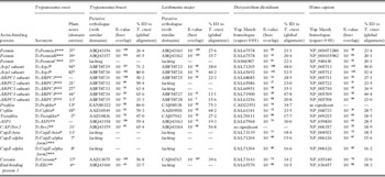
An extensive genome-wide search to identify the ubiquitous proteins regulating the nucleation, treadmilling, filament stability, and monomer sequestration (Carlier, Reference Carlier1998; Paavilainen et al. Reference Paavilainen, Bertling, Falck and Lappalainen2004; Disanza et al. Reference Disanza, Steffen, Hertzog, Frittoli, Rottner and Scita2005) in T. cruzi was carried out (Table 1). Some identified actin-binding proteins have been annotated in the genome project (El-Sayed et al. Reference El-Sayed, Myler, Bartholomeu, Nilsson, Aggarwal, Tran, Ghedin, Worthey, Delcher, Blandin, Westenberger, Caler, Cerqueira, Branche, Haas, Anupama, Arner, Aslund, Attipoe, Bontempi, Bringaud, Burton, Cadag, Campbell, Carrington, Crabtree, Darban, da Silveira, de Jong, Edwards, Englund, Fazelina, Feldblyum, Ferella, Frasch, Gull, Horn, Hou, Huang, Kindlund, Klingbeil, Kluge, Koo, Lacerda, Levin, Lorenzi, Louie, Machado, McCulloch, McKenna, Mizuno, Mottram, Nelson, Ochaya, Osoegawa, Pai, Parsons, Pentony, Pettersson, Pop, Ramirez, Rinta, Robertson, Salzberg, Sanchez, Seyler, Sharma, Shetty, Simpson, Sisk, Tammi, Tarleton, Teixeira, Van Aken, Vogt, Ward, Wickstead, Wortman, White, Fraser, Stuart and Andersson2005a, Reference El-Sayed, Myler, Blandin, Berriman, Crabtree, Aggarwal, Caler, Renauld, Worthey, Hertz-Fowler, Ghedin, Peacock, Bartholomeu, Haas, Tran, Wortman, Alsmark, Angiuoli, Anupama, Badger, Bringaud, Cadag, Carlton, Cerqueira, Creasy, Delcher, Djikeng, Embley, Hauser, Ivens, Kummerfeld, Pereira-Leal, Nilsson, Peterson, Salzberg, Shallom, Silva, Sundaram, Westenberger, White, Melville, Donelson, Andersson, Stuart and Hallb). However, our genome-wide search has expanded this analysis revealing new members and classifying those partially annotated. Once the actin-binding proteins were identified in T. cruzi, searches were extended to T. brucei and L. major, revealing an even greater number of conserved orthologues (Table 1). On the other hand, absent members in T. brucei and L. major included orthologues of ForminC and the 2 subunits of CapZ: CapZα and CapZβ. In addition, orthologues for EB1 and the subunits ARPC2 and ARPC3 of the Arp2/3 complex were not present in L. major (Table 1).
Actin-binding protein analysis
(The proteins analysed can be divided into 3 groups: those already annotated in the genome project (*) (El-Sayed et al. Reference El-Sayed, Myler, Bartholomeu, Nilsson, Aggarwal, Tran, Ghedin, Worthey, Delcher, Blandin, Westenberger, Caler, Cerqueira, Branche, Haas, Anupama, Arner, Aslund, Attipoe, Bontempi, Bringaud, Burton, Cadag, Campbell, Carrington, Crabtree, Darban, da Silveira, de Jong, Edwards, Englund, Fazelina, Feldblyum, Ferella, Frasch, Gull, Horn, Hou, Huang, Kindlund, Klingbeil, Kluge, Koo, Lacerda, Levin, Lorenzi, Louie, Machado, McCulloch, McKenna, Mizuno, Mottram, Nelson, Ochaya, Osoegawa, Pai, Parsons, Pentony, Pettersson, Pop, Ramirez, Rinta, Robertson, Salzberg, Sanchez, Seyler, Sharma, Shetty, Simpson, Sisk, Tammi, Tarleton, Teixeira, Van Aken, Vogt, Ward, Wickstead, Wortman, White, Fraser, Stuart and Andersson2005a, Reference El-Sayed, Myler, Blandin, Berriman, Crabtree, Aggarwal, Caler, Renauld, Worthey, Hertz-Fowler, Ghedin, Peacock, Bartholomeu, Haas, Tran, Wortman, Alsmark, Angiuoli, Anupama, Badger, Bringaud, Cadag, Carlton, Cerqueira, Creasy, Delcher, Djikeng, Embley, Hauser, Ivens, Kummerfeld, Pereira-Leal, Nilsson, Peterson, Salzberg, Shallom, Silva, Sundaram, Westenberger, White, Melville, Donelson, Andersson, Stuart and Hallb), those identified and annotated in this study (**), and those partially annotated in the genome project as a member of a family and/or subunit of a complex (***). Uppercase letters on the Pfam score column correspond to domains of the Pfam database: a – 02181, b – 00022, c – 04045, d – 04062, e – 05856, f – 04699, g – 00235, h – 00241, i – 01213, j – 01115, k – 01267, l – 08954, m – 03271. GenBank Accession numbers for Trypanosoma brucei and Leishmania major correspond with accuracy to the orthologues of the proteins identified in T. cruzi; the Accession numbers for Dictyostelium discoideum and Homo sapiens correspond to the sequences with the best overlaps but that are not necessarily the correct orthologues of the proteins identified in T. cruzi.)

Most eukaryotic cells employ the Arp2/3 complex as the major nucleator of actin polymerization, which regulates the propulsive force at the leading edge (Pollard and Beltzner, Reference Pollard and Beltzner2002; Weaver et al. Reference Weaver, Young, Lee and Cooper2003; Zigmond, Reference Zigmond2004). Arp2/3 complex is composed of 7 subunits, known as Arp2, Arp3, ARPC1/p41, ARPC2/p34, ARPC3/p21, ARPC4/p20, and ARPC5/p16 (Welch et al. Reference Welch, DePace, Verma, Iwamatsu and Mitchison1997; Cvrckova et al. Reference Cvrckova, Rivero and Bavlnka2004). Our genome-wide BLAST search revealed that T. cruzi encodes a canonical Arp2/3 complex with the presence of all subunits, appointed as TcArp2, TcArp3, TcARPC1, TcARPC2, TcARPC3, TcARPC4, and TcARPC5 (Table 1). Similar BLAST searches in T. brucei and L. major revealed the presence of orthologues of all the subunits in L. major, except for ARPC2 and ARPC3 (Table 1). Additional searches revealed the presence in L. major of an atypical ARPC (GenBank Accession number: ABF58726) which, by phylogenetic analyses, did not group with any ARPCs clades and was appointed as LmARPC-like (see Supplementary Fig. 3 – in Online version only). In addition, when compared with the subunits of the Arp2/3 complex of T. cruzi, the L. major subunits were much less conserved than their equivalents in T. brucei, as verified by BLAST E-values (Table 1).
Formins identified in T. cruzi were nominated in accordance with their phylogenetic grouping: ForminA and ForminB were conserved in the three trypanosomatids and ForminC was exclusive of T. cruzi (Table 1 and Supplementary figures – in Online version only). The main proteins performing actin treadmilling (polymerization/depolymerization) (Balcer et al. Reference Balcer, Goodman, Rodal, Smith, Kugler, Heuser and Goode2003; Paavilainen et al. Reference Paavilainen, Bertling, Falck and Lappalainen2004; Cvrckova et al. Reference Cvrckova, Rivero and Bavlnka2004) (Fig. 2) were also found in T. cruzi. The identified orthologues were: ADF/cofilin, twinfilin, profilin, AIP, CAP/Srv2, CapZα, and CapZβ (Table 1).
Comparative genomics also revealed that ubiquitous proteins responsible for filament bundling and cross-linking are not well distributed in T. cruzi. These proteins contain an actin-binding domain (ABD), a prototype that interacts with F-actin, composed of 2 calponin homology (CH) domains of 100aa each, arranged in tandem (Puius et al. Reference Puius, Mahoney and Almo1998; Stradal et al. Reference Stradal, Kranewitter, Winder and Gimona1998), however, only 2 entries related to proteins containing the ABD domain were present in T. cruzi: TcCoronin and TcEB1 (Table 1). Cross-linking proteins absent in T. cruzi include monomeric proteins like fimbrin, and dimeric proteins like α-actinin, β-spectrin, and filamin (Stradal et al. Reference Stradal, Kranewitter, Winder and Gimona1998). Notable absences also include gelsolin, a filament-severing protein, tropomyosin, a filament-stabilizing protein and spire, an actin nucleator. WH2 domain containing proteins, an actin monomer-binding motif, that includes β-thymosin, ciboulot, WASP, and N-WASP (Paunola et al. Reference Paunola, Mattila and Lappalainen2002) are also absent.
DISCUSSION
Previous studies describing subcellular localization of actin in T. cruzi were performed using antibodies produced against actin from other organisms (rabbit muscle by Mortara, Reference Mortara1989, and Entamoeba histolytica by De Souza et al. Reference De Souza, Meza, Martinez-Palomo, Sabanero, Souto-Padron and Meirelles1983). In contrast, in the present work antibodies were produced against the conserved N-terminal region present in all 3 T. cruzi actin alleles available from the GenBank.
The authors of those previous papers have only demonstrated that the immunofluorescence signal was not non-specific (due to direct binding of secondary antibody) because at that time they could not accurately test whether the primary antibody target was really actin.
Mortara (Reference Mortara1989) found a polypeptide of 43 kDa that remained associated with the insoluble fraction after detergent extraction. The identity of this protein was assayed using the actin property of binding to DNAse I, without success. The antibody used by De Souza and colleagues in1983 recognized T. cruzi flagella and did not show any signal in the parasite cell body, raising the hypothesis of the presence of actin at the paraflagellar rod (PFR). Subsequent work from many laboratories, including that of the authors, did not find actin in purified PFR fractions. Recently, a comprehensive proteomic analysis of T. brucei flagellum (Broadhead et al. Reference Broadhead, Dawe, Farr, Griffiths, Hart, Portman, Shaw, Ginger, Gaskell, McKean and Gull2006) also did not find actin.
By observing the structural prediction of T. cruzi actin, we can conclude that most amino-acid changes, when compared to other eukaryotic actin structures, are concentrated on the monomer surface, in most cases on the loops responsible for monomer-monomer interaction during oligomerization or interaction with actin-binding proteins. This peculiarity suggests the existence of species-specific differences that could be better deciphered by investigation of the subcellular localization and identification of the machinery responsible for actin cytoskeletal control in T. cruzi.
A similar pattern and localization, with patch-like structures distributed throughout the parasitic cytoplasm, was also present in other trypanosomatids, particularly among the procyclic forms of T. brucei (Garcia-Salcedo et al. Reference Garcia-Salcedo, Perez-Morga, Gijon, Dilbeck, Pays and Nolan2004). In Leishmania donovani promastigotes, actin was also equally distributed in patches throughout the cell (Sahasrabuddhe et al. Reference Sahasrabuddhe, Bajpai and Gupta2004). However, using detergent extracts and immunofluorescence we did not find TcActin associated with either subpellicular microtubules or with DNA in the nucleus or kinetoplast.
After overexpression of coronin, an actin-binding protein conserved in L. donovani, short filaments/bundles have been enriched (Nayak et al. Reference Nayak, Sahasrabuddhe, Bajpai and Gupta2005). In L. donovani, treatment with the non-ionic detergent NP-40 revealed that interactions between actin and coronin were unstable, weak and limited, after treatment, only to patches in the flagellar pocket (Nayak et al. Reference Nayak, Sahasrabuddhe, Bajpai and Gupta2005). In addition, the presence of actin only in the detergent-soluble fraction of T. cruzi corroborates with the actin-microtubule dissociation observed by immunofluorescence (data not shown).
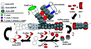
Genome-wide searches, carried out to identify the actin-biding proteins of trypanosomatids, revealed that, compared to the higher eukaryotes, trypanosomatids exhibit a set of actin-binding proteins that, while limited, is sufficient to theoretically carry out actin filament assembly and disassembly (Fig. 4). In particular, the main proteins responsible for actin treadmilling and actin nucleation were shown to be fully or partially conserved.
Actin treadmilling (polymerization/depolymerization) is performed by multiple coordinated proteins. The colours of the actin-binding proteins represent the conservation levels found in trypanosomatids. Signals represent the barbed end (+) and the pointed end (−) of the filaments. (1) AIP1 and cofilin physically co-operate to promote depolymerization. Cofilin preferentially binds to the actin-ADP of mature filaments by severing them and producing pointed ends. AIP1 associates with the new barbed ends on the filaments recently severed by cofilin, blocking the reannealing, thereby enhancing actin depolymerization. Twinfilin also binds to actin-ADP and provides a cytoplasmatic reservoir of monomers. (2) The severing promoted by cofilin rapidly leads to the accumulation of cofilin bound to actin-ADP monomers. (3) Profilin and CAP/Srv2 act during successive rounds of severing/depolymerization, enhancing the exchange of actin-bound ADP with ATP. CAP/Srv2 interacts with actin-ADP monomers and catalytically liberates them for nucleotide exchange by recycling cofilin and promoting barbed end depolymerization. (4) Nucleotide exchange, ATP for ADP, enhanced the binding of profilin to monomers. Profilin actin-ATP-bound monomers act at uncapped barbed ends during new rounds of actin assembly. (5) The capping protein CapZα/β binds to the barbed ends and restricts polymerization and filament length, thereby stabilizing the barbed ends. (6) The cross-linking proteins EB1 and coronin act out their role as bridges between microtubules.

When compared with the subunits of the Arp2/3 complex of T. cruzi, the L. major subunits were much less conserved than their equivalents in T. brucei, as verified by BLAST E-values, suggesting the possible absence of a functional Arp2/3 complex in L. major, that could be complemented by other actin nucleators, such as formins. All the formin homologues identified contained the characteristic juxtaposed domain arrangement of formins: the proline-rich FH1 domain and the FH2 domain related with the actin nucleation (data not shown). However, neither the FH3 nor the Rho-binding domains (GBD) in conjunction with the encompassed C-terminal Dia-autoregulatory domain (DAD), characteristic of the Diaphanous-related formins (DRFs), were found (data not shown). Rho GTPase binding to the GBD domain relieves intramolecular auto-inhibitory interaction (Zigmond, Reference Zigmond2004). The absence of these domains in trypanosomatids suggests either regulation by unknown motifs or the existence of constitutively active formins. As observed in other models, over-expression of formins lacking the GBD domain creates a constitutively active form (Evangelista et al. Reference Evangelista, Zigmond and Boone2003).
The presence of the major actin nucleators, formins and Arp2/3 complex, suggests that T. cruzi had somehow maintained its capacity to polymerize filaments during the life-cycle, either in a branched network at a 70° angle polymerized by the Arp2/3complex or in the actin linear bundles polymerized by formins (Paavilainen et al. Reference Paavilainen, Bertling, Falck and Lappalainen2004).
Together, the proteins responsible for actin treadmilling present conspicuous functional roles in other eukaryotes. Profilin is responsible for the recruitment of actin-ATP monomers for barbed ends and consequent polymerization. In previous work, T. brucei profilin complemented a profilin-deletion mutant of S. cerevisiae (Wilson and Seebeck, Reference Wilson and Seebeck1997). ADF/cofilins are implicated in actin turnover through filament severing, with pointed-end depolymerization and consequent cytoplasmatic sequestration of monomers (Fig. 4). CAP/Srv2 helps in the ADP/ATP nucleotide exchange between actin-ADP associated with ADF/cofilin and actin-ATP associated with profilin (Balcer et al. Reference Balcer, Goodman, Rodal, Smith, Kugler, Heuser and Goode2003; Paavilainen et al. Reference Paavilainen, Bertling, Falck and Lappalainen2004). Conversely, AIP1 stabilizes the recently severed filaments by ADF/cofilin, benefiting the disassembly mediated by ADF/cofilin (Ono, Reference Ono2003).
T. cruzi cofilin, in particular, contains divergent residues in the F-actin-binding region, as previously seen with respect to their orthologues in the apicomplexan parasites (Baum et al. Reference Baum, Papenfuss, Baum, Speed and Cowman2006). An unusual feature of T. cruzi cofilin is a conserved N-terminal serine-4 residue. In mammalian cells, ADF/cofilin serine-3 is phosphorylated by enzymes such as LIM-kinase, promoting deactivation and inhibition of its association with F-actin (Paavilainen et al. Reference Paavilainen, Bertling, Falck and Lappalainen2004). A probable LIM-kinase homologue is encoded by T. cruzi (GenBank Accession number: XP_813098). However, other regulators such as cofilin phosphatases (Huang, Reference Huang, DerMardirossian and Bokoch2006) are not encoded by T. cruzi. Capping proteins, CapZα/β, associate with barbed ends to control the access of monomers and filament length (Wear and Cooper, Reference Wear and Cooper2004). The conservation of CapZα/β only in T. cruzi suggests that in L. major and T. brucei an easier access of monomers to barbed ends could result in an increase of instability of the mature filaments, and favour actin turnover.
The reasons for the known difficulty in recognizing actin filaments in trypanosomatids like T. cruzi remain unknown. Our study suggested that alterations in actin treadmilling might be involved. First, in the event of highly active TcCofilin, it could sever and continuously depolymerize the filaments. Second, TcCapZ-α or TcCapZ-β, having different association properties with respect to barbed ends could possibly modify actin turnover, while a deficient TcProfilin might be blocking actin-ATP monomers available for polymerization.
Moreover, the existence in T. cruzi of 2 entries related with proteins containing the ABD domain, TcCoronin and TcEB1, that in higher eukaryotes act as molecular bridges between microtubules and microfilaments, suggests that actin in T. cruzi can exist in the filamentous form. The lack of the cross-linking protein EB1 in L. major could enhance coronin protein activity upon the cytoskeleton, overloading its role as a molecular bridge, otherwise played by both. This may explain the extremely rapid increase in filament-like structures that followed the over-expression of a coronin homologue in L. major (Nayak et al. Reference Nayak, Sahasrabuddhe, Bajpai and Gupta2005). This limited protein diversity with an ABD domain may favour poorly compacted and unstable filaments, which might be more susceptible to physical stress, making observation more difficult, as in fact is the case.
Other absences also include WH2 domain-containing proteins, an actin monomer-binding motif found in proteins such as β-thymosin, ciboulot, WASP, and N-WASP (Paunola et al. Reference Paunola, Mattila and Lappalainen2002), that suggest that the evolutionary dispersion of the WH2 domain subsequently occurred in animal and fungal lineages.
In brief, description in T. cruzi of monomer-binding proteins and F-actin-binding proteins, besides the actin nucleators, clearly reveals the existence of the machinery responsible for actin cytoskeleton control in these protozoa. Moreover, T. cruzi also takes advantage of species-specific proteins whose motifs and domains also remain unknown.
Conclusions
T. cruzi, L. major, and T. brucei actins present unusual features. F-actin was not observed, possibly due to filament instability, deficient assembly, and accentuated severing, events that are not yet clearly understood. Therefore, description of the molecular repertoire of actin-binding proteins by comparative genomics may aid the understanding of how this repertoire acts on the actin cytoskeleton, fomenting its unusual characteristics. In addition, adequate functional approaches to each key actin regulator are also in need of development. Future investigation of the upstream regulatory proteins coupling T. cruzi actin with its probable key regulator TcRho1, the Rho GTPase orthologue in T. cruzi (Nepomuceno-Silva et al. Reference Nepomuceno-Silva, Yokoyama, de Mello, Mendonca, Paixao, Baron, Faye, Buckner, Van Voorhis, Gelb and Lopes2001; De Melo et al. Reference De Melo, Nepomuceno-Silva, Sant'Anna, Eisele, Ferraro, Meyer-Fernandes, de Souza, Cunha-e-Silva and Lopes2004, Reference De Melo, Eisele, Nepomuceno-Silva and Lopes2006), will almost certainly disclose the pathways connecting the extracellular signals with actin dynamics, as occurs in higher eukaryotes by encoding Rho GTPases proteins. Additional assays with lineages of T. cruzi expressing mutants of this Rho orthologue are being done to allow for a better comprehension of the roles actin plays in T. cruzi. Moreover, novel targets for drug development are needed to control trypanosomiasis. Given the important roles of the actin cytoskeleton observed in other eukaryotes, it is probable that a blockade of key regulatory steps should give rise to severe phenotypes in these parasites.
This research received financial support from PRONEX, CNPQ, FAPERJ, and IAEA. The authors are grateful to Paulo C. da Silva and Antonio Bosco Carlos for their technical assistance, and to Judy Grevan for editing the text.