Implications
The male effect in sheep was first reported over 60 years ago, but our understanding of how this phenomenon works and in particular the socio-sexual stimuli involved remains limited. This study highlights the sensitivity of ewes to visual signals from rams and thus complete visual isolation, in addition to isolation from olfactory signals, is recommended for experiments and practical application of the male effect in sheep.
Introduction
The neuroendocrine response of sheep to a prospective mate is termed the ‘male effect’ in ewes and the ‘female effect’ in rams. In both cases, exposure to the opposite sex causes a dramatic increase in the secretion of luteinising hormone (LH), leading to stimulation of the reproductive system that, for females especially, can profoundly change their reproductive status (review; Walkden-Brown et al., Reference Walkden-Brown, Martin and Restall1999). Relatively little is known about the socio-sexual signals involved in the female effect (Gonzalez et al., Reference Gonzalez, Levy, Orgeur, Poindron and Signoret1991), but it is clear that the male effect is predominantly mediated by olfactory cues, with non-olfactory cues playing a synergistic or substitutive role (Gelez and Fabre-Nys, Reference Gelez and Fabre-Nys2004). Thus, the proportion of ewes ovulating in response to the male effect is maximised when ewes are exposed to the full complement of socio-sexual stimuli from rams (Pearce and Oldham, Reference Pearce and Oldham1988; Perkins and Fitzgerald, Reference Perkins and Fitzgerald1994) and anosmic ewes have an LH response to rams that is comparable to that observed in intact ewes (Cohen-Tannoudji et al., Reference Cohen-Tannoudji, Locatelli and Signoret1986).
Visual stimuli undoubtedly play an important role in the social structure of sheep flocks (Piggins, Reference Piggins1992). In fact, studies with projected visual images have shown that sheep use visual information to differentiate between familiar and unfamiliar conspecifics, the presence or absence of horns, and stressed or calm animals (review; Tate et al., Reference Tate, Fischer, Leigh and Kendrick2006). Importantly for the present study, female sheep are able to distinguish between the faces of male and female sheep and, when they are sexually receptive, actively select male images (Kendrick et al., Reference Kendrick, Atkins, Hinton, Broad, Fabre-Nys and Keverne1995). Auditory stimuli may also play a role in male–female communication because rams make a low-pitched vocalisation when courting an oestrous ewe (Banks, Reference Banks1964).
To determine the importance of audio–visual and visual stimuli in the ‘male’ and ‘female’ effects, we tested whether exposure of ewes and rams to visual or audio–visual images of a prospective mate would evoke an increase in LH secretion.
Material and methods
These experiments were carried out in accordance with the Australian Code of Practice for the Care and Use of Animals for Scientific Purposes (7th Edition, 2004) and were approved by the Animal Ethics Committee of The University of Western Australia (RA05/100/483).
Experiment 1: Auditory and/or visual stimuli and the male effect
Animals and experimental procedures
Adult Merino ewes that had been previously isolated from ram contact (not within 500 m for a minimum of 2 months) were housed in an animal facility at the University of Western Australia, Perth, Australia (31°58′S) during the non-breeding season. Ewes were studied during the non-breeding season to optimise the possibility of a detectable response. The ewes were fed at maintenance and housed in groups under natural light (9.5L : 14.5D). Blood was sampled twice weekly for 2 weeks prior to the experiment to monitor the concentration of progesterone.
Two weeks before the experiment began, groups of eight ewes were allocated to treatments, with each group balanced for age (6 years) and weight: Ram (55 ± 1.7 kg); Visual (50 ± 2.5 kg); Audio–visual (55 ± 1.1 kg) and Control (55 ± 1.9 kg). All the four groups were kept in the same animal house and exposed to their stimulus midway through a frequent blood-sampling regimen: the ‘Ram’ group was exposed to two adult, sexually experienced, Merino rams; the ‘Visual’ group was exposed to still images of Merino rams; the ‘Audio–visual’ group was exposed to a video of Merino ewes and rams mating. The ‘Control’ group remained isolated from ram stimuli for the duration of the experiment.
On the day of treatment, blood was sampled every 12 min for 6 h before and 6 h after exposure to the stimulus. The treatment days were staggered to ensure that no group was affected by stimuli from another group: Control ewes were sampled on Day 0; Visual ewes on Day 1; Audio–visual ewes on Day 4 and Ram ewes on Day 6. On the day of exposure to a chosen stimulus, the ewes that were not being treated were moved from the animal house to a paddock out of auditory and visual range (>500 m).
Means of exposure to stimuli
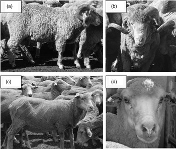
The still images of rams (n = 6) were loaded into presentation software and projected onto a 1.8 m × 1.5 m screen. The images included both frontal and side-on views of adult, Merino rams from the University Farm (Figure 1). Each image remained on the screen for 2 min and was scaled to ensure that it was life-size. The projected images rotated continuously for the second half of the blood-sampling regimen. The audio–visual stimulus was recorded at the University Farm using three sexually active Merino rams and two oestrous Merino ewes. The video was edited so that each 5 min loop contained an optimal combination of frequent displays of oestrous behaviour by the ewes and mating activity by the rams (about 2.5 mounts/min). After editing, the video was loaded onto a computer video player, sized and projected onto a screen as outlined above for the ‘Visual’ ewes. The auditory recording of rams was optimised using an external speaker system connected to the computer. The audio–visual stimulus played continuously for the second half of the blood-sampling regimen. The ewes did appear to be aware of both the audio and visual stimuli but no formal observations were made of their behaviour. For the positive controls, the Ram ewes, two adult, sexually experienced Merino rams, were introduced midway through the blood-sampling regimen and had full contact with the ewes for the remainder of the experiment. Control ewes remained isolated from all ram stimuli for the duration of the blood-sampling regimen.
Examples of the still images of rams used in Experiment 1 ((a) and (b)) and of ewes used in Experiment 2 ((c) and (d)).

Experiment 2: Audio–visual stimuli and the female effect
Animals and experimental procedures
During the breeding season, adult, sexually active, Merino rams (n = 14) and ewes (n = 8) were housed in an animal facility at the University of Western Australia, Perth, Australia (31°58′S). The rams were fed at maintenance in individual pens under a lighting regimen that paralleled the changes in day length occurring outside (11L : 13D). The rams were studied during the breeding season to optimise the probability of a detectable response. The ewes were also fed at maintenance in individual pens but kept in a separate room under the same lighting regimen as the rams.
Two weeks before the experiment, rams were allocated to two treatment groups that were balanced for age and live weight, and then housed in individual pens in separate rooms that prevented any auditory or visual contact between them. Exposure rams (2.5 ± 0.3 years; 66 ± 5.1 kg; n = 7) were subjected to two types of exposure midway through a frequent blood-sampling regimen. The first was still images of ewes accompanied by the sound of ewes and rams mating (audio–visual exposure). To optimise the likelihood of detecting a response, we used a combination of still images of ewes and sound used in Experiment 1. The rams did appear to be aware of the audio and visual stimuli but, as with Experiment 1, no formal observations were made of their behaviour. After a week of isolation from ewe stimuli, the Exposure rams underwent their second exposure period, where they were exposed to oestrous ewes (ewe exposure). Control rams (2.6 ± 0.3 years; 66 ± 5.5 kg; n = 7) remained isolated from ewe stimuli for the duration of the experiment. Blood was sampled every 15 min for 6 h before and 6 h after stimulus exposure. Control rams were sampled at the same frequency but remained isolated from ewe stimuli.
Image projection and ewe exposure
During the audio–visual exposure, rams were exposed simultaneously to still images of ewes and the sound of ewes and rams mating. We used this combination of stimuli to optimise the likelihood of detecting a response as the ewes in Experiment 1 did not appear to respond to a moving visual image. The images included both frontal and side-on views of Merino ewes (n = 6) selected from the breeding flock at the University Farm (Figure 1). The images were scaled to about life-size, loaded into presentation software and projected onto a 1.8 m × 1.8 m screen. Images were rotated continuously during the second half of the blood-sampling regimen, with each image remaining on the screen for 2 min. Each screened image was accompanied by 1 min of mating sounds followed by 1 min of silence. On the day of ewe exposure, each ram was given fence-line exposure for 2 h with at least one oestrous ewe. The Control rams remained isolated from ewe stimuli throughout the experiment.
Blood processing and immunoassay
All blood samples were centrifuged immediately at 3000 r.p.m. and plasma was decanted into duplicate plastic tubes, and stored at −20°C until analysis. Plasma LH was measured in duplicate by a double-antibody radioimmunoassay (Martin et al., Reference Martin, Oldham and Lindsay1980) using ovine LH (NIDDK-oLH-1-4 (AFP-8614B) for iodination and standards kindly supplied by A. Parlow, National Hormone and Pituitary Program, NIDDK, Torrance, CA, USA. The limit of detection of the assay was 0.06 ng/ml. Quality control samples for Experiment 1 (0.5, 0.9 and 1.6 ng/ml) and Experiment 2 (0.6, 1.0 and 1.9 ng/ml) were used to estimate intra-assay coefficients of variation (Experiment 1: 7.9%, 3.9% and 7.0%; Experiment 2: 8.6%, 9.9% and 3.8%).
Plasma progesterone (Experiment 1) was measured in duplicate using an active progesterone radioimmunoassay kit (Diagnostic Systems Laboratories Inc., Webster, TX, USA) as described elsewhere (Gray et al., Reference Gray, Bartol, Taylor, Wiley, Ramsey, Ott, Bazer and Spencer2000). All samples were included in one assay. The limit of detection of the assay was 0.1 ng/ml. Quality control samples (0.7 and 7.4 ng/ml) were used to estimate intra-assay coefficients of variation (11.7% and 8.70%).
Data analysis
LH pulses were detected using Munro, a modified version of the Pulsar algorithm (Merriam and Wachter, Reference Merriam and Wachter1982). Data for LH pulse frequency, LH pulse amplitude, mean and basal LH concentrations were subject to repeated measures ANOVA in Genstat 5 (Second Edition, Lawes Agricultural Trust, Rothamsted Experimental Station, UK) to assess the effect of stimulus, time relative to stimulus exposure and any time × stimulus interactions. For Experiment 1, progesterone concentrations on Day 0 were included in the repeated measures analysis as a covariate but had no effect on any parameters of LH secretion (P > 0.1). The basal concentration of LH was calculated as the mean of the lowest points during the period of observation as described elsewhere (Martin et al., Reference Martin, Scaramuzzi, Oldham and Lindsay1983). Where a significant effect of stimulus exposure was detected from repeated measures analysis, data were compared within treatment by paired t-test (Genstat) or between treatments by Student’s t-test (Genstat). Log10 transformation of mean LH concentrations was required to overcome skewness in the data. The time to the first LH pulse after stimulus exposure was subject to one-way ANOVA (Genstat 5) to determine whether stimulus exposure advanced the onset of the next LH pulse. If no LH pulse was detected, the maximum time from stimulus exposure to the end of the sampling period was used.
Results
Experiment 1: Auditory or visual stimuli and the male effect
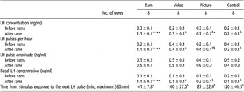
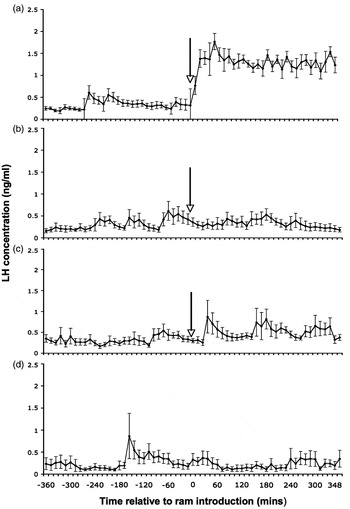
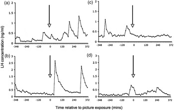
Time and type of stimulus had a significant effect on LH pulse frequency, mean and basal concentrations of LH (P < 0.05) with a time × type of stimulus interaction (P < 0.05). There was no effect of stimulus exposure on LH pulse amplitude (Table 1; P > 0.1). Specifically, ewes exposed to rams showed a rapid increase in LH pulse frequency and in the mean and basal concentrations of LH (Figure 2; P < 0.001). Exposure of ewes to still images of rams also increased the mean concentrations of LH (Table 1; P < 0.05) but only tended to increase LH pulse frequency (Table 1; P < 0.1). These increases were smaller than those observed in ewes exposed to rams (Figure 2; P < 0.05) and the presence or absence of a response varied among the group (Figure 3). As with the ewes isolated from all ram stimuli, exposure to a video of ewes and rams mating had no effect on any parameters of LH secretion (Table 1; P > 0.1). The time from stimulus exposure to the next LH pulse was shorter in the ewes exposed to rams than in all other groups (Table 1; P < 0.05), amongst which there was no difference (Table 1; P > 0.1).
Mean characteristics of LH release (±s.e.) of ewes exposed to different ram stimuli

Values differ within treatment: $P < 0.1; *P < 0.05; ***P < 0.001. Different superscripts indicate differences between stimulus exposure groups (at least P < 0.05).
Mean LH concentrations (±s.e.) before and after stimulus exposure of ewes exposed to rams ((a); Ram ewes), a video of ewes and rams mating ((b); Audio–visual ewes), still images of rams ((c); Visual ewes) or isolated from all stimuli of the ram for the duration of the experiment ((d); Control ewes). The arrow indicates the onset of stimulus exposure.

Representative profiles of LH secretion in ewes that appeared to respond ((a) and (b)) or were unaffected by ((c) and (d)) exposure to still images of rams. The arrow indicates the onset of stimulus exposure. Among the group, five ewes appeared to respond and three ewes appeared unaffected by still images of rams.

Experiment 2: Auditory and visual stimuli and the female effect
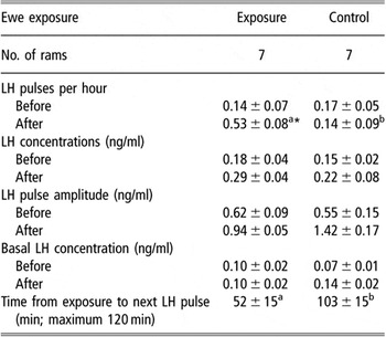
Exposure of rams to the combination of still images of ewes and the sound of ewes and rams mating (audio–visual) had no effect (P > 0.1) on LH pulse frequency (0.1 ± 0.0 v. 0.1 ± 0.1 pulses/h), LH pulse amplitude (0.6 ± 0.1 v. 0.8 ± 0.2 ng/ml) or basal concentrations of LH (0.1 ± 0.0 v. 0.1 ± 0.1 ng/ml). Exposure to audio–visual stimuli tended to increase LH concentrations (0.1 ± 0.0 v. 0.2 ± 0.1 ng/ml; P < 0.1) but had no effect on the time of onset of the next LH pulse, compared with rams isolated from ewe stimuli (214 ± 45 v. 212 ± 57 min; P > 0.1). There were no interactions between audio–visual stimulation and time on any measures of LH secretion (P > 0.1).
Exposure of rams to oestrous ewes increased LH pulse frequency (P < 0.05) and advanced the time to the next LH pulse (Table 2; P < 0.05). However, there was no significant effect of exposure to ewes on mean or basal concentrations of LH, or on LH pulse amplitude (Table 2; P > 0.1). There were also no interactions between exposure to ewes and time on any parameters of LH release (P > 0.1).
Mean characteristics of LH release (±s.e.) of rams exposed to oestrous ewes

Values differ within treatment: *P < 0.05. Different superscripts indicate differences between stimulus exposure groups (P < 0.05).
Discussion
The detectable LH response in ewes exposed to still images of rams supports our hypothesis that visual stimuli are involved in mediating the neuroendocrine response that underpins male-induced ovulation in sheep. However, the response was variable among ewes and, when present, the magnitude of the response was very small compared with that observed in ewes exposed to rams. Therefore, it seems likely that visual stimuli cannot substitute for the full complement of socio-sexual signals from rams. This is in contrast to olfactory stimuli, where the odour of rams’ wool can elicit an LH response similar to that observed in ewes exposed to rams (Cohen-Tannoudji et al., Reference Cohen-Tannoudji, Einhorn and Signoret1994) and even induce ovulation (Knight and Lynch, Reference Knight and Lynch1980).
The ability of ewes to recognise the projected images as conspecifics concurs with the wealth of literature on face recognition in sheep (review; Tate et al., Reference Tate, Fischer, Leigh and Kendrick2006). However, in this study, the ewes not only recognised the images as rams but also appeared to respond to these faces with a change in their reproductive endocrinology. Specifically, the images of rams appear to have activated the GnRH neurons and led to increased concentrations of LH. This physiological response concurs with Fabre-Nys et al. (Reference Fabre-Nys, Ohkura and Kendrick1997) who reported that exposure of oestrous ewes to the projected images of ram faces changed the pattern of neurochemical release in the medio-basal hypothalamus. Specifically, they found that exposure of ewes to male faces during early oestrus increased the concentrations of noradrenalin, glutamate, gamma-aminobutyric acid, dopamine and serotonin (Fabre-Nys et al., Reference Fabre-Nys, Ohkura and Kendrick1997). However, in the current study, exposure of ewes to a video of ewes and rams mating, combining audio and visual stimuli, had no effect on LH secretion. This is surprising, since audio–visual stimuli would be expected to be at least as effective as visual stimuli alone. It is unlikely that the ewes in this study were unable to recognise the animals in the video as sheep as a previous study showed that sheep will follow a moving, two-dimensional projection of a sheep down a raceway (Franklin and Hutson, Reference Franklin and Hutson1982). However, it is possible that they were not able to recognise the moving image as a ram. Direct eye contact is critical to individual recognition in sheep (Kendrick, Reference Kendrick1991) and the video used in this study showed predominantly side and rear images of rams. Therefore, we propose that obscuring the ram’s faces may have prevented an LH response to the visual stimuli.
Exposure of rams to oestrous ewes evoked an LH response that was absent when rams were exposed to still images of ewes combined with an audio clip of ram vocalisations. This indicates that, in contrast to the situation for ewes, neither auditory nor visual stimuli can even partially substitute for the full complement of socio-sexual signals that stimulate LH secretion in rams. Alternatively, this result may reflect a disparity between ewes and rams in their ability to recognise a prospective mate from a visual image. Studies in individual face recognition in sheep have typically used ewes or castrated males as their test subjects and not testis-intact males (e.g. Kendrick et al., Reference Kendrick, Atkins, Hinton, Broad, Fabre-Nys and Keverne1995 and Reference Kendrick, Atkins, Hinton, Heavens and Keverne1996). The ability of rams to identify ewes from projected images warrants further investigation.
Ram vocalisations had no effect on LH release in either ewes or rams, so auditory stimuli seem to be unable to even partially substitute for the other socio-sexual stimuli associated with a prospective mate. This is perhaps not surprising since sound does not enhance a sheep’s ability to discriminate between individual faces from a projected visual image (Kendrick et al., Reference Kendrick, Atkins, Hinton, Broad, Fabre-Nys and Keverne1995). However, the absence of a neuroendocrine response may also be a reflection of the lack of importance of auditory stimuli for communication in the Merino genotype. Kendrick et al. (Reference Kendrick, Atkins, Hinton, Broad, Fabre-Nys and Keverne1995) found a marked difference between Clun Forest and Dalesbred sheep in their ability to distinguish between human and sheep voices and attributed this divergence to differences in the social structure and native environment of the breed. Therefore, in Merino sheep, it seems that auditory stimuli play only a minor role in communication between the sexes.
In conclusion, audio–visual and visual signals are unable to completely substitute for the full complement of socio-sexual stimuli from a prospective mate. The importance or perception of the different signals appears to differ between the sexes, with visual stimuli being more important in mediating the neuroendocrine response of ewes to rams (male effect) than rams to ewes (female effect).
Acknowledgements
This research was supported under the Australian Research Council’s Discovery funding scheme (Project no. DP0558952) and Meat & Livestock Australia (Project no. B.MGS.0027). We thank B. Griffin and M. Gourlay for their assistance in the care and management of the animals, M. Blackberry for her assistance with the LH assays, and K. Vesterdorf and the numerous volunteers for their assistance in data collection.