INTRODUCTION
Morphological and functional differences between fish species are likely adaptational responses to changes in their habitat and environment. Though the ecological factors influencing the evolutionary terrestrialization of fish remain unknown, it seems plausible that predation, competition and food availability are potential evolutionary drivers (Sayer & Davenport, Reference Sayer and Davenport1991). Several examples of terrestrial fish morphologies stand out as being beneficial to ambulatory motion on land. Lobe-finned fish (subclass Sarcopterygii), for example, propel themselves out of the water using their strong and fleshy pectoral fins (Ahlberg & Clack, Reference Ahlberg and Clack2006). Certain terrestrial fishes conversely possess thinner, less fleshy pectoral fins (subclass Actinopterygii). One example in the Gobiidae family is the mudskipper. Mudskippers are sometimes considered modern-day models of extant transitional-amphibious fish, though this model is somewhat controversial. These fishes typically inhabit mangrove swamps, which are deemed analogous environmental conditions to those of early land plants (Kutschera & Elliott, Reference Kutschera and Elliott2013). Mudskippers have pelvic fins located more anteriorly than for example ancient tetrapods such as Ichtyostega. The pelvic fin is considered to be an advantageous adaptation (Cole et al., Reference Cole, Hall, Don, Berger, Boisvert, Neyt, Ericsson, Joss, Gurevich and Currie2011), by which means mudskippers are able to gain greater stability as these fins move antagonistically with the pectoral fins (Pierce et al., Reference Pierce, Hutchinson and Clack2013).
There are currently 10 identified genera of mudskipper, only four of which are able to remain out of water for longer periods (Murdy, Reference Murdy1989; Graham & Lee, Reference Graham and Lee2004). Of these, mudskippers from the genus Periophthalmus have been researched for their ability to climb. These fish use their pectoral fins antagonistically with their pelvic fins. The pectoral fins propel the fish forwards while the pelvic fins act as impaction pads that concurrently allow the fish to stick to climbing surfaces by means of Stefan adhesion (Wicaksono et al., Reference Wicaksono, Hidayat, Damayanti, Jin, Sintya, Retnoaji and Alam2016). According to You et al. (Reference You, Bian, Zan, Xu, Liu, Chen, Wang, Qiu, Li, Zhang and Sun2014), Periophthalmus has adapted to live in drier areas, whereas Periophthalmodons tend to live closer to the water at the high tidal zone. Boleophthalmus inhabits the intertidal zone between low tide and high tide, which is also where certain other genera of the Gobiidae family can be found. One example is Istigobius, which hunts small crustaceans, small fishes and eels (Fusianto & Tanjung, Reference Fusianto and Tanjung2011) but is unable to walk on land, unlike Boleophthalmus, which lives in the same tidal region. Some of these fishes have developed unfused pelvic fins that provide greater versatility in walking and climbing (Wicaksono et al., Reference Wicaksono, Hidayat, Damayanti, Jin, Sintya, Retnoaji and Alam2016), while other gobies retain a fused pelvic fin structure that in some cases act as a suction pad, allowing the fish to anchor itself to its substrate even in flowing water (Marsden & Jude, Reference Marsden and Jude1995).
Some Gobiidae are bottom dwellers or benthic fishes, and are known to be capable of both terrestrial and aquatic locomotion. Mudskippers rely on pectoral and pelvic fins movements to propel themselves forward. They do nevertheless need to have direct contact between their fins and the underlying substrate in order to achieve kinematic success. Gobies, such as the powerburst climbers Awaous guamensis and Lentipes concolor, and the inching climber Sycopterus stimpsoni are contrarily able to propel themselves vertically in a waterfall, which exhibits both aquatic and terrestrial interfaces. Awaous guamensis and L. concolor propel themselves by pectoral fin adduction coupled to an axial body powerburst, while S. stimpsoni alternates between oral and pelvic suction to attach to underlying substrates (Schoenfuss & Blob, Reference Schoenfuss and Blob2003). In short, both pectoral and pelvic fins appear to play important roles in terrestrial locomotion.
The mudskippers commonly display a form of locomotion called ambipedal progression or ‘crutching’. Crutching is named to form an analogy between the slow terrestrial, halted pectoral/pelvic fin movements exhibited by mudskippers, to human movement as aided by crutches. This term was originally coined by Harris (Reference Harris1960) and based on his observations of Periophtalmus koelreuteri (Pallas). The same style of motion was noted to occur in P. argentilineatus (Valenciennes) (Pace & Gibb, Reference Pace and Gibb2009), as well as in the aquatic mudskipper Boleophthalmus boddarti (Pallas) as reported by Clayton & Vaughan (Reference Clayton and Vaughan1988). Essentially, the mudskipper performs a pectoral fin stroke to propel itself forward. As a result, its body lifts slightly off the ground. As the body drops the pelvic fins extend first, after which the pectoral fins are pulled off the substrate, retracted anteriorly, and placed back onto the substrate during a recovery phase. In addition, the caudal region of the body arches slightly with the anal fin folded away from the substrate, presumably to prevent friction (Sayer, Reference Sayer2005). The fin is also reported to press firmly against the underlying substrate as a method by which mudskippers prevent body-slippage (Stebbins & Kalk, Reference Stebbins and Kalk1961).
Terrestrial locomotion in non-mudskipper fishes exhibit variant methods of propulsion to those of the mudskipper. The Pacific leaping blenny (Alticus arnoldorum) for example, combines its wide-rayed pectoral fins with bending of its body in the caudal area to move on an underlying substrate (Hsieh, Reference Hsieh2010). In blennids, although their body shapes are somewhat similar to that of mudskippers, their primary instrument of propulsion is the tail. The process of forward locomotion includes the bending of the tail to either side of the head, while the pectoral and pelvic fins are used to raise the fish prior to tail extension and ‘leaping’. This behaviour has also been observed in clinids such as M. macrocephalus, and labrisomids such as Dialommus fuscus. In climbing perches (Anabas sp.), the operculum or gill covers are specially modified for locomotion (Sayer, Reference Sayer2005). These perches have two parts to their gill covers, the first part is the main gill cover, the opercular, and the second part is the subopercular, which has a fin-like extension. The movements of the subopercular propel the fish forward as it makes contact with an underlying substrate (Davenport & Matin, Reference Davenport and Matin1990) and pushes the fish in a similar way to the way mudskipper pectoral fins propel the mudskipper forward. However, instead of these being synchronous on both sides of the fish, they cycle between the left and right organs, thereby exhibiting a form of ambulation.
This paper aims to characterize the physiological and biomechanical means by which terrestrial, tree-climbing fish effect pectoral-pelvic fin antagonism, and to compare those mechanisms against those of fish that are non-terrestrial and do not actively display pectoral–pelvic fin antagonism. The research herein focuses on in situ observations of terrestrial locomotion of mudskippers as compared with the aquatic ornate sandgoby (Istigobius ornatus). The musculature and biomechanics of these different fish species will also be compared and modelled using the finite element method. Biomechanical research into tree-climbing fishes naturally complements existing research on physiology, providing new insights on their functional morphology. Biomechanical information such as is presented in this paper, is moreover of technical benefit in cross-disciplinary fields such as biomimetic design and robotics.
MATERIALS AND METHODS
In situ fish movement observation
Three species of fish were captured using small fishing nets at three different locations on the island of Java, Indonesia. Two individuals were caught per species. Observations and procurement of the Gobiidae, I. ornatus took place at the Drini Coastline, Special District of Yogyakarta, Indonesia. The mudskipper, Periophthalmus gracilis was observed and procured in the Kulon Progo mangrove region, Special District of Yogyakarta, Indonesia. The mudskipper, Periophthalmus variabilis was observed and procured at the Mangkang near-coastline mangrove region of Semarang, Central Java, Indonesia. The fishes were placed into transparent glass aquaria and pectoral–pelvic movement was filmed and photographed during their locomotive efforts within the glass aquaria. The fishes were observed and filmed from multiple angles to observe and characterize their movements in dry terrestrial-like conditions using a frame rate of 50 fps. After observations were complete, the fishes were immobilized by slow cooling using ice and water, and subsequently, one of each species was preserved in 10% formalin solution, while the other of each species was preserved in 10% alcohol solution. The treatment and killing of the fishes complied with guidelines in the European Union Financial Instrument for Fisheries Guidance (FIFG) and the NIH Guide for the Care and Use of Laboratory Animals.
Morphological identification of fishes and barcoding
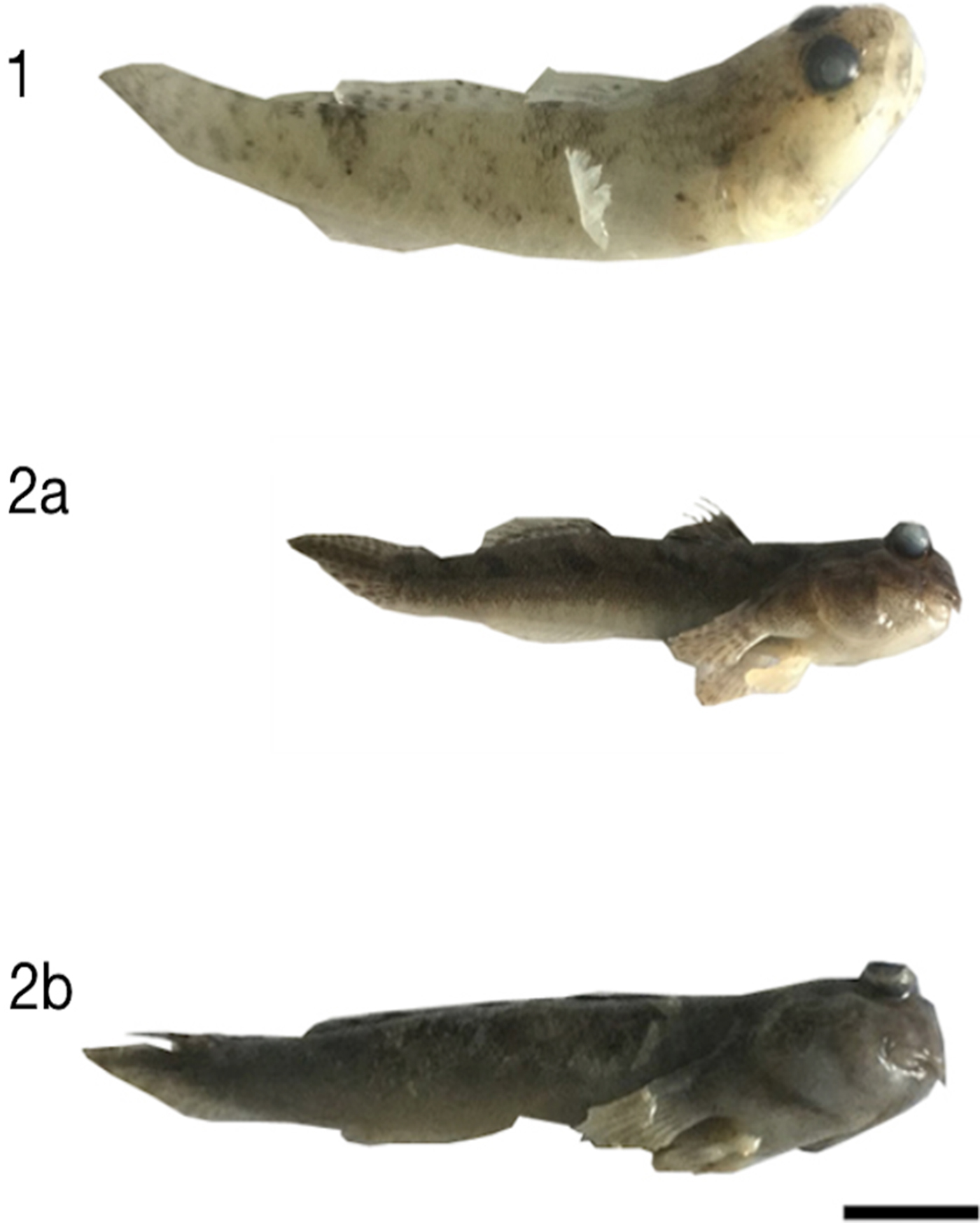
To ascertain the fish species, the colourations and patterns on the fish body surfaces were characterized alongside the morphological differences (Figure 1). For the mudskippers P. variabilis and P. gracilis, we used references from Murdy (Reference Murdy1989), Larson & Lim (Reference Larson and Lim1997) and Polgar (Reference Polgar2014). We then also checked against their recorded global distributions, which are mentioned in these references. For the Gobiidae fish, I. ornatus, we identified the species by comparing against the fish diversity records for Drini Beach according to Fusianto & Tanjung (Reference Fusianto and Tanjung2011). To morphologically characterize this fish, we compared against morphological characteristics reported by Capuli (Reference Capuli2016).
The formalin-preserved fish: (1) Ornate sandgoby (Istigobius ornatus), (2a) slender mudskipper (Periophthalmus gracilis), and (2b) dusky-gilled mudskipper (Periophthalmus variabilis). Scale bar = 1 cm.

Molecular barcoding was performed by amplifying the DNA of the three alcohol-preserved fishes by using primers specific to fish (Table 1) by polymerase chain reaction (PCR) as previously described (Sanka et al., Reference Sanka, Suyono, Rivero-Müller and Alam2016). The results from the PCR were subjected to electrophoresis and purified from the agarose gels to make sure that the DNA sequences were free from primer dimer contamination. The purified DNA from the three fishes had their concentrations checked using a Nanodrop, and then sequenced. The DNA sequences were analysed using the NCBI Nucleotide BLAST to compare species based on their highest identical percentages. The final results of molecular barcoding were rechecked and compared against the morphological results.
Primers used for fish DNA barcoding.

Fish dissection
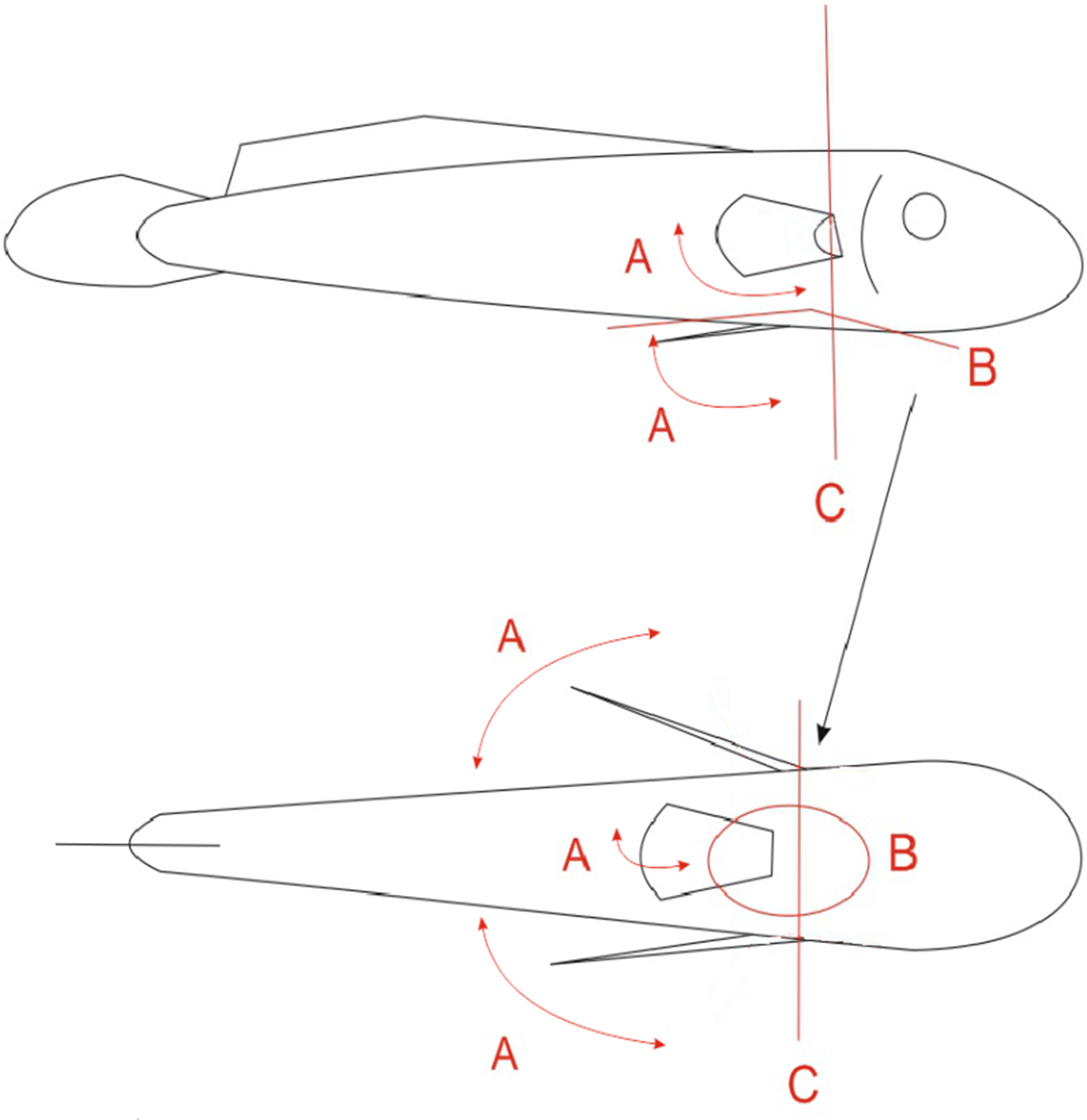
The fishes preserved in formalin solution were dissected to characterize the pectoral–pelvic fin musculatures with respect to physical location relative to the lateral muscles, and their antagonistic interactions. Prior to dissection, pectoral fins and pelvic fins were physically translated as shown in Figure 2 (see scalars A) in order to observe any antagonistic movement between the pelvic and pectoral fins. The fishes were subsequently dissected as shown in Figure 2 (planes B and C).
(A) The pectoral and pelvic fins were translated according to these scalars to observe whether they move antagonistically, synergistically or independently, (B) incision in this plane in the pelvic fin region to separate the pelvic muscle and (C) cross-sectional dissection of fish in this plane at the pectoral-pelvic fin boundary.

Finite element modelling
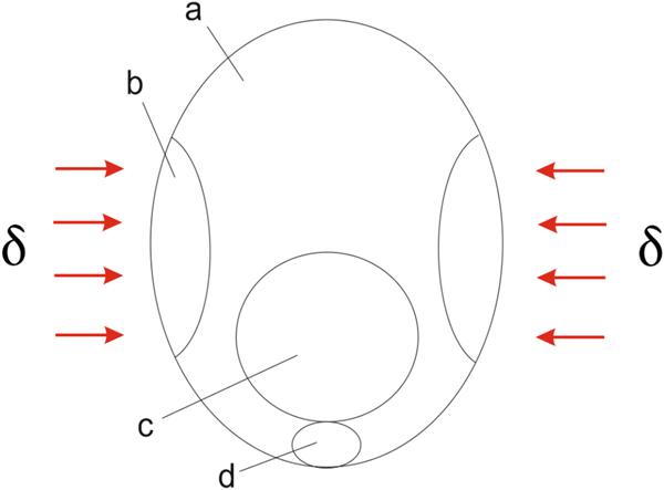
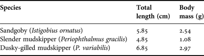
After characterizing the fishes' pelvic–pectoral fin musculatures, continuum mechanics finite element simulations of their cross sections were performed to more comprehensively discern and quantify the effects of pectoral fin motion and its relation to pelvic fin motion. The effect of lateral body muscle contraction and its subsequent compression on the pelvic fin muscle (Figure 3) was modelled for each fish species. The fish body musculature was used as a template for geometrical reconstruction in a two-dimensional plane. For each fish species, equal displacements were applied in compression from the mid-lateral side in accordance with its lateral/pelvic muscle positions and as determined by dissection (shown in Figure 3). The mode of displacement was applied to simulate the inward compression of the lateral body muscles during pectoral fin anterior-to-posterior movement, which was determined during dissection, movement of the pectoral fins and observation. The elliptical cross-section of the sandgoby was modelled as 1.15 cm high and 0.9 cm wide from the centre, while its pelvic muscle was also modelled as an ellipsoid located at the bottom of the body cross section with a height of 0.2 cm and a width of 0.35 cm from its centroid. Similar geometries were used for P. variabilis and P. gracilis but the dimensions of each was different. For P. variabilis the body cross section was modelled at 0.75 cm high and 0.55 cm wide, while its pelvic muscle was 0.2 cm high and 0.25 cm wide. For P. gracilis the body cross section was modelled at 1.25 cm high and 0.9 cm wide, while its pelvic muscle was similar to that of its smaller relative, P. variabilis and was modelled as 0.2 cm high and 0.25 cm wide. These values were measured and averaged in each species. We noted the muscles were by-and-large elliptical and found in each species, the height was approximately 50% longer than the width resulting in an elliptical shape approximation. Further measurements made included the total body length and the body mass. These are summarized as averages in Table 2.
Schematic representation showing direction and positions of prescribed displacements (δ) applied as an inward lateral compression arising from the anterior-to-posterior motion of the pectoral fins (red arrows). In the schematic are (a) body lateral muscles, (b) pectoral fin, (c) gastric cavity and (d) pelvic fin muscle.

Original measurements: total length from head to end of caudal tail and the body mass.

The Young's modulus of lateral body muscle is based on cod fish muscle reported in Nesvadba (Reference Nesvadba, Alasalvar and Taylor2002) and is set at 47 kPa, while the Poisson's ratio is set close to that of a near-incompressible material at 0.49. The pelvic muscle disc is notably stiffer on dissection and we recognize this is because this muscle also comprises cartilaginous matter alongside the normal fish muscle. Stiffness differences between relaxed and contracted muscles are well known to exist (Buttkus, Reference Buttkus1963). However, since there is a distinct lack of data availability on the stiffness of relaxed and contracted fish muscles, we approximated a stiffness increase using a Reuss model formulation (Reuss, Reference Reuss1929). In this model, a fractional ratio between bone (plate) and muscle in the pelvic disc was set as 0.65 : 0.35 and is based on image analysis using cross-sectional images of the fishes. The elastic modulus of the fish bone was taken as 6 GPa while the muscle was set at 47 kPa, using cod fish lateral body muscles as an analogue to the pelvic muscle (Nesvadba, Reference Nesvadba, Alasalvar and Taylor2002). Using the Reuss formulation we predicted the pelvic muscle in its contracted state would be ~3-fold higher than the surrounding body muscles, which were in a relaxed state. For the pelvic muscle disc, we therefore set the Young's modulus ~3-fold higher than the lateral body muscle, at 150 kPa while retaining a near-incompressible Poisson's ratio of 0.49.
The models were discretized using quadratic Langrangian elements as they are higher order polynomial elements that follow curvatures well. We concurrently used a plane stress formulation (equation (1) and (2)). In these equations E is the elastic modulus, σ is stress, ε is strain and ν the Poisson's ratio. The orthogonal Cartesian axes are represented by the subscripts 1−3. A system of asymmetrical sparse linear solvers (UMFPACK) formed the basis for the computation of solutions in the models.
 $$\sigma _{31} = \sigma _{13} = \sigma _{32} = \sigma _{23} = \sigma _{33} = 0$$
 $$\left( {\matrix{ {\sigma _{11}} \cr {\sigma _{22}} \cr {\sigma _{12}} \cr}} \right) = \displaystyle{E \over {1 - \nu ^2}}\left[ {\matrix{ 1 & \nu & 0 \cr \nu & 1 & 0 \cr 0 & 0 & {\displaystyle{{1 - \nu} \over 2}} \cr}} \right]\left( {\matrix{ {\varepsilon _{11}} \cr {\varepsilon _{22}} \cr {2\varepsilon _{12}} \cr}} \right)$$
RESULTS AND DISCUSSION
Identification results
The genera of the fishes were determined by genetic barcoding (see Supplemental Data). Due to the limited genomic data of the Periophthalmus genus, barcoding identification to the level of species was not possible and as such we relied on morphological identification as well as geographic distribution reports to determine the correct species.
By morphological identification, we determined the mudskipper species to be P. gracilis and P. variabilis, while the other fish was identified as I. ornatus (Fusianto & Tanjung, Reference Fusianto and Tanjung2011; Capuli, Reference Capuli2016). Periophthalmus gracilis and P. variabilis are commonly distributed along the coast of Java where we collected the fishes. Periophthalmus gracilis is distinguishable by its dorso-ventral white striation on the ventral part of the body, while P. variabilis has brown marks on the lateral sides of its body (Murdy, Reference Murdy1989; Larson & Lim, Reference Larson and Lim1997; Polgar, Reference Polgar2014), which is exactly the patterning that was observed on our collected fishes. Istigobius ornatus is characterized from other fish along the Drini shoreline by its ability to clamber laboriously outside of water (Fusianto & Tanjung, Reference Fusianto and Tanjung2011). The body colouration is pale grey with some brown spots situated along the entire length of the body (Capuli, Reference Capuli2016).
Terrestrial locomotion: observations
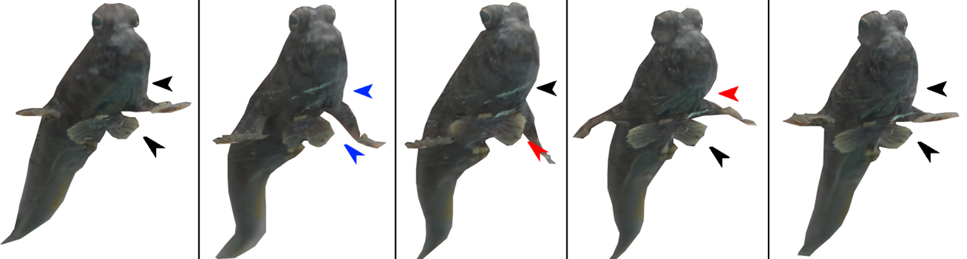
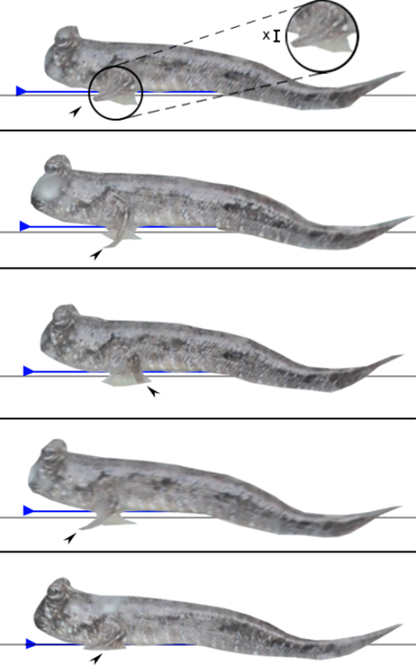
The ornate sandgoby (Istigobius ornatus) is aquatic and not well adapted to walking on drier land. During its walk within the glass aquarium, we observed that the sandgoby propels itself forward using its pectoral fins but that its pelvic fin remains motionless, fully expanded, and drags along the glass surface as the fish moves forward (Figure 4A). Contrarily, mudskippers were observed to use their pelvic fins as impact/suction pads walking on the glass and unlike the sandgoby, the pelvic fins were seen to be clearly retractable (Figure 4B, C) and working antagonistically with the pectoral fins. The mudskippers propel themselves forward, similarly to the sandgoby, using their pectoral fins. However, unlike the sandgoby, the mudskippers' bodies lift during forward propulsion as a response to a downward push by the pectoral fins. This is followed by a retraction of pelvic fins (Figures 5 & 6). Subsequently, as the body moves downwards, which occurs due to a forward (recovery stroke) swing of the pectoral fins, the pelvic fins expands almost instantaneously to absorb the impact force as described by Sayer (Reference Sayer2005) and to Stefan adhere the fish body to its underlying substrate as described in our previous work in Wicaksono et al. (Reference Wicaksono, Hidayat, Damayanti, Jin, Sintya, Retnoaji and Alam2016) – see Supplemental Video Data for motion sequences of all fishes from several angles.
(A) Ornate sandgoby (Istigobius ornatus), (B) slender mudskipper (Periophthalmus gracilis), and (C) dusky-gilled mudskipper (Periophthalmus variabilis) locomotion sequences as viewed from below. The black arrows identify the unchanged pelvic fin geometries while the blue arrows elucidate contraction of the pelvic fins. The picture of the locomotion sequence in Periophthalmus variabilis is reproduced and edited from our previous work by permission of Elsevier (from Wicaksono et al., Reference Wicaksono, Hidayat, Damayanti, Jin, Sintya, Retnoaji and Alam2016).

Front-bottom view of P. variabilis showing the downward pushing by pectoral fins during its walking sequence. The black arrows indicate a relaxed state, the blue arrows are indicative of upward movements, and the red arrows refer to downward movements.

Side view of the locomotion sequence of P. gracilis. During a horizontal walk, there is a down-thrust resulting in a physical upward movement of the body. The grey horizontal lines show the position of the flat surface upon which the fish is moving. The blue horizontal lines with arrows align with the lower surface of the fish body. The inset picture (encircled) highlights separation with × indicating a distance of separation.

Harris (Reference Harris1960) described the skeletal-muscular frameworks of the mudskipper using Periophtalmus koelreuteri samples where pectoral and pelvic fins musculatures were reported to control locomotion. In 2009, Pace and Gibb compared aquatic against terrestrial locomotion of Periophthalmus argentilineatus. They report that the caudal fin and body bend play individual roles in aiding aquatic propulsion, which is complemented by both pectoral fins. The pectoral fins were considered primarily responsible for locomotion, and the pelvic fins were considered as aiding this by stabilizing the body and absorbing impact during terrestrial movements (Pace & Gibb, Reference Pace and Gibb2009). Kawano & Blob (Reference Kawano and Blob2013), additionally compared the terrestrial locomotion ground reaction forces (GRF) of the mudskipper (Periophthalmus barbarus) and the salamander (Ambystoma tigrinum). Kawano & Blob report that the mudskipper has lower vertical magnitudes of GRF as a proportion of its body weight as compared with the salamander, and that the GRF is medially oriented. The mudskippers researched for this article (Periophthalmus gracilis and Periophthalmus variabilis) are similar in view of general locomotion. This is evidenced by the pectoral fins strokes, the deployment of pelvic fins, the positioning of the pectoral fins (comparable to the front limb of salamander in Kawano & Blob Reference Kawano and Blob2013), the stabilization function of the pelvic fins (similar to hind limb stabilization), their pectoral–pelvic musculatures (following Harris Reference Harris1960) and their pelvic disc muscles, which are similar to trout as reported by Standen (Reference Standen2010). We included the sandgoby (I. ornatus) in this study for comparison to the mudskipper. Similar to the mudskippers, the sandgoby moves its pectoral fins synchronously during terrestrial locomotion. The primary difference is that the pectoral fins of sandgoby are unable to provide a sufficient downward push to permit the elevation of the trunk of the body, unlike the mudskippers which are able to lift the majority of the main trunk of the body off the ground. As such, the single, fused pelvic fins of the sandgoby are dragged along the ground as it moves forward. Contrarily, the mudskippers P. gracilis and P. variabilis exhibit biomechanical means by which damage to the pelvic fins can be circumvented. Table 3 provides a summary of the measured average body lengths, and heights, the stride length from a pectoral fin push, the time taken for the stride and the elevation off the substrate during the pectoral stride. These were measured using our photographic and video sources.
Measured average values of the total body length, the body height, the pectoral stride length, the time taken for the pectoral stride and the elevation of the body from the substrate during the pectoral stride.

Pelvic and pectoral fin musculature
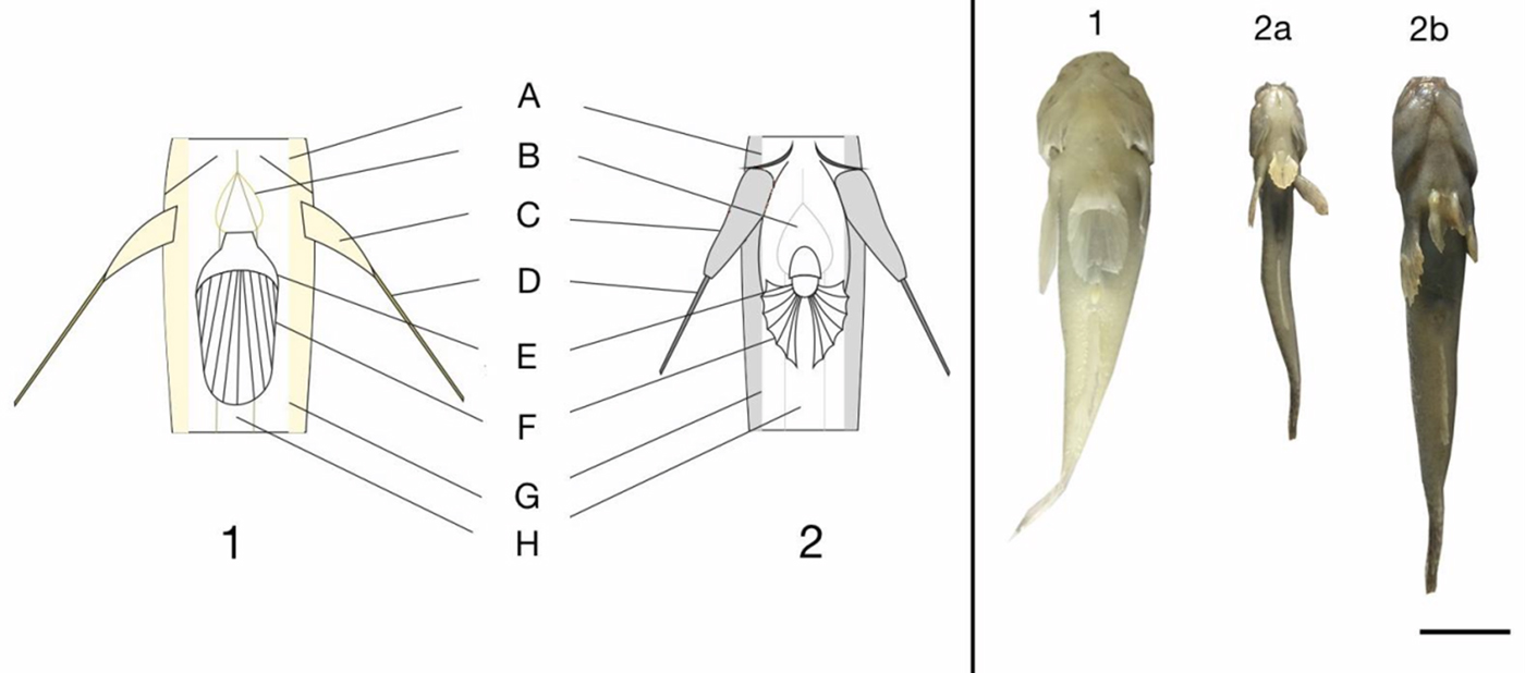
On dissection, we observed no physical connection between the muscles of the pectoral fins (radial and rays, Figure 7C) and the muscles of the pelvic fins (radial and rays, Figure 7E, F). Both were in fact noted to be separated by the lateral muscles of the body (Figure 7G).
Underside schematic representation of the fishes (left-hand side 1 (sandgoby) and 2 (mudskipper)) showing the locations of the gill chambers (A), the pelvic disc muscle (B), the pectoral fin radials (C), the pectoral fin rays (D), the pelvic fin radial bulk (E), the pelvic fin rays (F), the lateral muscles (G) and the visceral area/abdomen (H). On the right-hand side are images of I. gobius (1), P. gracilis (2a) and P. variabilis (2b). Scale bar = 1 cm.

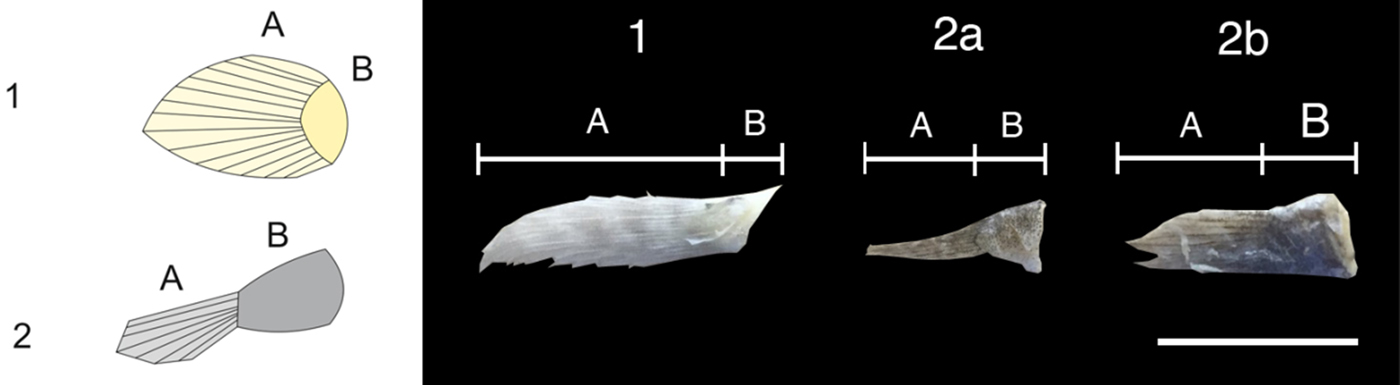
The pectoral fin consists of ray and radial parts at the macroscopic level (Figure 8A, B, respectively). Unlike the pectoral fins of other aquatic fishes e.g. in this case sandgoby (Figure 8-1), mudskipper pectoral fins (Figure 8-2) exhibit a radial part that protrudes and elongates from the body, resulting in two jointing systems; a body-to-fin (shoulder-like) joint and intra-ray jointing. This coupled to an inherent ventral mobility of the fin ray improves the intimacy of fish interaction and grip strength with a surface (Pace & Gibb, Reference Pace and Gibb2009). We furthermore observe that the pectoral fins of the mudskippers exhibit prolonged and more muscular radial sections than those of the sandgoby. Moreover, the pectoral fins tend to push down in the ventral direction with greater intensity and are considerably more bulky than the pectoral fins of the sandgoby (Figures 7C & 8). The bigger ray to radial ratios (Figure 8, ratio of B : A) of the pectoral fin muscle and fin indicates that mudskippers are more likely to exert a more forceful ventral direction push, the energy of which transfers to the fin during terrestrial locomotion. In the sandgoby (Figure 8-1), the B : A ratio is 1 : 4.5, while in P. gracilis (Figure 8-2a) has a B : A ratio of 1 : 1.2, and in P. variabilis (Figure 8-2b) the B:A ratio is 1 : 1.4.
Pectoral fin schematics and images of the sandgoby (1) and for mudskippers (2), showing larger radial (B) to ray (A) ratios in the mudskippers than in the sandgobby. Fish ID: Istigobius ornatus (1), Periophthalmus gracilis (2a) and Periophthalmus variabilis (2b). Scale bar = 1 cm.

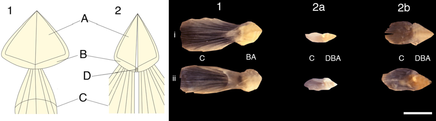
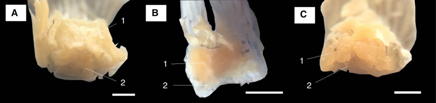
The pectoral fin radial thickness of the sandgoby is closest to the radial thickness of the largest mudskipper in this study (P. variabilis) (Figure 9). In all fishes, the pectoral fins consisted of intraradial ray fin bones (lepidotrichia) (Figure 9-2) sandwiched by abductor (distal, facing outward, pushing fin outward; Figure 9-1) muscles and adductor (proximal, facing inward, pulling fin inward; Figure 9-3) muscles.
Pectoral radial muscle cross-sections, showing the abductor muscles (1), ray fin lepidotrichia bones (2), and adductor muscles (3). Fish ID: Istigobius ornatus (A), Periophthalmus gracilis (B) and Periophthalmus variabilis (C). Scale bar in each slide = 1 mm.

The pelvic disc consists of semi-ossified basal plates (os basipterygium) (Figure 10A), which are framed by the radial bone of the ray (Figure 10B) and upon which the pelvic ray is attached (Figure 10C). The os basipterygium is split into two halves that are separated by the processus symphysicus anterior. Both adductor and abductor muscles control the pelvic fins' small movements and are bound by the triangularly shaped basal plates (Standen, Reference Standen2010). The sandgoby has fused pelvic fins and its disc is broader and flatter than the discs of either of the mudskipper pelvic fins (Figure 11). The mudskipper nevertheless exhibits bulkier pelvic disc muscles than the sandgoby (Figure 11A). These larger muscles structures of the mudskippers plausibly exist to absorb mechanical energy since the mudskipper actively utilizes its pelvic fins during land walking (unlike the sandgoby). Mudskipper pelvic discs (Figure 11B, C) are of a more pyramidal geometry than those of the sandgoby. The mudskipper abductor muscles pull the fish outward while adductor muscles pull it inward.
Left-hand side: Anatomical diagram of the pelvic disc of the sandgoby (1) and of mudskippers (2) showing the semi-ossified basal plate or os basipterygium (A), the radial bone (B), the pelvic fin rays (C) and the connective tissue connecting the os basipterygium, called facies symphysica tuberculum (D). To differentiate the parts we referred to Standen (Reference Standen2010). Right-hand side: actual images of the isolated pelvic disc and rays musculature as viewed from its (i) dorsal and (ii) ventral faces. Here (1) is I. ornatus, (2a) P. gracilis and (2b) P. variabilis. Scale bar = 5 mm.

Pelvic disc cross-section, showing the adductor muscles (1) and abductor muscles (2). Fish ID: Istigobius ornatus (A), Periophthalmus gracilis (B) and Periophthalmus variabilis (C). Scale bars = 1 mm.

While observing the musculature and testing for the mechanism of antagonistic movement, agonistic movement or no movement, we note that there are no muscles that connect the pelvic and pectoral fins in any of the fish species. Rather, the pelvic fins were noted to move in the mudskipper (not sandgoby) via a piston-like forced compression by the lateral body muscles during anterior-to-posterior pectoral fin movements. When physically moving (by hand) either the pelvic or pectoral fins of the mudskippers, antagonistic motion was noted to occur. This was not evident during the physical manipulation of the sandgoby.
Coupled pelvic–pectoral motion
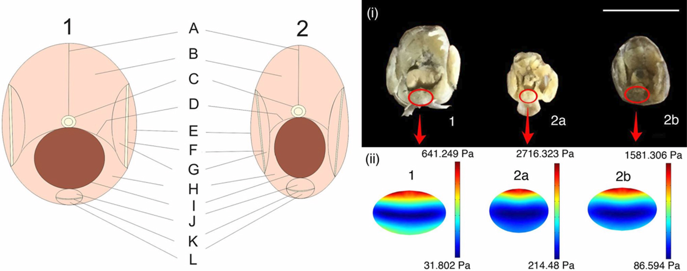
The lateral body muscle for all fishes can be divided into two parts in the dorsoventral orientations: epaxial (upper, dorsal side) and hypaxial (lower, ventral) muscles. Both body muscles are separated by a horizontal septum (Genten et al., Reference Genten, Terwinghe and Danguy2009; Roberts, Reference Roberts2012). The sandgoby body is broader than the body of either mudskipper species (Figure 12 left-hand side and plate (i)). There is no muscular connection observed between muscle in pectoral fin radials and the pelvic muscle disc that would give rise to any form of synergistic or antagonistic control (i.e. there is no abductor-adductor muscle relationship). Nevertheless, we previously noted that in the mudskippers pectoral and pelvic fins move antagonistically relative to one another (refer to Figure 4 and Supplementary Video Data). To understand how the antagonistic mechanism might function from a mechanical perspective, we refer to our finite element models (Figure 12 plate (ii)). Here, we note that the bulky pectoral fin radials of the two mudskipper species push the pelvic fin disc downwards much like a mechanically driven piston. As the pectoral fins move from anterior to posterior positions, the lateral muscles of the body compress inwards and these muscles generate a squeezing force upon the dorsal end of the pelvic fin muscle, driving the pelvic fin in a ventral direction. The stress exerted upon the dorsal side of the pelvic muscle is highest in P. gracilis, followed by P. variabilis and the lowest stress is computed to occur in the sandgoby. We postulate therefore, that the observed antagonistic movement of the pelvic fins with the pectoral fins is to some extent controlled by a mechanical piston-like system that forces the pelvic fin towards an underlying surface, at which point Stefan adhesion allows the fish to stick to a surface. In the case of P. gracilis and P. variabilis the dorsal stresses are higher than for I. ornatus and are entirely localized to the dorsal side of the pelvic muscle. As such, we suggest the location and magnitude of stress in these mudskipper species is sufficient for a ventral direction thrust of the pelvic fin giving rise to Stefan adhesion of the pelvic fin to a substrate (Wicaksono et al., Reference Wicaksono, Hidayat, Damayanti, Jin, Sintya, Retnoaji and Alam2016). In the case of I. ornatus however, the magnitude of stress at the dorsal side of the pelvic fin muscle is considerably less and in fact, there is also a relatively strongly opposing stress at the ventral side of the pelvic fin muscle, which overall reduces the likelihood that this fish can thrust its pelvic fin towards a substrate antagonistically with its pectoral fins and with sufficient force to develop Stefan adhesion between the fin and an underlying substrate. This fits well with our observations, in that the mudskipper species climb trees and show clear pelvic-pectoral fin antagonism, while the sandgoby is unable to climb trees or crawl effectively and moreover does not show any clear pelvic-pectoral fin antagonism (refer to Figure 4 and Supplemental Video).
Left-hand side: Cross-sectional schematic representation of pectoral-pelvic boundary at the posterior of the head in the sandgoby (1) and in mudskippers (2). Parts: vertical septum (A), epaxial body muscle (B), vertebral column (C), horizontal septum (D), abductor muscle of pectoral (E), intraradial ray part (F), adductor muscle of pectoral (G), hypaxial body muscle (H), gastric cavity (I), adductor muscle of pelvic disc (J), os basipterygium/ossified base of pelvic disc (K) and abductor muscle of pelvic disc (L). Right-hand side: comparison of dissected cross-sections of the pectoral-pelvic boundaries (i) and of the von Mises stresses experienced in the pelvic muscles of each. Fish ID: Istigobius ornatus (1), Periophthalmus gracilis (2a) and Periophthalmus variabilis (2b). Scale bar = 1 cm.

According to Harris's study (Harris, Reference Harris1960) on Periophtalmus koelreuteri, there is a connection between the pelvic and pectoral fins system, more specifically in the skeletal frame. The anterior end of the pelvic fin bone is attached to the pectoral frame in the condyle of the cleithrum, the anterior ventral part of the pectoral bone. Though the skeletal frames between pelvic and pectoral fins are connected, their specific musculatures are individual. There are sets of muscles controlling the pectoral fin movements. These include the superficial flexor muscles (superficial abductor muscles) and a depressor radiorum muscle (profundal abductor), in combination with an adductor muscle (the coraco-radial muscle) and the superficial levator, which raises the pectoral fin rays and two extensors (medial and ventral) that in turn extend the fins. Regarding the pelvic fins, the muscles are primarily attached to the pelvic disc bone, a pelvic protractor that pulls the pelvic fins to the anterior side, a dorsal and main (ultimus) retractor (pulling to the posterior side), and a levator, which pulls the pelvic fins upward. Apart from the pelvic disc, a cleithro-hyoid muscle connecting the ceratohyal bone to the cleithrum (where the pectoral and pelvic bones attach) serves to stabilize the over-rotation of the pelvic disc that would otherwise overstretch the protractor muscle. These sets of muscles in both the pelvic and pectoral regions have no connection in between, unlike the skeletal frames.
We hypothesized herein that the inward compression of the muscular frameworks through the anterior-to-posterior motion of the pectoral fins may aid in the extension of the pelvic fins through a piston-like action. We suggest that the lateral muscles of the body push the pelvic disc in a ventral direction, causing the protractor or cleithro-hyoid muscles to contract, and lowering the pelvic fin. Our hypothesis is deemed complementary to the muscular deployment hypothesis previously suggested by Harris (Reference Harris1960) as it does not negate the involvement of other larger muscle involvement in pushing the pelvic fins in a ventral direction.
A mechanical piston-like system for pelvic-pectoral fin antagonism as noted for the mudskippers should theoretically be less energetically demanding than a case of pure muscular antagonism as the piston can aid pelvic fin motion. As such, a simple system like this may act as a viable means by which mudskippers are able to be mobile on land without using too much energy. From a perspective of biomimetics, a piston-like system such as has been noted in the mudskipper species, which allows antagonistic ambulation of robot legs with a single locomotor, would also be less energetically demanding. It would perhaps moreover be more instantaneous and with fewer electronic parts, decreasing thus the statistical likelihood of electronic part failure. Mudskipper motion shows great potential in biomimetic robotics and further research is needed to more comprehensively characterize and elucidate the biomechanics of their systems of motion.
CONCLUSIONS
The musculatures and biomechanical systems of motion were characterized for three different fish. Two were mudskippers that are able to climb trees and walk with relative fluidity on land, and the third was a sandgoby that requires an aqueous environment or surface in order to be mobile. All three fish species appear to have unconnected pectoral and pelvic fin muscles. However, there appears to be a physical piston-like system of pelvic–pectoral fin antagonism in the mudskipper species, which presumably helps them to ambulate on land and climb trees. This system is not present in the sandgoby studied herein, and we hypothesize that a system like this not only aids with the pelvic fin down-stroke, but is also a good model for energy efficient ambulation that could be applied to biomimetic robots.
SUPPLEMENTARY MATERIAL
The supplementary material for this article can be found at https://doi.org/10.1017/S0025315417001722
ACKNOWLEDGEMENTS
We thank CIMO, Finland for their financial support. We are indebted to the Cell Fate Laboratory at the Turku Centre for Biotechnology for allowing us to use their laboratory.