Introduction
Obsessive compulsive disorder (OCD) is the fourth most common psychiatric disorder, affecting 2–3% of the general population. OCD is defined by the combination of (a) obsessions, which are recurrent and distressing ideas, and (b) compulsions, behaviours that the subject feels compelled to perform to prevent or reduce the anxiety associated with the obsessions. Alongside medications, cognitive behavioural therapy (CBT) represents a well-validated first line of treatment for OCD (Gava et al. Reference Gava, Barbui, Aguglia, Carlino, Churchill, De Vanna and McGuire2007). Despite the ever-growing knowledge on haemodynamic modifications in OCD observed with positron emission tomography and functional magnetic resonance imaging (fMRI), the neurocognitive processes underlying the action of CBT remain poorly understood. Indeed, few studies have investigated CBT-induced anatomo-functional changes (Linden, Reference Linden2006). According to anatomo-functional models for OCD (Baxter et al. Reference Baxter, Saxena, Brody, Ackermann, Colgan, Schwartz, Allen-Martinez, Fuster and Phelps1996; Schwartz, Reference Schwartz1998), the cognitive part of CBT is designed to correct the erroneous cognitive patterns at the origin of obsessions. This would target cortical processes, mainly in the orbitofrontal cortex (OFC) and anterior cingulate cortex (ACC), known to be also involved in the cognitive control of emotion (Clark & Beck, Reference Clark and Beck2010). The behavioural component of the therapy – exposure and prevention of response – would reinstate goal-directed, cortical control on the dysfunctional, automated behaviours stored in the basal ganglia (Graybiel & Rauch, Reference Graybiel and Rauch2000; Gillan et al. Reference Gillan, Papmeyer, Morein-Zamir, Sahakian, Fineberg, Robbins and de Wit2011).
OCD patients compared with matched controls present a haemodynamic hyperactivity in the OFC and ACC, as well as in subcortical structures (Menzies et al. Reference Menzies, Chamberlain, Laird, Thelen, Sahakian and Bullmore2008; Rotge et al. Reference Rotge, Guehl, Dilharreguy, Cuny, Tignol, Bioulac, Allard, Burbaud and Aouizerate2008b ; Haynes & Mallet, Reference Haynes and Mallet2010). A few longitudinal studies have employed on/off medication or pre-/post-treatment designs to show that the abnormal activities of the ACC, OFC and basal ganglia were corrected after a successful treatment (pharmacotherapy/psychotherapy) (Nakatani et al. Reference Nakatani, Nakgawa, Ohara, Goto, Uozumi, Iwakiri, Yamamoto, Motomura, Iikura and Yamagami2003; Whiteside et al. Reference Whiteside, Port and Abramowitz2004; Nakao et al. Reference Nakao, Nakagawa, Yoshiura, Nakatani, Nabeyama, Yoshizato, Kudoh, Tada, Yoshioka, Kawamoto, Togao and Kanba2005; Linden, Reference Linden2006; Nabeyama et al. Reference Nabeyama, Nakagawa, Yoshiura, Nakao, Nakatani, Togao, Yoshizato, Yoshioka, Tomita and Kanba2008). Treatment of OCD could, therefore, be effective through an effect on these prefrontal and subcortical regions. However, the questions of when and how these changes occur have not been carefully addressed. Indeed, little seems to have been published on the dynamics of the neural changes underlying effective treatments of OCD [but see Schiepek et al. (Reference Schiepek, Tominschek, Karch, Lutz, Mulert, Meindl and Pogarell2009) for an exploratory single-case study].
To address this issue, we used fMRI before, during, at the end and after the therapy. To sensitize the detection of fine changes in cerebral activity, we used an exposure task also known as a symptom-provocation task (Rauch et al. Reference Rauch, Jenike, Alpert, Baer, Breiter, Savage and Fischman1994; Linden, Reference Linden2006). Provocation of symptoms is achieved by presenting obsession-inducing images to patients. We used images of each patient's own symptom-triggering situation. Based on the literature, we expect the latter type of stimuli to produce even greater activations of ACC and OFC metabolism (Menzies et al. Reference Menzies, Chamberlain, Laird, Thelen, Sahakian and Bullmore2008; Rotge et al. Reference Rotge, Guehl, Dilharreguy, Cuny, Tignol, Bioulac, Allard, Burbaud and Aouizerate2008b ). We decided to use neutral stimuli instead of the resting state in order to avoid the bias of uncontrolled cognitive processing such as autobiographic or episodic memory which may differ across patients (Morcom & Fletcher, Reference Morcom and Fletcher2007). Generic obsession-inducing images were all checking-related (e.g. stove, door), as this was the dominant feature in the symptomatology of the patients, and was demonstrated to evoke clearer neural responses (Rauch et al. Reference Rauch, Jenike, Alpert, Baer, Breiter, Savage and Fischman1994; Mataix-Cols et al. Reference Mataix-Cols, Wooderson, Lawrence, Brammer, Speckens and Phillips2004). Moreover, the use of thematically tailored obsession-inducing stimuli demonstrated that state-related changes in OCD patients overlap anatomically with the trait-related changes measured between OCD and non-OCD populations (Adler et al. Reference Adler, McDonough-Ryan, Sax, Holland, Arndt and Strakowski2000). This strategy remains limited by its reliance on average group effects: it overlooks the idiosyncratic nature of symptom-triggering stimuli, even within a given OCD subtype (Abramowitz et al. Reference Abramowitz, Franklin, Schwartz and Furr2003; Schienle et al. Reference Schienle, Schafer, Stark, Walter and Vaitl2005). Therefore, we used personalized obsession-inducing stimuli in addition to the generic ones. These were, for each patient, photographs taken by the patient himself of symptom-triggering situations in his or her usual environment. Finally, we expected these pictures to induce stronger symptoms (Schienle et al. Reference Schienle, Schafer, Stark, Walter and Vaitl2005) and, thus, maximize our ability to detect OCD-related fMRI activities and their potential changes.
Methods
Participants
A total of 66 OCD patients (age 18–65 years) were screened after referral by the French association of OCD patients (Association Française des personnes atteintes de Trouble Obsessionnel Compulsif; AFTOC) and the out-patient clinic of the Pitié-Salpêtrière Teaching Hospital (Paris, France), to participate in a CBT trial. Patients underwent a detailed structured diagnostic interview [Mini-International Neuropsychiatric Interview (M.I.N.I. 5.0.0); Sheehan et al. Reference Sheehan, Lecrubier, Sheehan, Amorim, Janavs, Weiller, Hergueta, Baker and Dunbar1998] and a clinical interview by a clinical psychologist to establish the proper diagnosis according to the Diagnostic and Statistical Manual of Mental Disorders, fourth edition, text revision (DSM-IV-TR). Severity of OCD symptoms was rated using the standard Yale–Brown Obsessive Compulsive Scale (YBOCS; Goodman et al. Reference Goodman, Price, Rasmussen, Mazure, Fleischmann, Hill, Heninger and Charney1989). Symptoms were identified and categorized by the interviewing clinical psychologist, with the help of the YBOCS check-list and the Padua Inventory (Sanavio, Reference Sanavio1988). We included only moderately severe patients (YBOCS between 16 and 25) with predominant checking symptoms and a stable pharmacological treatment for the 2 months preceding inclusion (treatments for each patient are reported in Supplementary Table S1). This led to a study sample of 35 patients. In all, four patients did not complete the course of the CBT and were not included in the analysis, yielding a final sample of 31 patients. Additional psychopathology was investigated using the Beck Depression Inventory (BDI; Beck & Beamesderfer, Reference Beck and Beamesderfer1974) and the State-Trait Anxiety Inventory (STAI-A and -B; Spielberger et al. Reference Spielberger, Gorsuch, Lushene, Vagg and Jacobs1983). Exclusion criteria included any co-morbid DSM IV-TR Axis I disorder, pharmacological treatment stabilized for less than 2 months and repeated no-shows at CBT sessions (two consecutive absences or three over the course of the therapy). The study was reviewed and approved by the local ethics committee. All patients gave written, informed consent.
Study design
Patients were assessed using clinical interviews and fMRI at four time points: immediately before CBT, at mid-therapy (i.e. about 1.5 months/6 weeks after baseline), at the end of the therapy (3 months after baseline) and 6 months after the end of the therapy (i.e. 9 months after the first time point). Each session consisted of a psychiatric evaluation (comprising YBOCS assessment) performed by a clinical psychologist who was not involved in the CBT, followed on the same day by MRI acquisitions (structural and functional during the exposure task, see below).
CBT
Four experts, experienced in the treatment of OCD, conducted the CBT which consisted of 15 individual, weekly 45-min sessions. Patients were randomly assigned to one therapist. Treatment plans were based on a reference manual (Bouvard, Reference Bouvard2006) and were individually tailored to address patient-specific symptoms, according to current standards (Koran et al. Reference Koran, Hanna, Hollander, Nestadt and Simpson2007). The aim of the cognitive therapy is for the patient to identify his or her symptoms better, and to learn to differentiate obsessions from standard thoughts. Once identified, the obsessions are then discussed with the therapist in order to expose their irrational nature and replace them with constructed, logical ideas. An example is to discuss with the patient what another person would do in his or her symptom-triggering situations. The behavioural therapy is centred on exposure and response prevention. Patients are instructed to refrain from performing some of their rituals (starting with the less severe ones), or reduce the number of iterations (e.g. ‘try to check your door only three and not seven times this week’). Instructions were given during the weekly sessions but the exercises were performed at home. To reduce inter-therapist variability, several meetings were organized prior to the start of the trial to ensure that all participating therapists had the same understanding of the procedures to be used.
This study was conducted as part of a clinical trial assessing the interest of adding a computer-assisted psychopedagogic tool (CAPT) to classical CBT (described above). The CAPT used a previously published checking task based on a delayed matching-to-sample (Rotge et al. Reference Rotge, Clair, Jaafari, Hantouche, Pelissolo, Goillandeau, Pochon, Guehl, Bioulac, Burbaud, Tignol, Mallet and Aouizerate2008a ) under the supervision of the therapist. Patients saw two pictures one after the other, and had to decide whether they matched or not (match to sample). After their response, they had the possibility to ‘check’, i.e. to see again the two pictures and change their answer. They could check as many times as they wished before going on to the feedback. The therapists used the task as a psychopedagogic tool to teach the patients the cognitive processes involved in their checking compulsion and thus help the patient resist their compulsions in a virtual setting. The length of CBT was identical for each patient. The pictures used in the CAPT were different from those used in the MRI exposure task (see ‘Behavioural paradigm’ section). At baseline, patients were randomly assigned to one of the CBT + CAPT versus CBT-only treatment arms. Statistical analyses showed no difference between the groups, whether in terms of initial severity (YBOCS score, CBT + CAPT: 26.2, s.d. = 5.0; CBT-only: 24.6, s.d. = 4.4; t 29 = 0.95, p = 0.34, d = 0.48) or of clinical improvement (improvement of YBOCS scores, CBT + CAPT: 45%; CBT-only: 48%; pooled s.d. 24%; p = 0.36, d = 0.12). Therefore, patients from both randomization arms were pooled together.
Behavioural paradigm: exposure task
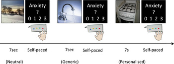
In order to probe OCD-specific circuits, patients performed an exposure task in the MRI scanner. It involved presenting obsession-inducing stimuli to the patients while measuring event-related fMRI activations. Following each stimulus, patients had to rate the induced anxiety on a scale of 0 (no anxiety) to 3 (maximum anxiety) (Fig. 1). In all, three types of images were used: neutral, generic obsession-inducing and personalized obsession-inducing (photographs taken by the patient himself of scenes pertaining to his or her major symptoms). Generic obsession-inducing images were all checking-related (e.g. stove, door). All the neutral and generic obsession-inducing images were selected by a clinical psychologist (M.M.) and validated by an independent group of patients. Stimuli were normalized for luminance and contrast. Each was presented for 7 s onscreen, followed by an unlimited rating time, and then a 1.5–3.5 s blank screen. Two runs were performed. Each included 15 images of each type, in a random order (the same images were used in both runs, although in a different order).
Behavioural paradigm. Images were presented in two blocks of 15. In all, three types of images were presented, from left to right in the figure: neutral, generic obsession-inducing and personalized obsession-inducing. These images were presented for 7 s. Patients were then asked to rate their level of anxiety in response to the images between 0 and 3.

Image acquisition
Each MRI session comprised structural and functional MRI acquired on a 3-T Trio Tim Siemens scanner with a 12-channel phased-array head coil at the Centre de neuro-imagerie de recherche (CENIR) imaging facility of the Brain and Spine Institute Research Centre. Functional images of blood oxygenation-level dependent (BOLD) contrast were collected during the two runs of the exposure task using a T2*-weighted echo planar sequence using parallel imaging [Integrated Parallel Acquisition Techniques (iPAT) = 2]; repetition time = 2290 ms; echo time = 25 ms; flip angle = 75°; image matrix = 96 × 96 × 40; isotropic voxels = 2 × 2 × 2 mm (+1 mm interslice gap)]. The field of view (192 mm × 192 mm × 120 mm) covered the whole brain and was tilted to optimize functional sensitivity in the OFC (Weiskopf et al. Reference Weiskopf, Hutton, Josephs and Deichmann2006). For each run, about 240 volumes (range: 209–355) were collected, depending on the time needed for the participant to complete the task. Standard high-resolution structural scans were collected using a three-dimensional (3D) T1-weighted magnetization prepared rapid acquisition gradient echo (MPRAGE) sequence with the following parameters: inversion time = 900 ms; repetition time = 2300 ms; echo time = 4.18 ms; flip angle = 9°; image matrix = 256 × 256 × 176; isotropic voxels = 1 × 1 × 1 mm (no gap); 1 volume.
Data analysis
Clinical scores and image ratings were processed using Excel (Microsoft, USA) and R software (http://CRAN.R-project.org) for parametric statistical analyses. Our primary measure of clinical improvement was the percentage reduction in YBOCS score relatively to the YBOCS score before therapy (ΔYBOCS, as used by Whittal et al. Reference Whittal, Thordarson and McLean2005). In the absence of a consensual cut-off to distinguish responding from non-responding OCD patients in clinical trials (varying from 20% to 50%; Tolin et al. Reference Tolin, Abramowitz and Diefenbach2005), we decided to base our criteria on the present sample, separating low- and high-responders using a median split. Clinical data were submitted to parametric statistical procedures (analysis of variance; ANOVA) to assess the effect of time (four levels: before, mid-therapy, after and at the end of the therapy) and group (two levels: high- versus low-responders to treatment). For behavioural data from the exposure task we also included the image type factor (representing the three types of stimuli: neutral, generic and personalized).
Functional data were pre-processed and analysed using the SPM8 toolbox (University College London, UK; http://www.fil.ion.ucl.ac.uk) and MATLAB (The Mathworks Inc., USA). In brief, pre-processing included: realignment to compensate for head motion, normalization using the structural scan and smoothing (6 mm full-width half-maximum) to meet the hypotheses of the SPM approach. High-pass filtered (128-s cut-off) functional time series were then entered in an event-related general linear model (GLM) using the canonical haemodynamic response function convolved with boxcar functions matched to stimulus duration in order to reveal activations associated with the presentation of each image type. The model also included six realignment parameters as covariates of no interest to capture residual movement-related artifacts and an autoregressive model was applied to account for temporal autocorrelation.
Based on the literature, we hypothesized that successful treatment is underlain by and drives a correction of ACC, OFC, caudate and thalamus hyperactivity. Therefore, we decided to focus our analyses on regions of interest (ROIs) within these areas. These ROIs were functionally defined at baseline through a second-level analysis conducted on individual smoothed (6 mm) contrasts (personalized stimuli versus neutral stimuli, before therapy, thresholded at p < 0.001 voxel-wise uncorrected, size threshold: 100 voxels, one-tailed) (for a similar approach, see Beutel et al. Reference Beutel, Stark, Pan, Silbersweig and Dietrich2010). Parameter estimates (β values) for each image type averaged over all voxels in each functional ROI were extracted for each participant and submitted to statistical analysis using within-subject parametric tests (t tests, ANOVA and Pearson's correlation) along with behavioural and clinical data and submitted to random-effects second-level analyses. Anatomical coordinates are given in Montreal Neurological Institute (MNI) coordinates.
Data presented here are therefore reported for the 27 fMRI patients who completed the whole study, i.e. excluding those unable to attend the last fMRI session (n = 3) and one patient with artifacted data at the first session.
The registry name of the study is: ‘Modification of Cerebral Activity of Obsessive Compulsive Disorder (OCD) Patients During Cognitive and Behavioral Therapy (TOC TOC)’ (http://clinicaltrials.gov/ct2/show/NCT01331876).
Results
Patients and clinical results
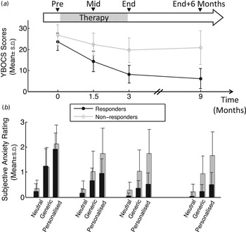
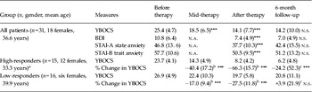
Demographic, clinical scores at inclusion and pharmacological treatment data are provided for each individual patient in Supplementary Table S1. CBT was seen to improve OCD symptoms. Indeed YBOCS scores after therapy were below the clinical threshold for OCD (i.e. YBOCS <16). Scores at each time point are reported in Table 1. The average response over the course of the therapy was 46.3% (s.d. = 23.9%) [Cohen's d = 1.76, 95% confidence interval (CI) 1.17–2.34]. Mean YBOCS score showed a main effect of time (F 3,90 = 65.7, p < 0.001), that is, symptoms had already started decreasing by mid-therapy, were even lower at the end of therapy [mid versus end of therapy: 29.5% (s.d. = 20.2%) reduction; t 29 = 5.9, p < 0.001] and stabilized at this level [end of therapy versus end + 6 months: 8.2% (s.d. = 39.8%) reduction; t 29 = 5.9, p > 0.05, d = 0.28] (Fig. 2 a). Considering the progress already seen at mid-therapy, we tested the relationship between this intermediate improvement and the final outcome measured after therapy or at the 6-month after follow-up. For both outcomes, we measured a strong correlation (respectively, r = 0.82, 95% CI 0.65–0.91 and r = 0.76, 95% CI 0.55–0.88, both p < 0.001) with the mid-therapy improvement (Supplementary Fig. S1).
Clinical (a) and behavioural (b) changes over the course of cognitive behavioural therapy. Data were acquired at four time points: before therapy, mid-therapy, end of therapy and end + 6 months (0, 1.5, 3 and 9 months after inclusion, respectively). (a) Change in symptom severity [Yale–Brown Obsessive Compulsive Scale (YBOCS) scores] in the responder (>45% clinical improvement) and low-responder (<45% clinical improvement) groups. (b) Change in subjective anxiety ratings induced by neutral, generic obsession-inducing and personalized obsession-inducing images in the responders and low-responders. Values are means, with standard deviations represented by vertical bars.

Patient demographics and clinical data at time points during the study, with significance of the changes

YBOCS, Yale–Brown Obsessive Compulsive Scale; n.s., non-significant; BDI, Beck Depression Inventory; n.a., not applicable; STAI, State-Trait Anxiety Inventory.
Data are given as mean (standard deviation).
a Two patients from the high-responder group did not attend the 6-month follow-up session.
b Change from before therapy.
c Change from end of therapy.
*** p < 0.005.
We then defined two groups of patients according to a median split on the degree of their clinical improvement (as indicated by the percentage reduction in YBOCS). These were: the high-responder group (n = 15), which was composed of the patients who had improved by 45% or more at the end of the therapy; and the low-responder group (n = 16), whose improvement on YBOCS was less than 45% (including non-responders). Change in mean YBOCS scores for both groups is represented in Fig. 2 a. The detailed clinical characteristics of both groups are presented in Table 1. Initial YBOCS scores were the same in each group. Although these two groups did not differ with respect to their depressive symptoms before therapy (BDI scores for high-responders = 10.2/low-responders = 11.4, p = 0.60), they differed after therapy (4.2/10.4, p < 0.001); similarly, state anxiety and trait anxiety were also significantly different after therapy (STAI-A: before 46.6/46.9, p = 0.94, after: 32.9/42.2, p = 0.009; STAI-B: before 56.7/58.6, p = 0.63, after: 46.4/54.4, p = 0.02).
Exposure task
There was a main effect of the image-type on anxiety ratings during the exposure task, in both the high-responder and low-responder groups (F 2,52 = 69.5, p < 0.001). Before therapy, personalized images [mean rating (s.d.): 2.12 (0.72)] were rated as more anxiogenic than generic ones [1.31 (0.75)], which were themselves more anxiogenic than neutral ones [0.31 (0.33)] (Fig. 2 b). A time × image type interaction (F 6,156 = 15.89, p < 0.001) showed that the change in anxiety ratings changed differently depending on image type. In fact, as the three-way interaction (time × image type × group: F 6,156 = 6.15, p < 0.001) indicates, the interaction effect of time × image type reached significance in the high-responder group (F 6,72 = 21.0, p < 0.001) but not in the low-responder group (F 6,84 = 1.82, p = 0.1); while both groups reported similar ratings to each image before therapy (main effect of image type: F 2,52 = 94.2, p < 0.001; no group × image type interaction: F 2,52 = 0.48, p = 0.60), ratings of anxiety decreased for all types of images over the course of the therapy in the high- but not the low-responder group [post-therapy ratings for personalized, generic and neutral, respectively, for high/low responders (s.d.): 0.58/1.82 (0.47/0.97), 0.39/1.06 (0.32/0.82), 0.08/0.27 (0.13/0.32); group × image type interaction: F 2,52 = 9.84, p < 0.001].
Brain imaging
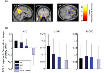
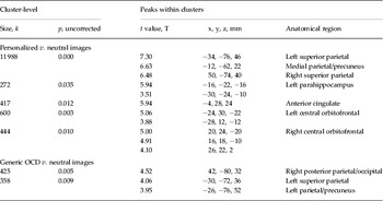
Neuroimaging results are presented in Fig. 3 and Table 2. Before therapy, fMRI revealed a strong main effect of the image type on the BOLD responses from extended regions within the parietal and frontal regions. Indeed, personalized stimuli yielded stronger activations than neutral ones in a bilateral dorsal parietal cluster as well as in a dorsal, anterior cingulate (ACC), a left, central orbitofrontal (OFC), and a right, central OFC cluster (Table 2, Fig. 3 a). However, the generic images compared with neutral ones only triggered significantly stronger activations in two bilateral parietal clusters (all included in the parietal activations observed with the personalized images) (Table 2).
Neural regions activated by personalized symptom exposure and effect of cognitive behavioural therapy. (a) Personalized obsession-inducing images versus neutral images (p < 0.001, uncorrected, see text for details) led to the activation of bilateral orbitofrontal clusters and of an anterior cingulate cluster, as shown on a sagittal section [x = −4, Montreal Neurological Institute (MNI) coordinates], a coronal section (y = +28) and an axial section (z = −22). Significance values are given using the colour scheme indicated. (b) Changes in activations induced by personalized obsession images during the course of the therapy, averaged over voxels within the anterior cingulate cortex (ACC), left orbitofrontal cortex (L OFC) and right orbitofrontal cortex (R OFC) clusters. BOLD, Blood oxygenation-level dependent.

Significant differences in BOLD responses to personalized v. neutral images before therapy

BOLD, Blood oxygenation-level dependent; OCD, obsessive compulsive disorder.
As the latter three clusters are the most commonly reported hyperactive regions in OCD patients and are theorized to be at the core of OCD pathophysiology (Rotge et al. Reference Rotge, Guehl, Dilharreguy, Cuny, Tignol, Bioulac, Allard, Burbaud and Aouizerate2008b ), they were used to define group-level ROIs. BOLD responses to personalized images in each of these ROIs were then compared between the three image types across the four time points through a three-way within-subject ANOVA (ROI × image type × time). This analysis showed a marginal effect of ROI (F 2,52 = 3.075, p = 0.055), with a strong main effect of the image type (F 2,52 = 11.38, p < 0.001) and no main effect of time (F 3,78 = 0.60, p = 0.62, n.s.). However, neither the three-way (F 12,312 = 1.45, p = 0.141, n.s.) nor the ROI × time (F 6,156 = 1.67, p = 0.133, n.s.) interaction was significant for both ROI × image type (F 4,104 = 2.70, p = 0.034) and image type × time (F 6,156 = 4.04, p < 0.001).
We further qualified these effects by conducting two-way ANOVA (image type × time) in each of the three ROIs. To account for multiple testing across the three ROIs, we applied Bonferroni-correcting factors yielding a p < 0.016 significance threshold. Activity in the ACC was influenced by the type of stimuli (main effect of image type: F 2,52 = 9.51, p < 0.001), with a greater activation for personalized compared with generic or neutral images. There was no main effect of time (F 3,78 = 1.43, p = 0.24) but there was an interaction between time and image type (F 6,156 = 3.34, p = 0.006). The same pattern was also observed for the left OFC cluster (main effect of image type: F 2,52 = 8.18, p < 0.001; main effect of time: F 3,78 = 0.17, p = 0.91, n.s.; interaction: F 6,156 = 3.97, p = 0.001). In these regions, and for personalized stimuli, cerebral activity decreased following the same time course as symptom reduction, with a continuous improvement along the three time points (Fig. 2 a v. Fig. 3 b). The right OFC showed only an effect of image type (F 2,52 = 8.99, p < 0.001), whereas time and the interaction between time and image type were not significant (respectively, F 3,78 = 1.11, p = 0.34, n.s.; F 6,156 = 1.90, p = 0.09, n.s.) (Fig. 3 b). Notably, no significant difference of activation existed between right OFC and either left OFC (p = 0.12) or ACC (p = 0.09) responses before therapy
The comparison of high- versus low-responders in each ROI, using three-way ANOVA (image type x time x group), did not reveal significant differences between the two groups in the dynamics of the BOLD response (F 6,150 = 0.87, p = 0.52, n.s.). BOLD responses before therapy were not different between the two groups, whatever the ROI considered (F 2,50 < 2.66, p > 0.079, n.s. across all ROIs). Despite the concomitant changes we observed in brain activations and symptom severity at the group level, we failed to observe across-patient significant correlations between changes in left or right OFC nor ACC activations and clinical improvement (all r 2 < 0.02, p > 0.45). When looking for biomarkers of response predictors, we failed to identify any cluster across the whole brain showing significant correlation between functional activations to personalized pictures before therapy and later clinical improvement (ΔYBOCS).
Discussion
We evaluated a cohort of OCD patients before, halfway through, at the end of a CBT course, as well as 6 months after. These evaluations included a full clinical assessment and a behavioural task performed during fMRI. To investigate the obsessive dimension of the disorder, we asked patients to rate the anxiety induced by three types of stimuli: neutral, generic obsession-inducing and personalized obsession-inducing images. Personalized stimuli were based on photographs taken by each patient of situations triggering his or her major symptoms. All patients reported higher ratings for personalized stimuli than for generic ones, than for neutral ones. The task activated a large parietal cluster and smaller ones in the ACC and both OFCs. Activities in these prefrontal regions were more important for the personalized than the generic and neutral stimuli, mirroring the differences in ratings. The course of CBT was effective on OCD symptoms. Nonetheless, we were able to distinguish high- versus low-responder subgroups. The change in clinical characteristics in the high-responder group was matched by a decrease of sensitivity to both types of obsession-inducing stimuli, and the related cortical activities.
Our results tend to confirm that CBT is an effective therapeutic strategy for OCD (Gava et al. Reference Gava, Barbui, Aguglia, Carlino, Churchill, De Vanna and McGuire2007; Abramowitz et al. Reference Abramowitz, Taylor and McKay2009). Effect sizes and long-term improvement were similar to those reported in studies with comparable populations and interventions. Indeed, recent meta-analyses report pre–post effect sizes ranging from 1.20 to 1.52 (Eddy et al. Reference Eddy, Dutra, Bradley and Westen2004; Butler et al. Reference Butler, Chapman, Forman and Beck2006; Stewart et al. Reference Stewart, Stack, Tsilker, Alosso, Stephansky, Hezel, Jenike, Haddad, Kant and Jenike2009), while long-term improvement ranged from 41.5% (Whittal et al. Reference Whittal, Thordarson and McLean2005) to 48.9% on YBOCS scores (Eddy et al. Reference Eddy, Dutra, Bradley and Westen2004) and remained stable in the long term (Abramowitz & Foa, Reference Abramowitz and Foa1998; Jaurieta et al. Reference Jaurieta, Jimenez-Murcia, Alonso, Granero, Segalas, Labad and Menchon2008).
These results cannot be directly applied to the common CBT settings for everyday patients because of the constraints of randomized controlled studies (Laurenceau et al. Reference Laurenceau, Hayes and Feldman2007). Indeed, patients in this study were clinically homogeneous, without co-morbidity, and with predominant checking symptoms. This differs from the typical OCD population, wherein 50% patients (lifetime prevalence) have at least one co-morbidity (Steketee et al. Reference Steketee, Eisen, Dyck, Warshaw and Rasmussen1999; Abramowitz et al. Reference Abramowitz, Taylor and McKay2009), and up to 75–82% have at least one major depressive episode (Foster & Eisler, Reference Foster and Eisler2001). In spite of this, another cross-sectional study showed that 54% of ‘mainstream’ OCD patients had neither Axis I nor Axis II co-morbidity (Denys et al. Reference Denys, Tenney, van Megen, de Geus and Westenberg2004). This suggests that there is little bias in our patient sample.
There was no control group (i.e. without treatment) in our study which was initially designed to test a psychopedagogic addition to CBT (see Introduction) while CBT itself has already been demonstrated effective (Gava et al. Reference Gava, Barbui, Aguglia, Carlino, Churchill, De Vanna and McGuire2007). While lacking a control group may be considered a methodological limitation regarding the neurobehavioural outcomes, we considered that it was ethically impossible to withhold – or delay – effective treatment from symptomatic patients for such a long duration (9 months).
Mid-therapy improvement was strongly predictive of final outcome and subsequent states. This suggests that CBT success is conditioned by its early effects, and prompts us to investigate the underlying mechanisms. Unfortunately, only one study has been published on the dynamics of a course of composite psychotherapies in a single patient (Schiepek et al. Reference Schiepek, Tominschek, Karch, Lutz, Mulert, Meindl and Pogarell2009).
We designed a personalized exposure task, using both generic and personalized obsession-inducing stimuli. Our behavioural results demonstrate a much higher sensitivity to personalized images, thus validating their use in this task. While stimuli were built considering the constraints of any experimental psychology paradigm, personalized stimuli could only be adjusted and controlled for luminance and contrast, not spatial frequency. Also, obsessions and compulsions in OCD patients often depend on their immediate surroundings. Thus, photographs of their triggers may be insufficient to induce the obsession. While it would be difficult to scan patients in front of their own door, one could consider the possibility of using computerized 3D environments [virtual reality (Kim et al. Reference Kim, Kim, Cha, Park, Rosenthal, Kim, Han, Kim and Kim2010) or augmented reality (Breton-Lopez et al. Reference Breton-Lopez, Quero, Botella, Garcia-Palacios, Banos and Alcaniz2010)]. Indeed, a highly realistic 3D scene in an immersive device could create a stronger experience of ‘presence’ and induce stronger obsessions. Avoidance strategies may have led patients not to select photographs related to their most severe obsessions; however, given the present difference in effect between personalized and generic stimuli, one dare say that such a bias was of no significant influence in our study.
As the same stimuli were used at each session, the decrease in some patients of the anxiety ratings over the course of the therapy could have been caused by habituation effects. However, the decrease was observed only in the high-responder group, and matched symptom reduction, thereby suggesting that unspecific habituation is not the likeliest cause. Although this contrast between high- and low-responders does not provide the strongest level of proof, having an OCD control group appeared ethically unfeasible for the aforementioned reasons.
Before the therapy, the exposure task activated regions known for their hyperactivity in OCD (ACC, OFC), thereby supporting the use of our paradigm. Indeed, a number of imaging studies have shown alterations in these regions (Menzies et al. Reference Menzies, Chamberlain, Laird, Thelen, Sahakian and Bullmore2008; Rotge et al. Reference Rotge, Guehl, Dilharreguy, Tignol, Bioulac, Allard, Burbaud and Aouizerate2009; Hoexter et al. Reference Hoexter, de Souza Duran, D'Alcante, Dougherty, Shavitt, Lopes, Diniz, Deckersbach, Batistuzzo, Bressan, Miguel and Busatto2012), sometimes correlated with symptoms (Adler et al. Reference Adler, McDonough-Ryan, Sax, Holland, Arndt and Strakowski2000; Mataix-Cols et al. Reference Mataix-Cols, Wooderson, Lawrence, Brammer, Speckens and Phillips2004), and corrected after successful therapy (Nakatani et al. Reference Nakatani, Nakgawa, Ohara, Goto, Uozumi, Iwakiri, Yamamoto, Motomura, Iikura and Yamagami2003; Huyser et al. Reference Huyser, van den Heuvel, Wolters, de Haan, Boer and Veltman2013). A few exposure experiments have activated the same circuit (Linden, Reference Linden2006; Rotge et al. Reference Rotge, Guehl, Dilharreguy, Cuny, Tignol, Bioulac, Allard, Burbaud and Aouizerate2008b ). Moreover, in our study, activity decreased over the course of the CBT, confirming that our task makes an adequate probe of the OCD network.
The roles of the OFC and ACC in the psychopathological process underlying OCD have been understood in the context of behavioural, cognitive and emotional regulation (Menzies et al. Reference Menzies, Chamberlain, Laird, Thelen, Sahakian and Bullmore2008; Clark & Beck, Reference Clark and Beck2010), through maladaptive habit learning of putative ‘cognitive patterns’, analogous to ‘fixed action patterns’ in the motor domain (Graybiel & Rauch, Reference Graybiel and Rauch2000; Graybiel, Reference Graybiel2008). The ACC and OFC are critical nodes of behavioural control through their inputs to the ventral and basal ganglia (Everitt & Robbins, Reference Everitt and Robbins2005). Their hyperactivity during symptom exposure could, therefore, reflect unsuccessful attempts to implement goal-directed action plans over compulsive habit-like rituals (Gillan et al. Reference Gillan, Papmeyer, Morein-Zamir, Sahakian, Fineberg, Robbins and de Wit2011). Indeed, CBT is sometimes associated with extinction. The reduction in ACC activation observed in relation to obsession-inducing stimuli might also reflect the concomitant change in the emotional response (i.e. anxiety ratings) and/or the improvement of emotional regulation over the course of CBT (Ressler & Mayberg, Reference Ressler and Mayberg2007; Milad & Rauch, Reference Milad and Rauch2012).
Activities in the left OFC and ACC continued to decrease after therapy. This could reflect slow modifications triggered by the therapeutic intervention. Nevertheless, one can also suggest that this continued disappearance of symptoms keeps driving neural plasticity towards the re-establishment of a ‘normal’ profile. Unlike the left OFC and ACC and despite the clinical efficacy, right OFC hyperactivity was not reduced by CBT. We are unaware of discussions on such asymmetries in the literature; thus, we suggest that the right OFC participates in a ‘trait’ network that would make one more vulnerable to the development of OCD symptoms, whereas the left OFC and ACC could participate to a ‘state’ symptom modulating symptom intensity. In this framework, complete recovery would be accompanied by the disappearance of the ‘trait’ network involving the right OFC. This ‘trait’ circuit may be linked to previously reported functional alterations in the OFC of OCD relatives (Chamberlain et al. Reference Chamberlain, Menzies, Hampshire, Suckling, Fineberg, del Campo, Aitken, Craig, Owen, Bullmore, Robbins and Sahakian2008) and considered to be endophenotypes associated with increased vulnerability for the disorder (Chamberlain & Menzies, Reference Chamberlain and Menzies2009). A similar dissociation between trait and state circuits has already been evidenced in bipolar disorder (Blumberg et al. Reference Blumberg, Leung, Skudlarski, Lacadie, Fredericks, Harris, Charney, Gore, Krystal and Peterson2003).
Unfortunately, as others, we did not identify a BOLD signal predictive of response to treatment; nor did we observe a significant difference in brain responses between high- and low-responder groups (Brody et al. Reference Brody, Saxena, Schwartz, Stoessel, Maidment, Phelps and Baxter1998; Saxena et al. Reference Saxena, Brody, Ho, Zohrabi, Maidment and Baxter2003; Apostolova et al. Reference Apostolova, Block, Buchert, Osen, Conradi, Tabrizian, Gensichen, Schroder-Hartwig, Fricke, Rufer, Weiss, Hand, Clausen and Obrocki2010). However, the prediction of final clinical outcome by mid-therapy prompts us to investigate with a finer resolution the early stages (i.e. the 6 weeks before mid-therapy evaluation) of the CBT; this would identify the processes separating the high-responder from the low-responder group.
A number of studies have shown treatment-related changes in the ACC, OFC and other brain regions (Nakatani et al. Reference Nakatani, Nakgawa, Ohara, Goto, Uozumi, Iwakiri, Yamamoto, Motomura, Iikura and Yamagami2003). However these changes were not always necessarily concomitant with clinical improvement (Saxena et al. Reference Saxena, Brody, Ho, Alborzian, Maidment, Zohrabi, Ho, Huang, Wu and Baxter2002; Nakao et al. Reference Nakao, Nakagawa, Yoshiura, Nakatani, Nabeyama, Yoshizato, Kudoh, Tada, Yoshioka, Kawamoto, Togao and Kanba2005), and may reflect the fact that brain dynamical processes may be occurring but remain undetected by clinical instruments. Van Calker et al. (Reference van Calker, Zobel, Dykierek, Deimel, Kech, Lieb, Berger and Schramm2009) suggest that this type of phenomenon may explain the delay seen in the onset of the therapeutic effect, in particular with psychotherapeutic treatment. However, since we could not associate the BOLD changes with clinical improvement, we cannot exclude that habituation not only to anxiogenic stimuli but also to the scanner environment (Schunck et al. Reference Schunck, Erb, Mathis, Jacob, Gilles, Namer, Meier and Luthringer2008), occurring across scanning sessions, might partly result in the blunting of activations in the ACC and OFC (Chapman et al. Reference Chapman, Bernier and Rusak2010). Future studies using a waiting-list group of OCD patients would allow controlling for such habituation effects.
In conclusion, our results indicate that our exposure task is a valid means of exploring the neural correlates of obsessions in OCD. Related haemodynamic modifications in the ACC and left OFC have a similar time-course to behavioural and clinical variables, while right OFC abnormalities are unaffected by successful CBT. Our results point out the importance of the earliest stages of the CBT process on the final outcome. Future studies should, therefore, study the earlier stages of CBT with finer temporal resolution.
Supplementary material
For supplementary material accompanying this paper visit http://dx.doi.org/10.1017/S0033291713002237.
Acknowledgements
We thank Christophe Demonfaucon and members of AFTOC for providing help and support during all the steps of the study. We also thank Vincent Trybou, Anna Maestre, Corine Lobjoie and Romina Bianco for having conducted the CBT sessions with the patients, as well as Aurélie Saulton and Christian Gaudeau for having performed some of the MRI procedures. We thank Bruno Falissard for his help in designing the study and Vincent Morice for the randomization procedure, as well as Jérôme Yelnik for his thoughful comments on the manuscript.
Declaration of Interest
A.H.C. and W.I.H. received a Ph.D. fellowship from the French Ministry for Research (Ministère de la Recherche). M.M. received a fellowship from the Région Ile de France included in a Partenariat Institution Citoyen pour la Recherche et l'Innovation (PICRI) project. Part of this work is supported by grants from the Direction de Recherche Clinique d'Ile-de-France (Contrat d'Interface AP-HP INSERM), and from the Agence Nationale pour la Recherche (ANR).