Introduction
As green biocatalysts, enzymes exhibit exceptional catalytic efficiency, substrate selectivity, and they offer mild operational conditions, and environmental benignity, enabling broad industrial applications (Mu et al., Reference Mu, Meng, Ju and Li2023; Chen et al., Reference Chen, Huang, Wang, Pang, Lou, Li, Yang and Qiu2024; Yang et al., Reference Yang, Su, Wang, Zhang, Jing, Meng, Zhang, Huang, Guo, Wang and Gao2024). In particular, β-glucosidases (BG, EC 3.2.1.21) play a pivotal role in the bioconversion of lignocellulosic biomass into high-value chemicals and biofuels (Verma et al., Reference Verma, Gupta, Keerti and Dubey2018; Magwaza et al., Reference Magwaza, Amobonye and Pillai2024). However, the industrial potential of free BG is severely hindered by its sensitivity to harsh processing conditions (e.g. pH fluctuations, elevated temperatures) and the difficulty of recovery, leading to high operational costs (Yang et al., Reference Yang, Fan, Zhou, Zhou, Yan, Ju and Li2020; Mu et al., Reference Mu, Meng, Ju and Li2023). Enzyme immobilization has emerged as a standard solution to enhance enzyme robustness and recyclability. Among various carriers, halloysite nanotubes (HNTs) have attracted significant attention due to their biocompatibility, abundance, and unique tubular nanostructure, which provides a large surface area for enzyme loading and protection against aggregation (Pandey et al., Reference Pandey, Munguambe, Tharmavaram, Rawtani and Agrawal2017; Balci et al., Reference Balci, Rosales, Pazos, Sofuoglu and Sanroman2023).
HNTs are aluminosilicate clay minerals (chemical formula of Al2Si2O5(OH)4·nH2O), which offer advantages over carbon nanotubes (CNTs) due to their abundance, low cost, and biocompatibility (Pandey et al., Reference Pandey, Munguambe, Tharmavaram, Rawtani and Agrawal2017; Sahiner et al., Reference Sahiner, Demirci and Sahiner2022; Balci et al., Reference Balci, Rosales, Pazos, Sofuoglu and Sanroman2023). From a surface-chemistry perspective, HNTs demonstrate a distinct spatial segregation of chemical properties. The outer surface is characterized by a siloxane (Si-O-Si) network with exposed silanol (Si-OH) functional groups, whereas the lumen features an aluminol (Al-OH) arrangement analogous to that of gibbsite (Ma et al., Reference Ma, Wu, Higaki and Takahara2018; Pandey et al., Reference Pandey, Tharmavaram, Khatri and Rawtani2023). This spatial chemical heterogeneity allows for the selective functionalization of either the external or internal surfaces, facilitating the design of tailored biocatalytic systems. To functionalize the chemically inert surface of HNTs for enzyme attachment, traditional methods often rely on organosilanes (e.g. APTES) (Lima et al., Reference Lima, Boemo, de Araújo and de Oliveira2021; Tharmavaram et al., Reference Tharmavaram, Pandey, Bhatt, Prajapati, Rawtani, Sooraj and Ranjan2021). While effective, these processes are energy-intensive and involve toxic organic solvents. Alternatively, bio-inspired polydopamine (PDA) offers a mild, eco-friendly surface modification strategy (Sun and Davis, Reference Sun and Davis2022).
As a natural modifier, dopamine (DA) undergoes oxidative self-polymerization in mildly alkaline media to form an adherent PDA coating. The PDA can adhere tightly to diverse surfaces (metals, ceramics, polymers) through hydrogen bonding, π–π interactions, and metal coordination (Sun and Davis, Reference Sun and Davis2022; Kimmins et al., Reference Kimmins, Henríquez, Torres, Wilson, Flores, Pio, Jullian, Urbano, Braun-Galleani, Ottone, Muñoz, Claros and Urrutia2024; Zhang et al., Reference Zhang, Chen, Feng, Li and Liu2025). Crucially, the PDA framework is rich in hydroxyl, amino, and quinone groups, which not only provide hydrophilicity and biocompatibility but also enable the covalent or non-covalent anchoring of proteins via their terminal amino or carboxyl groups (Chen et al., Reference Chen, Sun, Lv, Li, Wang and Wang2018; Sahiner et al., Reference Sahiner, Demirci and Sahiner2022; Sillu et al., Reference Sillu, Agnihotri and Reddy2022). Although PDA-modified HNTs are known, current research often overlooks the role of the substrate’s surface state in determining the quality and robustness of the PDA coating. To address this gap, this study introduces a novel alkali-activation pre-treatment step to engineer the HNT surface prior to PDA modification (Zhang et al., Reference Zhang, Guo, Yuan, Li, Wang, Deng and Liu2020; Ferrante et al., Reference Ferrante, Bertini, Ferlito, Lisuzzo, Lazzara and Duca2023; Lisuzzo et al., Reference Lisuzzo, Bertini, Lazzara, Ferlito, Ferrante and Duca2023). We postulate that controlled alkali etching will increase surface roughness and expose a higher density of hydroxyl groups (Al-OH and Si-OH). These changes are expected to facilitate stronger hydrogen bonding and π-π stacking interactions between the HNT surface and the PDA layer (Feng et al., Reference Feng, Fan, Zha, Wang and Jin2016), ultimately resulting in a more stable microenvironment for the immobilized BG.
The present study focuses therefore on constructing a robust Alkali-HNTs-PDA support (D-AHNTs) for the high-efficiency immobilization of β-glucosidase (D-AHNTs/BG). The specific objectives are to: (1) evaluate the impact of alkali activation on the physicochemical properties of HNTs and the subsequent PDA coating; (2) immobilize BG onto the optimized carrier and systematically characterize its loading capacity, retained activity, and optimal catalytic parameters (including optimal temperature and pH); and (3) comprehensively evaluate the thermal stability, pH stability, operational stability, and re-usability of the immobilized enzyme in comparison with the free enzyme, thereby validating the potential of this strategy in addressing the bottlenecks associated with its industrial application.
Collectively, this work focuses on constructing a hierarchically optimized carrier, D-AHNTs, via controlled alkali activation and PDA modification to address the stability and recyclability issues of β-glucosidase. Ultimately, this study not only provides a promising immobilized biocatalyst solution for the high-value application of β-glucosidase but also expands the utilization of halloysite nanotubes in the field of enzymatic catalysis.
Materials and methods
Halloysite (sourced from Shiyan, Hubei Province, China; white powder) exhibits a nanoscale hollow tubular morphology. β-Glucosidase (from almonds; specific activity: 10–30 U mg–1, enzyme units per milligram of protein) and 4-nitrophenyl β-D-glucopyranoside (p-NPG) were purchased from Macklin Reagent (Shanghai, China). Dopamine hydrochloride (C8H11NO2·HCl, purity ≥98%) was supplied by RiboBio (Guangzhou, China). N-(3-Dimethylaminopropyl)-N′-ethylcarbodiimide hydrochloride (EDC; C8H17N3·HCl, 98%) and sodium phosphate dibasic (Na2HPO4, 99%) were procured from Aladdin Reagent Co., Ltd, Shanghai, China. Other analytical-grade reagents were obtained from Chengdu Kelong Chemical Co. (Chengdu, China) and used as received.
Characterization studies
The mineral phases of HNTs, AHNTs, D-AHNTs, and D-AHNTs/BG were analyzed by XRD using an Ultima IV diffractometer (Rigaku Corporation, Japan) with a D/teX Ultra detector and small-angle accessory. Measurements were performed with CuKα radiation at 40 kV and 40 mA. The scanning range was 2°–80° (°2θ) with a scan speed of 2° min–1. The FTIR spectra were acquired using a Spectrum One spectrometer (PerkinElmer, USA). The solid sample was mixed with potassium bromide (KBr) at a mass ratio of 1:100, ground into a fine powder, and pressed into a pellet. All spectra were collected in the range of 4000 to 400 cm–1 at a resolution of 4 cm–1. X-ray photoelectron spectroscopy (XPS) spectra were recorded using a Thermo Scientific K-Alpha X-ray photoelectron spectrometer (USA). Survey scans were recorded over a binding energy range of 0–1350 eV with a pass energy of 100 eV and a step size of 1 eV. High-resolution regional scans for the C 1s, O 1s, and N 1s core levels were acquired with a pass energy of 30 eV and a step size of 0.1 eV. The number of scans was set to 3–5 for C 1s and O 1s, and 10–20 for N 1s to ensure adequate signal-to-noise ratio. Surface morphology, microstructure, and elemental composition were characterized by scanning electron microscopy (SEM; Sigma 300, Carl Zeiss, Germany). The specimens were mounted on carbon conductive tape, sputter-coated with a 5–10 nm layer of gold/palladium (Au/Pd), and then imaged at an accelerating voltage of 5 kV. Zeta potentials of samples were measured using a zeta potential analyzer (Zeta 90Plus, Brookhaven). The sample was dispersed in 0.1 M citrate-phosphate buffer (pH 3–8) at a final concentration of 0.2 mg mL–1. The particle-size measurements of the AHNTs, D-AHNTs, and D-AHNTs/BG were performed using the laser particle size analyzer (90Plus, Brookhaven). The sample was dispersed in ultrapure water to a final concentration of 0.2 mg mL–1. The specific surface area and pore-size distribution were analyzed by N2 physisorption using a Micromeritics ASAP 2460 instrument (USA). Measurements were performed at liquid nitrogen temperature (–196°C). Prior to analysis, the sample was degassed at 393 K for 24 h on the degas port. The total pore volume was derived from the adsorbed amount of N₂ at a relative pressure of 0.97. Thermogravimetric analysis (TGA) was performed using a TG 309 Libra Supreme instrument under a nitrogen flow of 40 cm3 min–1, from room temperature to 800°C at a heating rate of 10°C min–1.
Functionalization of halloysite
To enhance the defect density on the outer surface and the surface hydroxyl density of HNTs, sodium hydroxide (50 mL of 2 M NaOH solution) was employed to activate the HNTs (Liu et al., Reference Liu, Lv, Sun, Tan, Dong, Golubev, Nie, Kotova, Liu and Wang2022). Specifically, 5 g of HNTs were added to the NaOH solution and stirred at 450 rpm in a 50°C water bath for 2 h. The mixture was then centrifuged at 6000 rpm (4950×g), and the supernatant was discarded. The precipitate was washed repeatedly with a 1:1 (v/v) ethanol/water mixture until the supernatant reached neutral pH. The resulting alkali-activated product was designated as AHNTs in this experiment. The preparation of polydopamine-functionalized HNTs was initially done by Feng et al. (Reference Feng, Fan, Zha, Wang and Jin2016). At room temperature, 0.6 g of dried AHNTs was dispersed in 300 mL of a 2 mg mL–1 dopamine solution (in 10 mM Tris-HCl buffer, pH 8.5). The mixture was then subjected to magnetic stirring at 500 rpm for 12 h under ambient air (to ensure oxygen availability) and in the dark. The black reaction product was isolated via centrifugation at 10,000 rpm (13,750×g) for 20 min. The solid collected was subsequently subjected to multiple washings until the supernatant reached neutrality (pH≈7.0) and then vacuum-dried at 50°C. The final product was referred to as D-AHNTs.
Immobilization of β-glucosidase
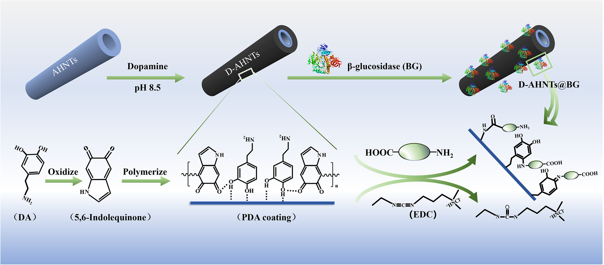
Vacuum-dried D-AHNTs (60 mg) was dispersed in 20 mL of the 0.25% (w/v) EDC solution. Subsequently, the mixture was stirred magnetically at 200 rpm for 30 min at room temperature. After this stirring step, the suspension was centrifuged at 10,000 rpm (13,750×g) for 10 min. Next, the activated D-AHNTs were dispersed in 5 mL of β-glucosidase solution (2 mg mL–1 in 0.1 M citrate-phosphate buffer, pH 5.0). The mixture was then stirred magnetically at 200 rpm for 3 h at ambient temperature. Following centrifugation at 10,000 rpm (13,750×g) for 10 min, the resulting pellet was rinsed three times with 10 mM phosphate buffer (pH 7.0). Finally, the halloysite-based biocatalyst, designated as D-AHNTs/BG, was stored at 4°C under dark conditions. The stepwise immobilization procedure and underlying mechanism are illustrated in Fig. 1.
Schematic diagram of β-glucosidase immobilization on polydopamine-functionalized halloysite nanotubes.

Figure 1. Long description
From left to right, the diagram begins with a blue halloysite nanotube labeled A H N Ts. An arrow labeled Dopamine, p H 8 point 5, points to a black nanotube labeled D-A H N Ts. Below, dopamine (D A) is shown oxidizing to 5 comma 6-indolequinone, then polymerizing to form a polydopamine (P D A) coating on the nanotube. The next arrow, labeled beta-glucosidase (B G), points to a nanotube with attached enzyme molecules, labeled D-A H N Ts at B G. Below, the P D A coating reacts with a carboxyl-amine group via E D C, forming a covalent bond. Chemical structures for dopamine, indolequinone, P D A, and the coupling reaction are depicted. The flow illustrates the sequential functionalization and enzyme immobilization steps.
β-glucosidase activity assay
β-glucosidase activity assay was conducted in a reaction system containing 100 μL of 50 mM pNPG as the substrate (Ramírez-Ramírez et al., Reference Ramírez-Ramírez, Zazueta-Alvarez, Fileto-Pérez, Reyes-Jáquez, Núñez-Núñez, De la Rosa, López-Miranda and Vázquez-Ortega2022). The reaction was initiated by adding 100 μL of β-glucosidase solution (2 mg mL-1, dissolved in 10 mM phosphate buffer with pH 7.0), using either free or immobilized enzyme. The quenched solution was vortexed thoroughly and equilibrated for 5 min at room temperature. Absorbance at 400 nm was measured using a UV-2800A spectrophotometer (UNICO, Shanghai, China) with a 1 cm pathlength cuvette. One enzyme unit (U) was defined as the amount producing 1 μmol of p-nitrophenol per minute under standard assay conditions (50°C, pH 5.0). A standard curve (Fig. S1, Supplementary material) was generated with p-nitrophenol solutions (0–60 μM).
β-Glucosidase assay
The amount of β-glucosidase immobilized on D-AHNTs was determined indirectly by subtracting the residual protein concentration in the supernatant from the initial protein concentration (Jung et al., Reference Jung, Shin, Song, Kim and Kim2012). Protein concentrations (both initial and residual in the supernatant) were quantified using the Bradford method (Bradford, Reference Bradford1976), with bovine serum albumin (BSA) as the standard.
Physicochemical environment tests
To determine the optimal pH and temperature, free β-glucosidase and D-AHNTs/BG were pre-incubated for 10 min across the pH range of 3.0–8.0 (using 100 mM citrate-phosphate buffer) and a temperature range of 30–80°C, respectively. To assess pH stability, the samples were incubated in 100 mM citrate-phosphate buffer (pH 3.0–8.0) at 25°C for 1 h with continuous shaking. In the thermal stability test, free β-glucosidase and D-AHNTs/BG were incubated in 100 mM citrate-phosphate buffer (pH 5.0) at 30–80°C for 1 h with shaking.
Michaelis–Menten kinetics
Initial reaction rates (ν ₀) were measured in 100 mM citrate-phosphate buffer (pH 7.0) using 0.5–10.0 mM pNPG at 50°C. Michaelis–Menten kinetics (Pota et al., Reference Pota, Salerno, Costantini, Silvestri, Passaro and Califano2022) (Eqn 1) was applied to analyze the substrate concentration dependence for D-AHNTs/BG. Kinetic parameters (Km and V max) for free β-glucosidase and D-AHNTs/BG were derived from Lineweaver-Burk plots (Eqn 2):


where V max is the maximum specific activity (mM L–1 min-1), [S]0 denotes the substrate concentration (mM L–1), and Km is the Michaelis–Menten constant (mM L–1), defined as the substrate concentration at half-maximal reaction velocity.
Stability and re-usability of immobilized β-glucosidase
For storage stability evaluation, the enzymes were stored in a 10 mM phosphate buffer (pH 7.0) at 4°C for 30 days, and their activity was measured at 5-day intervals. Relative activity is defined as the percentage of residual enzyme activity relative to the maximum enzyme activity observed within each experimental set. The re-usability of D-AHNTs/BG biocatalyst was assessed through six consecutive hydrolytic cycles using pNPG as substrate under standard assay conditions (50°C, pH 5.0 in 100 mM citrate-phosphate buffer). Each cycle was followed by centrifugation of the reaction mixture at 10,000 rpm (13,750×g) for 10 min to isolate D-AHNTs/BG. The recovered biocatalyst was washed three times with 10 mL of phosphate buffer (10 mM, pH 7.0) via centrifugation. A fresh reaction mixture (100 μL of 50 mM pNPG, and 2.9 mL of citrate-phosphate buffer) was introduced to initiate the subsequent cycle. The activity measured in the first cycle was defined as 100% for relative activity calculation in subsequent cycles.
Results and Discussion
Preparation of matrix and β-glucosidase immobilization
Characterization of matrix and the immobilized β-glucosidase
SEM and EDS characterization
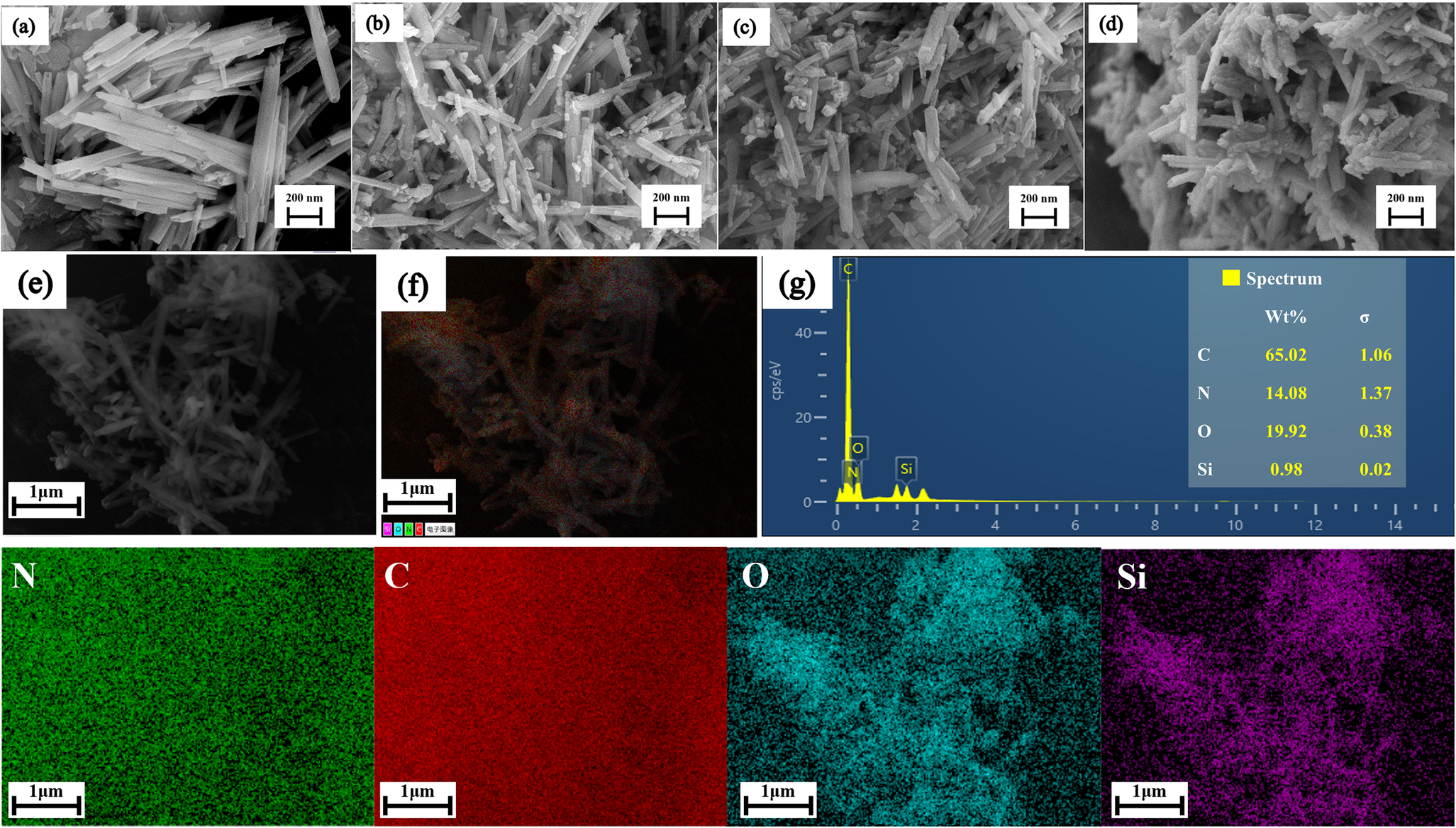
The surface morphologies and microstructural features of HNTs, AHNTs, D-AHNTs, and D-AHNTs/BG were characterized using SEM (Fig. 2a–d). Pure HNTs exhibited tubular structures with uniform width (Fig. 2a) (Das et al., Reference Das, Ganguly, Bhawal, Remanan, Mondal and Das2018; Sahiner et al., Reference Sahiner, Demirci and Sahiner2022). Following alkali activation and PDA coating, HNTs still retain the tubular morphology (Fig. 2b,c) (Das et al., Reference Das, Ganguly, Bhawal, Remanan, Mondal and Das2018). Figure 2e,f presents the EDS spectrum of D-AHNTs, in which characteristic signals of five primary elements – C, N, O, Si, and Al – were clearly detected. In the corresponding elemental mapping image (Fig. 2g), the N signal (14.08 wt.%) emerged and the C content increased significantly to 65.08 wt.%. Further elemental distribution analysis indicated that the characteristic elements (N and C) exhibited a high degree of co-localization and uniform distribution with the substrate elements (Si and Al). This suggested that PDA did not form a simple physical mixture or agglomeration; instead, it generated a uniform and continuous polymer coating on the surface of the alkali-etched HNTs, thereby confirming the successful preparation of the D-AHNTs.
SEM images of the outer surfaces of (a) HNTs, (b) AHNTs, (c) D-AHNTs, and (d) D-AHNTs/BG; (e) and (f) EDS elemental mapping of D-AHNTs confirming distribution of C, N, O, and Si; (g) the corresponding elemental mapping image of D-AHNTs.

Figure 2. Long description
The composite contains eleven panels. Top row, left to right: four S E M micrographs labeled a, b, c, d, each showing clusters of elongated tubular structures with scale bars of 200 nanometers, representing H N T s, A H N T s, D-A H N T s, and D-A H N T s forward slash B G, respectively. Middle row, left to right: two grayscale E D S elemental mapping images labeled e and f, both showing the same nanotube cluster at 1 micrometer scale, with f overlaying color-coded elemental signals. The third panel, g, is an E D S spectrum with a yellow line plot showing counts per electron volt on the y axis and energy from 0 to 14 kilo electron volts on the x axis. Four peaks are labeled C, N, O, Si, with a table listing weight percentages: C 65.02, N 14.08, O 19.92, Si 0.98, and corresponding standard deviations. Bottom row, left to right: four elemental distribution maps for N (green), C (red), O (cyan), and Si (magenta), each showing a uniform or dispersed signal across the nanotube region at 1 micrometer scale.
TEM and specific surface area characterization
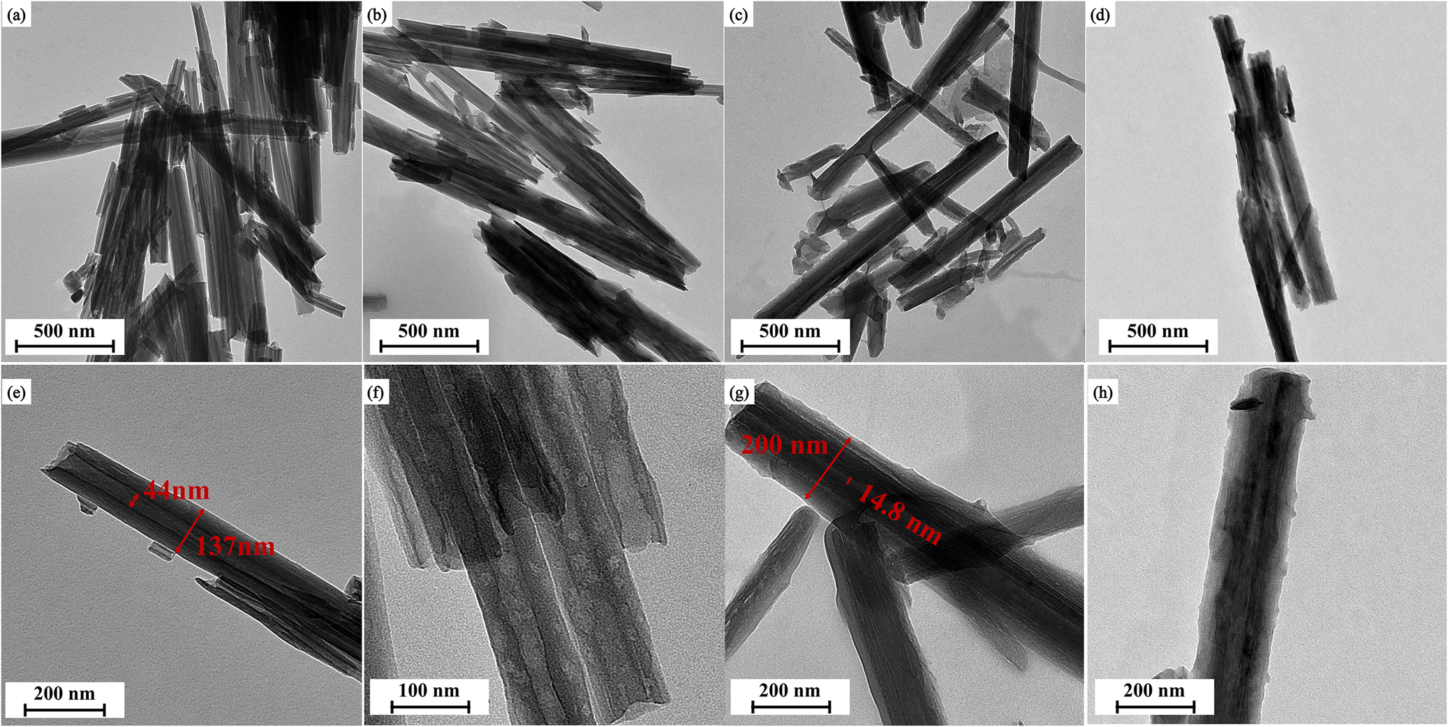
The morphological evolution of the samples at different preparation stages was investigated via TEM. Figure 3a and 3e display the typical morphology of pristine HNTs, which exhibited a characteristic hollow nanotubular structure with a high aspect ratio. Upon alkali activation, the overall tubular integrity of AHNTs was well-preserved without structural collapse (Fig. 3b and 3f). As shown in Fig. 3g, the outer diameter of D-AHNTs increased to ~200 nm. Compared with pristine HNTs (~137 nm), this increment of 63 nm corresponded to a PDA coating thickness of roughly 31.5 nm on each side. The formation of this substantial polymer layer confirmed that the alkali-activated surface effectively facilitated the robust deposition of PDA. Upon immobilization of β-glucosidase, the resulting D-AHNTs/BG (Fig. 3d and 3h) retained the characteristic tubular morphology and the distinct polymer shell observed in D-AHNTs. No structural collapse or significant aggregation was observed, verifying the mechanical stability of the composite carrier under the immobilization conditions.
TEM images of (a, e) HNTs, (b, f) AHNTs, (c, g) D-AHNTs, and (d, h) D-AHNTs/BG.

Figure 3. Long description
There are eight panels arranged in two rows and four columns. Top row, from left to right: panel a shows clustered tubular structures labeled H N T with a 500 nanometer scale bar; panel b displays similar but slightly less dense tubes labeled A H N T, also with a 500 nanometer scale bar; panel c presents D hyphen A H N T tubes, again with a 500 nanometer scale bar; panel d shows a single bundle of tubes labeled D hyphen A H N T slash B G, with a 500 nanometer scale bar. Bottom row, from left to right: panel e is a close-up of H N T with red lines marking 44 nanometers and 137 nanometers across the tube wall and diameter, scale bar 200 nanometers; panel f is a higher magnification of A H N T, showing wall layering, scale bar 100 nanometers; panel g is a close-up of D hyphen A H N T with red lines marking 200 nanometers and 14.8 nanometers, scale bar 200 nanometers; panel h is a close-up of D hyphen A H N T slash B G, showing a single tube, scale bar 200 nanometers. The panels highlight differences in tube morphology, wall thickness, and aggregation among the four sample types.
The specific surface areas of HNTs, AHNTs, and D-AHNTs were determined via N2 adsorption-desorption isotherms (Fig. S2, Table S1; see Supplementary material). After alkali treatment, the specific surface area of AHNTs increased slightly from 37.788 m2 g–1 (pristine HNTs) to 38.094 m2 g–1, while the average pore diameter expanded from 22.613 nm to 23.282 nm. This trend suggested that moderate alkali treatment removed amorphous silica impurities from the lumen or surface and unclogged blocked pores, thereby enlarging the effective pore size (Boro et al., Reference Boro, Priyadarsini and Moholkar2022). Upon the introduction of the PDA coating, the pore parameters of D-AHNTs underwent significant changes. Specifically, the specific surface area decreased notably from 38.094 m2 g–1 (AHNTs) to 31.721 m2 g–1, and the average pore diameter was reduced from 23.282 nm to 21.645 nm. This reduction was attributed to the PDA polymer layer covering the outer surface of the halloysite and potentially causing partial blockage at the tube openings. This occupation of effective mesoporous volume restricted the surface area accessible to N2 molecules, which confirmed the successful construction of the functional polymer layer.
Thermogravimetric analysis
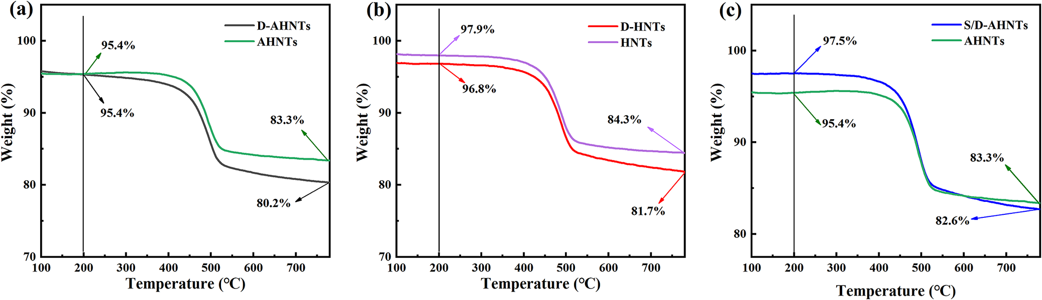
Thermogravimetric analysis quantified the PDA loading efficiency (Fig. 4a–c), with the baseline curves for HNTs and AHNTs shown in Fig. S3 (see Supplementary material). At 150°C, HNTs and AHNTs exhibited mass losses of 2.0% and 4.7%, respectively. This increased weight loss was likely attributed to the formation of surface hydroxyls via alkaline hydrolysis, where the generated silanol groups served as active sites to adsorb water molecules through hydrogen bonding. Both substrate materials (AHNTs and HNTs) exhibited a characteristic mass loss step in the temperature range of 450–550°C, which corresponds to the dehydroxylation of structural aluminol groups within the halloysite lattice (Yuan et al., Reference Yuan, Southon, Liu and Kepert2012; Ferrante et al., Reference Ferrante, Bertini, Ferlito, Lisuzzo, Lazzara and Duca2023). Compared with the D-HNTs, which showed a PDA loading of ~1.1% (Fig. 4b), the D-AHNTs prepared under weakly alkaline conditions (pH 8.5) exhibited an increased loading of 2.9% (Fig. 4a). This enhancement suggested that the alkali-induced surface hydroxyl groups and defects provided favorable sites for the anchoring and in situ polymerization of dopamine. To examine pH effects, S/D-AHNTs were prepared under acidic conditions (pH 6.5 Tris-HCl). TGA results revealed a lower loading (~2.6%) compared with the D-AHNTs (2.9%). This reduction is tentatively attributed to the electrostatic repulsion induced by the simultaneous protonation of surface hydroxyls and dopamine amino groups, which likely hinders the formation of a robust PDA layer (Ball et al., Reference Ball, Frari, Toniazzo and Ruch2012; Sahiner et al., Reference Sahiner, Demirci and Sahiner2022).
TGA curves of the unmodified and modified halloysite nanotubes: (a) D-AHNTs vs AHNTs, (b) D-HNTs vs HNTs, and (c) S/D-AHNTs vs AHNTs.

Figure 4. Long description
From left to right, panel a compares D dash A H N T s (black) and A H N T s (green). Both curves start near 100 percent weight at 100 degrees Celsius. At 300 degrees, both are at 95.4 percent. By 700 degrees, D dash A H N T s drops to 80.2 percent and A H N T s to 83.3 percent. Panel b compares D dash H N T s (red) and H N T s (purple). At 300 degrees, D dash H N T s is at 97.9 percent and H N T s at 96.8 percent. At 700 degrees, D dash H N T s is at 81.7 percent and H N T s at 84.3 percent. Panel c compares S slash D dash A H N T s (blue) and A H N T s (green). At 300 degrees, S slash D dash A H N T s is at 97.5 percent and A H N T s at 95.4 percent. At 700 degrees, S slash D dash A H N T s is at 82.6 percent and A H N T s at 83.3 percent. All panels show a two-step weight loss with modified samples generally retaining less weight at higher temperatures.
XPS characterization
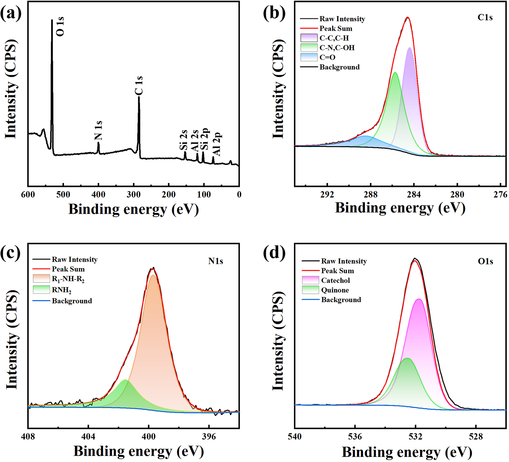
XPS measurement was conducted on D-AHNTs to further confirm the presence of a PDA coating layer on AHNTs. The XPS survey spectra of D-AHNTs show O, N, C, Si, and Al (Fig. 5a), while the complicated nature of polydopamine on D-AHNTs is revealed by the different chemical states of C, N, and O in deconvoluted core-level spectra (Fig. 5b–d). In the C 1s spectrum (Fig. 5b), the characteristic peaks at 284.2, 285.7, and 287.9 eV correspond to the C-C/C-H bonds (relative peak area = 45.64%), C-N/C-OH bonds (relative peak area = 31.33%), and C=O bonds in the dopamine polymer (relative peak area = 13.03%), respectively. The N 1s spectrum is shown in Fig. 5c. The main peak at 399.8 eV indicates the presence of R₂NH (secondary amine, relative peak area = 78.17%) structures, while the peak at 401.8 eV is attributed to RNH₂ (primary amine, relative peak area = 21.83%) structures. The O 1s high-resolution spectrum of D-AHNTs (Fig. 5d) can be peak-fitted into two components corresponding to catechol groups (531.9 eV) and quinone structures (532.7 eV) in PDA (Feng et al., Reference Feng, Fan, Zha, Wang and Jin2016; Choi et al., Reference Choi, Chaudhari, Shin, Cho, Lee, Shon, Nam and Park2022).
The survey of XPS spectra of (a) D-AHNTs and high-resolution scanning of (b) C 1s, (c) N 1s, and (d) O 1s for D-AHNTs.

Figure 5. Long description
Panel a, top left, displays a survey X P S spectrum with binding energy from 600 to 0 electron volts and intensity in counts per second. Major peaks are labeled O one s at about 532, N one s at about 400, C one s at about 285, Si two s, Al two s, Si two p, and Al two p at lower energies. Panel b, top right, shows a high-resolution scan of C one s with binding energy from 292 to 276 electron volts. The raw intensity (black), peak sum (red), and fitted components for C dash C, C dash H (magenta), C dash N, C dash O H (green), C dash O (blue), and background (gray) are shown, with the main peak near 284.5 electron volts. Panel c, bottom left, presents N one s with binding energy from 408 to 396 electron volts. Raw intensity (black), peak sum (red), R one dash N H dash R two (magenta), R N H two (green), and background (blue) are plotted, with the main peak near 400 electron volts. Panel d, bottom right, shows O one s with binding energy from 540 to 528 electron volts. Raw intensity (black), peak sum (red), catechol (magenta), quinone (green), and background (blue) are plotted, with the main peak near 532 electron volts. All panels use consistent color coding for fitted components as indicated in their legends.
XRD characterization
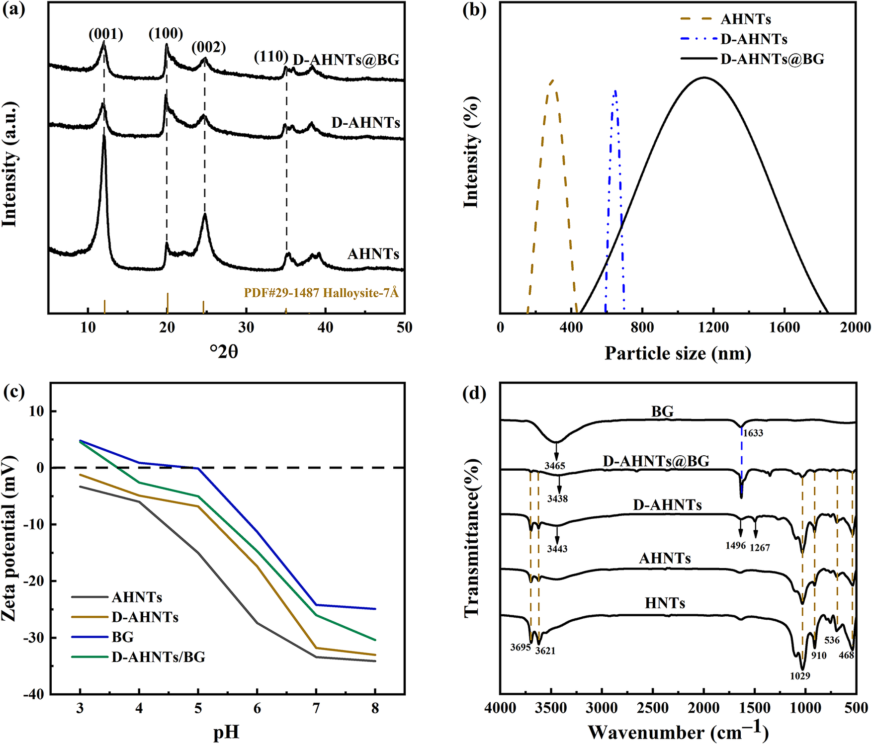
X-ray diffraction analysis (Fig. 6a) was employed to characterize the crystalline structures of AHNTs, D-AHNTs, and D-AHNTs/BG. The XRD pattern of pristine, unmodified halloysite (as-received) is provided in Fig. S3 (see Supplementary material). In the AHNTs, D-AHNTs, and D-AHNTs/BG samples, the set of four characteristic diffraction reflections of HNTs at 12.1, 20.1, 24.6, and 35.0°2θ, corresponding to the d 001, d 100, d 002, and d 110 values of HNTs, is clearly observable (Meng et al., Reference Meng, Ju, Wang, Cui, Liu, Ma, Wang and Yang2025). Compared with AHNTs, the unchanging peak positions of D-AHNTs indicated that the crystalline structure of HNTs remained intact. However, a noticeable attenuation in peak intensity was observed. This was attributed to the mass dilution and shielding effects of the amorphous PDA coating (Sahiner et al., Reference Sahiner, Demirci and Sahiner2022; Massaro et al., Reference Massaro, D’Acunzi, Paganelli, Alfieri, Liotta, Lopez-Galindo, de Melo Barbosa, Piccolo, Sánchez-Espejo, Viseras and Riela2025). Similarly, for the enzyme-immobilized D-AHNTs/BG, the diffraction pattern exhibited no discernible changes regarding peak position or shape compared with D-AHNTs. This verified that the mild immobilization process did not compromise the tubular framework.
(a) XRD patterns of AHNTs, D-AHNTs, D-AHNTs/BG; (b) particle-size distribution of AHNTs, D-AHNTs, D-AHNTs/BG; (c) zeta potentials of D-AHNTs, free β-glucosidase (BG), D-AHNTs/BG; (d) FTIR spectra of BG, HNTs, AHNTs, D-AHNTs, and D-AHNTs/BG.

Figure 6. Long description
Top left panel a shows X R D patterns with intensity on the y axis and two theta on the x axis, displaying three stacked traces for A H N Ts, D-A H N Ts, and D-A H N Ts at B G. Peaks are labeled at 001, 100, 002, and 110, with reference lines for Halloysite-7A. Top right panel b plots particle size distribution with intensity percent on the y axis and particle size in nanometers on the x axis. Three traces are shown: A H N Ts (dashed brown, peak near 400 nanometers), D-A H N Ts (dashed blue, peak near 600 nanometers), and D-A H N Ts at B G (solid black, broad peak centered near 1200 nanometers). Bottom left panel c presents zeta potential in millivolts on the y axis versus p H on the x axis, with four traces: A H N Ts, D-A H N Ts, B G, and D-A H N Ts at B G. All traces show a decrease in zeta potential as p H increases from 3 to 8, crossing from positive to negative values. Bottom right panel d displays F T I R spectra with transmittance percent on the y axis and wavenumber in inverse centimeters on the x axis. Five traces are shown for B G, D-A H N Ts at B G, D-A H N Ts, A H N Ts, and H N Ts. Key peaks are labeled at 3465, 3438, 3443, 3695, 3621, 1633, 1496, 1267, 1029, 910, 536, and 468 inverse centimeters.
Particle-size and zeta-potential analysis
Sequential size increases from pristine AHNTs (282±3.10 nm) through polydopamine-functionalized D-AHNTs (650±21.5 nm) to enzyme-immobilized D-AHNTs/BG (1088±33.7 nm) in aqueous media (Fig. 6b) validate the hydrodynamic radius expansion mechanism (Cui et al., Reference Cui, Fu, Zhou, Zhang, Zhang, Yang and Fu2025). Light-scattering data confirm successful enzymatic loading via carrier-surface modification. Figure 6c displays the zeta-potential curves of free enzyme (BG), D-AHNTs, and the immobilized enzyme (D-AHNTs/BG). Variations in zeta potential arise primarily from differences in particle morphology, dimensions, and surface functional groups. Over pH 3–8, AHNTs exhibited the most negative zeta potential across the tested pH range. Upon PDA modification, the zeta potential of D-AHNTs shifted toward less negative values compared with AHNTs. This trend confirms that the deposition of the PDA layer effectively shields the inherent negative charge of the AHNTs surface through electrostatic neutralization (Sahiner et al., Reference Sahiner, Demirci and Sahiner2022; Taş et al., Reference Taş, Gundogdu and Ünal2022). Immobilization enriched BG molecules on the carrier surface, and shifted the zeta potential of D-AHNTs/BG toward less negative values relative to D-AHNTs alone. Free β-glucosidase exhibits an isoelectric point (pI) of 4.8, indicating a net zero charge at this pH (Reshmi and Sugunan, Reference Reshmi and Sugunan2013). At pH 5 (greater than the pI value for BG), partial protonation of carboxyl groups confers weak negative charge on the enzyme. Although electrostatic repulsion occurs with the negatively charged carrier, this does not prevent enzyme-carrier binding. Successful immobilization is probably mediated by covalent between surface amino groups of the carrier and carboxyl residues of the enzyme (Coutinho et al., Reference Coutinho, Rojas, Tardioli, Paris and Farinas2018).
FTIR characterization
The FTIR spectrum (Fig. 6d) exhibits distinct absorption bands characteristic of HNTs. Specifically, the peaks at 3695 cm–1 and 3621 cm–1 are attributed to the -OH- stretching vibrations of Al-OH groups situated on the inner surface of the halloysite, while the band at 910 cm–1 is assigned to Al-OH bending modes. Additionally, the spectral regions 1000–1100 cm–1 (e.g. 1029 cm–1 Si-O-Si) and 450–550 cm–1 (e.g. 536 cm–1 Al-O-Si, 468 cm–1 Si-O-Si) originate from Si-O stretching and Si-O bending vibrations, respectively (Jiao et al., Reference Jiao, Feng and Chen2023). The broad absorption band at 3443 cm–1 in alkali-activated HNTs is attributed to hydrogen-bonded hydroxyl groups, which may overlap with the N-H stretching vibration arising from the -NH₂ groups of polydopamine. Concomitantly, the emergence of a new absorption peak at 1496 cm–1, assignable to the coupled stretching vibration of aromatic rings and N-H deformation, and at 1267 cm–1 assigned to C-O stretching from phenolic groups (Zangmeister et al., Reference Zangmeister, Morris and Tarlov2013; Jiao et al., Reference Jiao, Feng and Chen2023), validates further the successful synthesis of PDA on the surfaces of HNTs (Das et al., Reference Das, Ganguly, Bhawal, Remanan, Mondal and Das2018; Ou et al., Reference Ou, Wu, Han, Kong, Song, Chen and Su2022). The amide I band (1633 cm–1) persists in both free and immobilized β-glucosidase, confirming the successful immobilization of β-glucosidase (Jiao et al., Reference Jiao, Feng and Chen2023).
Enzyme secondary structures analysis
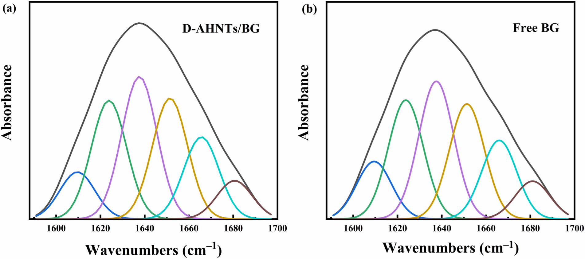
The amide I band arises from overlapping vibrational modes corresponding to fundamental enzyme secondary structures, including α-helix, β-sheet (parallel and anti-parallel), β-turn, and random coil (Sheng et al., Reference Sheng, Dong, Coffin, Myrold and Kleber2022). Owing to its sensitivity to secondary structural alterations, the amide I band serves as a primary probe for monitoring protein conformational dynamics. Deconvolution of FTIR/ATR spectra quantified the secondary structures of native β-glucosidase and D-AHNTs/BG, with comparative results visualized in Fig. 7a,b. Table 1 summarizes the compositional distribution of secondary structures derived from spectral fitting for both enzyme forms. Immobilization on D-AHNTs preserved the secondary structural profile of β-glucosidase. This covalent immobilization strategy effectively mitigated enzyme leaching while maintaining the enzyme’s structural integrity, demonstrating the successful fabrication of the D-AHNTs/BG biocatalyst.
Deconvolution analysis of the FTIR/ATR spectroscopy amide I band for (a) D-AHNTs/BG and (b) free β-glucosidase (BG).

Figure 7. Long description
Panel a, labeled D dash A H N T s forward slash B G, and panel b, labeled Free B G, each show a main black curve representing total absorbance with five colored component peaks underneath. The x-axis is labeled Wavenumbers in centimeters super minus 1, ranging from 1600 to 1700. The y-axis is labeled Absorbance. In both panels, the colored peaks (blue, green, purple, yellow, cyan, brown) are distributed from left to right, with maxima at different wavenumbers. The sum of these colored peaks forms the overall amide I band envelope. The peak positions and relative intensities differ slightly between the two panels, indicating changes in secondary structure composition between D dash A H N T s forward slash B G and free B G.
A comparison of enzyme secondary structures from FTIR/ATR Amide I band deconvolution analysis

Table 1. Long description
From left to right, the first column lists sample names: Free B G and D dash A H N Ts slash B G. The next six columns display protein secondary structure percentages. For Free B G: Aggregated strands 10.8, beta-sheet 21.7, Random coil 25.2, alpha-helix 21.0, beta-turn 14.4, beta-anti-parallel 6.91. For D dash A H N Ts slash B G: Aggregated strands 10.4, beta-sheet 21.2, Random coil 25.4, alpha-helix 21.6, beta-turn 14.6, beta-anti-parallel 6.86. Each value is presented as a percentage, enabling direct comparison between the two samples across all structural categories.
Immobilization studies
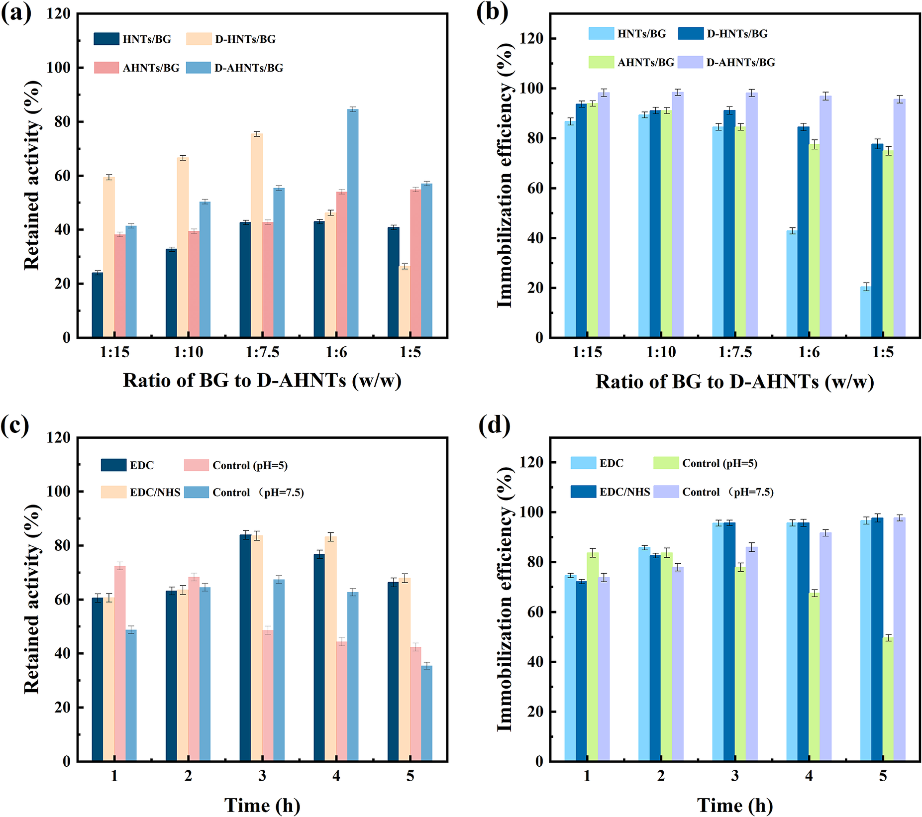
Carbodiimide (EDC), acting as a cross-linker, activates carboxyl groups under acidic conditions (pH 4.5–5.5) to react with primary amines (Ricco et al., Reference Ricco, Doherty and Falcaro2014; Wang et al., Reference Wang, Sun, Ma, Dong, Huo, Li, Zhao, Yu and Huang2019a; Zhang et al., Reference Zhang, Sun, Lv, Golubev, Wang, Dong, Kotova and Kotova2021). This mechanism facilitates the formation of amide bonds between the carboxyl groups of β-glucosidase and the amino groups on the PDA layer (Fig. 1). Figure 8a,b illustrates the retained activity and immobilization efficiency of β-glucosidase immobilized on various supports (HNTs, AHNTs, D-HNTs, and D-AHNTs) via EDC activation across different loading densities. The control groups (HNTs and AHNTs) relying on physical adsorption exhibited lower efficiency and stability. D-HNTs showed improved performance but was limited by the lower PDA loading capacity of the pristine surface. In contrast, D-AHNTs exhibited the greatest retained activity and immobilization efficiency. At the enzyme to D-AHNTs ratio of 1:6, D-AHNTs/BG achieved a maximum activity recovery of 84.57%, while maintaining an immobilization efficiency of 96–98% with an enzyme loading of ~167 mg g–1 support.
Retained activity and immobilization efficiency of β-glucosidase: (a, b) effect of support type (HNTs, AHNTs, D-HNTs, and D-AHNTs) and ratio of BG to D-AHNTs; (c, d) effect of coupling strategies (EDC, EDC/NHS, and different pH conditions) on D-AHNTs.

Figure 8. Long description
Panel a, top left, plots retained activity percentage on the y axis against ratio of B G to D dash A H N T s on the x axis, with five ratios from 1 to 15 to 1 to 5. Four bar groups per ratio represent H N T s slash B G, A H N T s slash B G, D dash H N T s slash B G, and D dash A H N T s slash B G. D dash A H N T s slash B G shows the highest retained activity, peaking at 1 to 5 ratio. Panel b, top right, shows immobilization efficiency percentage on the y axis versus the same x axis ratios and bar groups as panel a. D dash A H N T s slash B G consistently achieves the highest efficiency, exceeding 100 percent at higher ratios. Panel c, bottom left, plots retained activity percentage on the y axis against time in hours from 1 to 5 on the x axis. Four bar groups per time point represent E D C, E D C slash N H S, control at p H 5, and control at p H 7.5. E D C slash N H S and control at p H 7.5 show higher retained activity at later time points. Panel d, bottom right, shows immobilization efficiency percentage on the y axis versus time in hours on the x axis, with the same four bar groups as panel c. E D C slash N H S and control at p H 7.5 maintain the highest immobilization efficiency across all time points.
Generally, EDC-mediated coupling requires the addition of N-hydroxysuccinimide (NHS) to enhance reaction efficiency and stabilize the O-acylisourea intermediate (Zhang et al., Reference Zhang, Xu, Yuan, Qi, Liu and He2012; Li et al., Reference Li, Wang, Jiang, Ye, Zhang and Zhang2015). Furthermore, residual quinone groups within the PDA coating can react with nucleophiles under weakly alkaline conditions (pH 7.5–8.5), allowing for covalent coupling with the amino groups of the enzyme via Michael addition or Schiff base formation (Lin et al., Reference Lin, Xia, Huang, Zhu, Cai, Wei and Liao2022; Kimmins et al., Reference Kimmins, Henríquez, Torres, Wilson, Flores, Pio, Jullian, Urbano, Braun-Galleani, Ottone, Muñoz, Claros and Urrutia2024). Figure 8c,d display the retained activity and immobilization efficiency of β-glucosidase immobilized on D-AHNTs using different strategies (EDC, EDC/NHS, and without EDC/NHS at pH 5.0 or 7.5). The results indicated that the alkaline coupling strategy yielded lower retained activity and required longer reaction times to achieve comparable immobilization efficiency. Prolonged stirring under weakly alkaline conditions (pH 7.5) during the spontaneous coupling process likely induced partial conformational changes, leading to a loss of intrinsic enzymatic activity. Our XPS analysis revealed a relatively low content of quinone species on the D-AHNTs surface. The deficiency of reactive quinone sites limited the efficiency of Schiff base formation or Michael addition under alkaline conditions. The results demonstrated that D-AHNTs/BG prepared via EDC activation without NHS (pH 5.0, loading of 167 mg g–1support, 3 h) exhibited superior performance, where the retained activity and immobilization efficiency were 84.6% and 98%, respectively.
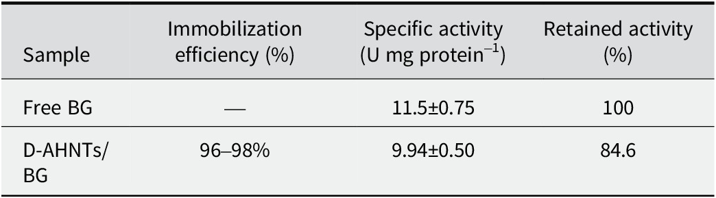
Covalent immobilization of the enzyme onto the PDA matrix, combined with the efficient utilization of carrier surface sites, effectively prevented enzyme leaching while preserving catalytic functionality. A consistently high immobilization efficiency (>96%) was maintained across all loading capacities, while carrier modification enabled a threefold increase in enzyme loading without compromising the specific activity of β-glucosidase (9.94 U mg protein–1) (Table 2). In addition, enzyme leaching from D-AHNTs was evaluated by repeatedly washing the immobilized biocatalyst with deionized water and measuring residual activity in the wash solution (Table S2; see Supplementary material). Negligible activity was detected in the wash effluents, confirming minimal leaching.
Immobilization efficiency and catalytic performance of free vs immobilized β-glucosidase (167 mg g–1support)

Table 2. Long description
Column headers from left to right are Sample, Immobilization efficiency (percent), Specific activity (U per mg protein), and Retained activity (percent). The first row lists Free B G with immobilization efficiency not applicable, specific activity 11.5 plus or minus 0.75, and retained activity 100 percent. The second row lists D dash A H N Ts slash B G with immobilization efficiency 96 to 98 percent, specific activity 9.94 plus or minus 0.50, and retained activity 84.6 percent.
Physicochemical environment tests
Effects of pH and temperature
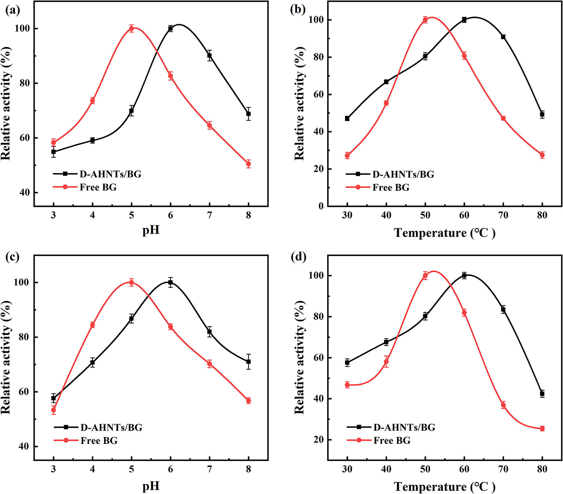
To maximize the catalytic performance of both D-AHNTs/BG and free β-glucosidase, the influence of pH and temperature on their activities was investigated systematically. As illustrated in Fig. 9a, free β-glucosidase exhibited optimal activity under slightly acidic conditions (pH 5.0), whereas the immobilized counterpart reached maximum activity at pH 6.0. Upon immobilization, the interaction between the enzyme and the carrier may have selectively stabilized a specific conformational state, leading to a shift of the optimal pH towards the alkaline region (Reshmi and Sugunan, Reference Reshmi and Sugunan2013; Cui et al., Reference Cui, Fu, Zhou, Zhang, Zhang, Yang and Fu2025). Moreover, the free enzyme showed a significant and rapid decline in activity when pH exceeded 6.0 or fell below 5.0. In contrast, D-AHNTs/BG maintained over 80% relative activity across pH 5.5–7.0, demonstrating broader pH adaptability.
Effects of pH and temperature on enzymatic activity and stability. (a) Optimal pH for activity; (b) optimal temperature for activity; (c) pH stability profiles; (d) thermal stability profiles (free BG: free β-glucosidase; immobilized BG: D-AHNTs/BG).

Figure 9. Long description
Panel a, top left, plots relative activity percent on the y-axis versus p H from 3 to 8 on the x-axis. D dash A H N T s slash B G peaks at p H 6, Free B G at p H 5. Panel b, top right, plots relative activity percent versus temperature from 30 to 80 degrees Celsius. D dash A H N T s slash B G peaks at 70 degrees, Free B G at 60 degrees. Panel c, bottom left, shows p H stability with both lines peaking at p H 5, but D dash A H N T s slash B G maintains higher activity at higher p H. Panel d, bottom right, shows thermal stability with D dash A H N T s slash B G peaking at 70 degrees and Free B G at 60 degrees, with D dash A H N T s slash B G retaining more activity at higher temperatures. Legends indicate black squares for D dash A H N T s slash B G and red circles for Free B G. Error bars are present on all data points.
The temperature-dependent activity profiles of free β-glucosidase and D-AHNTs/BG were evaluated across 30–80°C (Fig. 9b). The free β-glucosidase attained peak activity at 50°C, beyond which a rapid deactivation occurred. In contrast, the D-AHNTs/BG maintained near-maximum activity at 60°C prior to gradual decline. When temperature was adjusted to 30°C or 80°C, the catalytic activity of free β-glucosidase declined to 23.0% and 25.0% of its peak value, respectively. In contrast, the D-AHNTs/BG retained significantly greater activity than the free enzyme under identical conditions. An improvement in the optimum temperature of D-AHNTs/BG over free β-glucosidase probably stems from increased rigidity of the enzyme’s peptide structure post-immobilization, thereby restricting enzyme conformational transitions at elevated temperatures (Wang et al., Reference Wang, Zhi, Wan, Han, Li and Wang2019b; Sillu et al., Reference Sillu, Agnihotri and Reddy2022).
pH and temperature stability
The D-AHNTs/BG and free β-glucosidase were incubated separately in 100 mM citrate-phosphate buffer solutions (pH ranging from 3.0 to 8.0) at 25°C for 1 h with continuous shaking. The pH stability (Fig. 9c) of D-AHNTs/BG in neutral to slightly alkaline environments (pH 7.0–8.0) was enhanced compared with free enzymes. In acidic and alkaline environments with pH values of 3.0 and 8.0, the catalytic activity of free β-glucosidase decreased significantly, maintaining only 53.3% and 56.8% of its maximum catalytic activity, respectively. This is attributed to the conformational changes of enzyme protein molecules under non-optimal pH conditions, leading to the disruption of the active center structure. In contrast, the D-AHNTs/BG exhibits stronger acid-base stability. Under the same conditions of pH 3.0 and pH 8.0, the β-glucosidase in this clay-based enzyme composite system can retain 57.7% and 71.0% of its relative catalytic activity, respectively. This is mainly attributed to the unique nano-confined effect of AHNTs, as well as the stable composite structure formed by the cross-linking between the D-AHNTs and β-glucosidase, which effectively mitigates the destructive effect of extreme pH on the conformation of enzyme proteins.
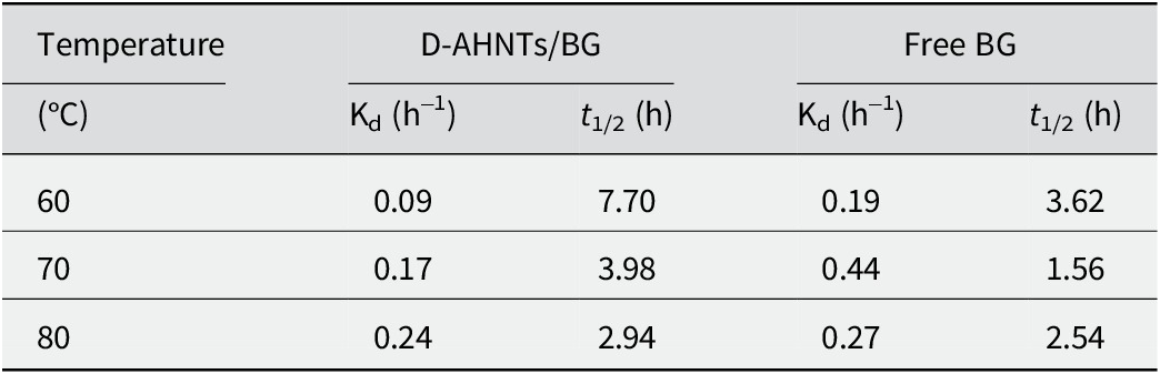
The relative activities of free enzyme and D-AHNTs/BG clay-based enzyme biocatalyst were analyzed after incubation at 30–80°C for 1 h (Fig. 9d). The D-AHNTs/BG demonstrated enhanced thermal stability at elevated temperatures (60–80°C), retaining >70.0% relative activity compared with <40.0% for free enzyme. The enhanced thermal stability following enzyme immobilization is further corroborated by Table 3. Compared with the free β-glucosidase, the β-glucosidase covalently bonded to D-AHNTs demonstrates an extended half-life when exposed to temperatures within the 60–80°C range. This stabilization arose from synergistic conformational restraint and microenvironmental hydration mechanisms; enzyme-support interactions suppress polypeptide chain mobility and thermal vibration (Cao et al., Reference Cao, Yang, Tian, Liu, Fan and Wu2023) while the hydrophilicity of PDA coating maintains water molecules to preserve the enzyme’s spatial conformation (Xu et al., Reference Xu, Liu, Wang, Li, Xu, Sun and Suo2024).
The deactivation thermal constants (Kd) and half-life (t 1/2) of both free and D-AHNTs/BG biocatalyst

Table 3. Long description
The table has five columns. The first column lists temperature in degrees Celsius: 60, 70, and 80. The next two columns are for D-A H N T s forward slash B G: K sub d in hours to the minus one and t sub 1 over 2 in hours. The last two columns are for Free B G: K sub d in hours to the minus one and t sub 1 over 2 in hours. At 60 degrees Celsius, D-A H N T s forward slash B G has K sub d 0.09 and t sub 1 over 2 7.70, while Free B G has K sub d 0.19 and t sub 1 over 2 3.62. At 70 degrees Celsius, D-A H N T s forward slash B G has K sub d 0.17 and t sub 1 over 2 3.98, Free B G has K sub d 0.44 and t sub 1 over 2 1.56. At 80 degrees Celsius, D-A H N T s forward slash B G has K sub d 0.24 and t sub 1 over 2 2.94, Free B G has K sub d 0.27 and t sub 1 over 2 2.54.
Michaelis−Menten kinetics
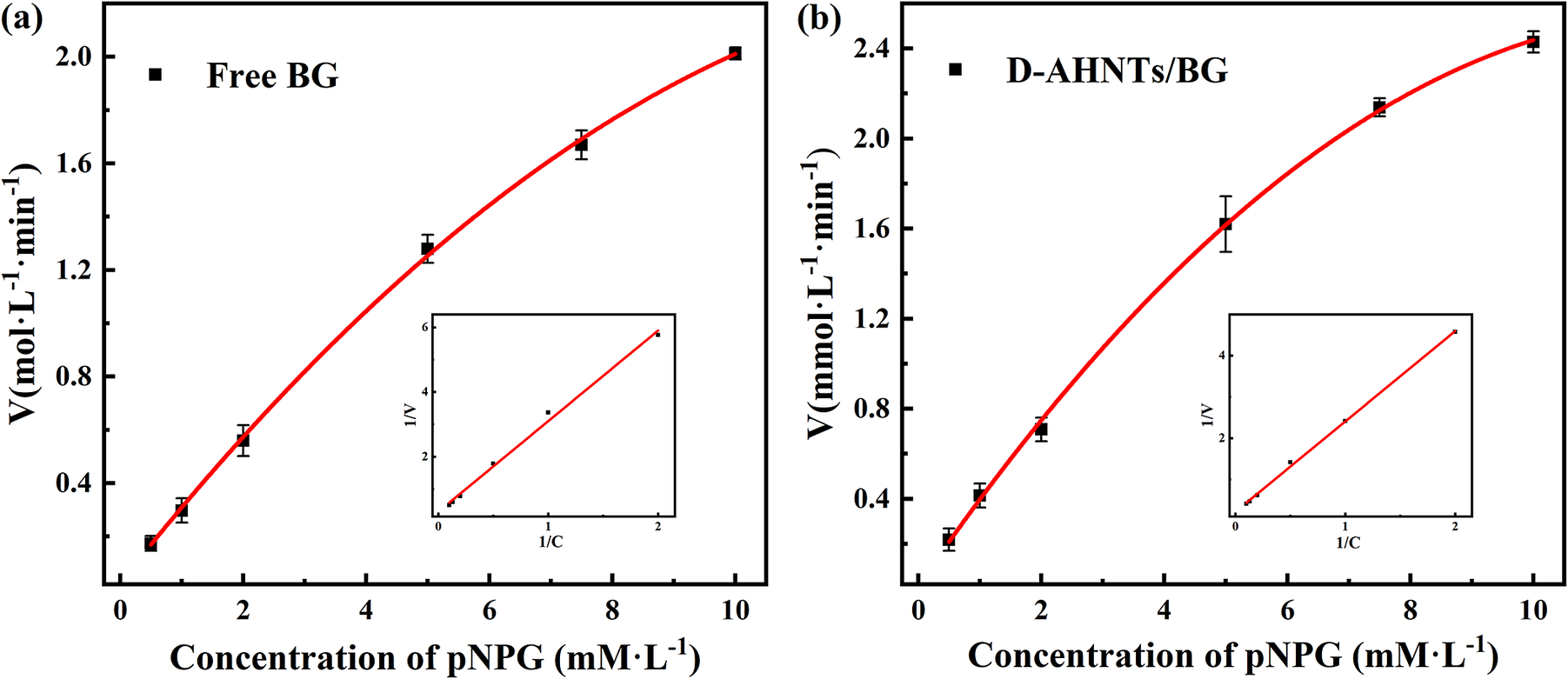
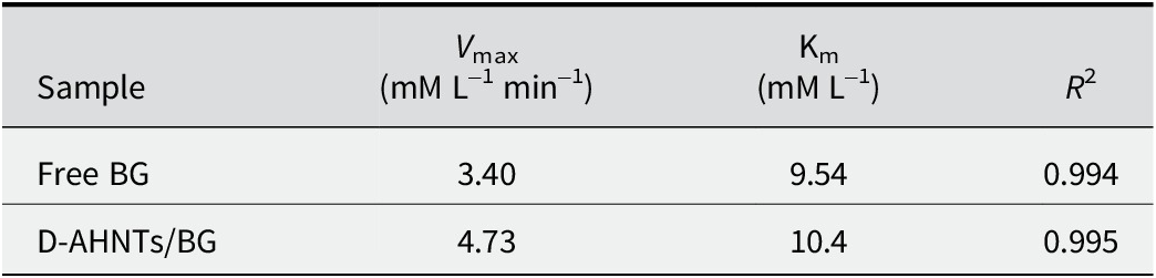
Changes in kinetic parameters post-immobilization reflect immobilization efficacy. The Michaelis constant (Km) indicates enzyme-substrate affinity, where greater values denote reduced affinity, while the maximum reaction rate (V max) represents catalytic efficiency. The catalytic activity curves and Lineweaver–Burk curves for free β-glucosidase and D-AHNTs/BG are shown in Fig. 10; the changes in V max and Km of immobilized enzymes are essentially the synergistic outcomes of conformational rigidity and mass-transfer resistance. Lineweaver–Burk analysis (Table 4) quantified V max at 3.40 mM L–1 min–1 and Km at 9.54 mM L–1 for free β-glucosidase, versus 4.73 mM L–1 min–1 and 10.4 mM L–1 for D-AHNTs/BG. The V max of D-AHNTs/BG was 39% higher than that of free β-glucosidase. This enhancement may be attributed to a combination of favorable interfacial effects. While the porous structure and PDA layer inevitably impose diffusion limitations, other factors such as the potential accumulation of substrate within the hydrophilic PDA layer or the rigidification of the enzyme in an active conformation might exert positive influences. (Reshmi and Sugunan, Reference Reshmi and Sugunan2013). The calculated Km value of the free β-glucosidase was slightly less than that of D-AHNTs/BG. The increase in the Km of the immobilized enzyme may be attributed to steric hindrance induced by the support, which reduces substrate accessibility. Although covalent immobilization altered the kinetic behavior of the free enzyme, the accessibility of the substrate to the active sites was not significantly compromised, and the enzyme-substrate affinity was well preserved.
Michaelis−Menten kinetics of free and immobilized β-glucosidase. (a) Catalytic activity and Lineweaver−Burk plot of free β-glucosidase at varying pNPG concentrations; (b) catalytic activity and Lineweaver−Burk plot of D-AHNTs/BG biocatalyst at varying pNPG concentrations.

Figure 10. Long description
Panel a, at the left, is labeled Free B G. The x-axis is Concentration of pN P G in milli molar per liter, ranging from 0 to 10. The y-axis is V in mol per liter per minute, ranging from 0 to 2.0. Black squares with error bars show data points at approximately 0, 2, 4, 6, 8, and 10 milli molar per liter. A red curve rises nonlinearly, showing increasing catalytic activity with higher substrate concentration. The inset at lower right is a Lineweaver-Burk plot with x-axis labeled 1 over C and y-axis labeled 1 over V, showing a straight line with positive slope. Panel b, at the right, is labeled D-A H N T s/B G. The x-axis is Concentration of pN P G in milli molar per liter, ranging from 0 to 10. The y-axis is V in milli mol per liter per minute, ranging from 0 to 2.4. Black squares with error bars show data points at similar intervals. The red curve also rises nonlinearly, reaching a higher maximum than panel a. The inset at lower right is a Lineweaver-Burk plot with x-axis labeled 1 over C and y-axis labeled 1 over V, showing a straight line with positive slope. Both panels demonstrate Michaelis-Menten kinetics, with the immobilized enzyme (panel b) showing higher maximum velocity.
Kinetic parameters of free β-glucosidase and D-AHNTs/BG biocatalyst

Table 4. Long description
Column headers from left to right are Sample, V max in milli molar per liter per minute, K m in milli molar per liter, and R squared. The first row lists Free B G with V max 3.40, K m 9.54, R squared 0.994. The second row lists D-A H N Ts slash B G with V max 4.73, K m 10.4, R squared 0.995. All values are aligned horizontally under their respective columns.
Storage stability and reusability
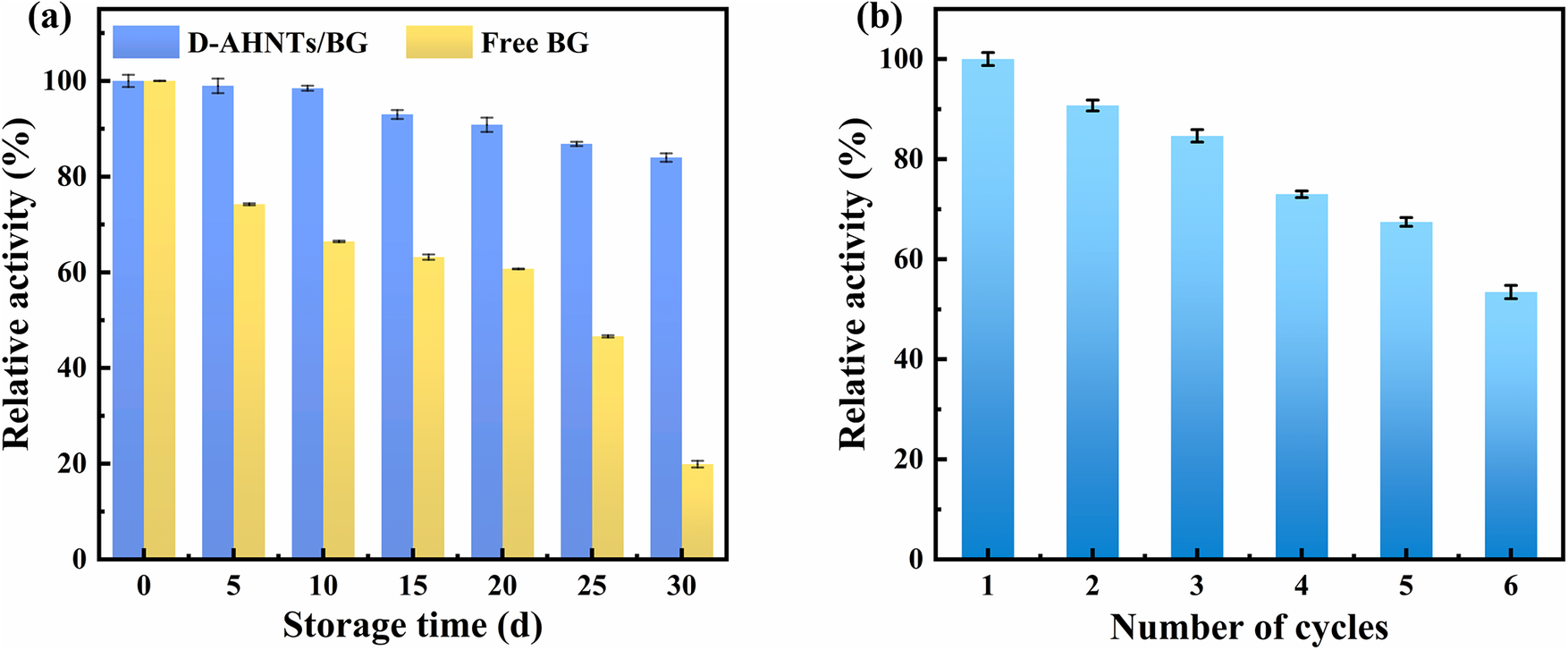
The storage stability of D-AHNTs/BG significantly outperformed that of free β-glucosidase, as illustrated in Fig. 11a. Following storage at 4°C for 30 days, the immobilized β-glucosidase retained ~84% of its relative activity. In stark contrast, the free β-glucosidase retained merely 20% of its initial activity. This notable improvement can be attributed predominantly to the D-AHNTs serving as an enzyme-immobilizing matrix, which reinforces the rigidity of the enzyme and effectively protects its native conformation. This conformational protection mechanism not only improves the storage stability of the enzyme but also lays a solid foundation for its efficient performance in practical applications.
Storage stability (a) of free β-glucosidase and D-AHNTs/BG at 4°C; and (b) re-usability of D-AHNTs/BG in pNPG hydrolysis.

Figure 11. Long description
Panel a, at left, plots storage time in days on the x-axis from 0 to 30 and relative activity percent on the y-axis from 0 to 100. Blue bars represent D-A H N T s slash B G, yellow bars represent free B G. At day 0, both show near 100 percent activity. Over 30 days, D-A H N T s slash B G retains higher activity than free B G, with D-A H N T s slash B G above 80 percent at day 30 and free B G dropping below 40 percent. Panel b, at right, plots number of cycles from 1 to 6 on the x-axis and relative activity percent from 0 to 100 on the y-axis. Blue bars show D-A H N T s slash B G activity decreases with each cycle, from near 100 percent at cycle 1 to about 60 percent at cycle 6.
In the re-usability assessment, the immobilized β-glucosidase maintained 53% of its initial catalytic activity even after six successive cycles of pNPG hydrolysis, as depicted in Fig. 11b. The minor activity reduction during cycling resulted from solid loss during washing. Nevertheless, this recyclable biocatalyst maintained its functional activity across multiple hydrolysis cycles. This phenomenon not only implies its potential application value in industrial biocatalysis processes, but also provides valuable references for the subsequent optimization of immobilized enzyme systems.
Conclusions
The AHNTs functionalized with PDA offer a robust and sustainable platform for high-efficiency BG immobilization. The synergistic modification strategy of alkali activation and PDA functionalization presented achieved very efficient enzyme loading (167 mg g–1support) with 96% immobilization efficiency and 83% retained activity. The immobilized biocatalyst of D-AHNTs/BG exhibited superior catalytic performance at pH 6.0 and 60°C, conditions that are harsher than those optimal for free BG (pH 5.0, 50°C). Kinetic evaluation via Lineweaver–Burk analysis confirmed that the catalytic efficiency of β-glucosidase was well preserved. The clay-based biocatalyst demonstrated remarkable stability, attributed to a dual stabilization mechanism: robust covalent immobilization via PDA suppressed enzyme leaching and conformational denaturation, while the nanoconfined environment of alkali-activated HNTs mitigated pH and temperature-induced unfolding. Collectively, this work provides a facile and effective surface engineering strategy for HNTs, establishing D-AHNTs as a versatile and robust platform for high-value enzymatic catalysis applications.
Supplementary material
The supplementary material for this article can be found at http://doi.org/10.1017/cmn.2026.10030.
Acknowledgments
This work was supported by the National Natural Science Foundation of China (grant number 42372056).
Financial support
The present study was supported by the National Natural Science Foundation of China (grant number 42372056), Sichuan Provincial International Science and Technology Innovation Cooperation Project (grant number 2024YFHZ0262).
Competing interests
The authors declare that they have no known competing financial interests or personal relationships that could have appeared to influence the work reported in this paper.
Data availability statement
Data will be made available on reasonable request to the authors.
Authors contribution
Yuqi Yao: Conceptualization, Data curation, Investigation, Methodology, Writing - original draft and Writing - review & editing; Shiyong Sun: Conceptualization, Writing - review & editing, Supervision, and Funding acquisition; Xingyun Yang: Data curation and Investigation; Pan Zhou: Data curation and Investigation; Haoming Tang: Data curation and Investigation; Shanshan Pu: Data curation and Investigation.