Intraoperative ultrasound has been reported to be a useful tool in the assessment of the immediate results after paediatric cardiac surgery.1–3
The provision of detailed morphologic and haemodynamic information contributes to the immediate recognition of significant problems after the surgical procedures, which can be addressed during a second period of cardiopulmonary bypass. In this way, the technique helps to improve the early results of surgery. Our purpose was critically to assess our use of intra-operative ultrasound, and to compare our experience in earlier years, as well as with recently published experiences.
Material and methods
Data was obtained retrospectively from January 1997 until December 2001, and prospectively from January 2002 until October 2002. We chose these periods because of the excellence of the documentation of data from surgical interventions and findings using intraoperative ultrasound. During the year of 2002, our team was supported by an additional member of staff to facilitate prospective evaluation. Since 2002, our approach to intraoperative ultrasound has remained the same. For this reason, and because of the lack of appropriate continued funding for this service, we were unable to include data relative to the years 2003 through 2005.
Data was acquired in the first time period using patient records, operation notes, and ultrasound reports, and by prospective collection in the second time-period. In cases of inconsistent information, video recordings of examinations were reviewed.
Throughout the entire period of study, we used epicardial as well as transoesophageal echocardiography, depending on the age of the patient, and on the type of surgery. For example, epicardial echocardiography was preferred in neonates and infants, whereas transoesophageal echocardiography was preferred to obtain optimal views of the atrioventricular valves or the atrial septum. In 2002, we routinely examined patients undergoing complex cardiac surgery, such as the first stage of the Norwood sequence, procedures for functionally single ventricle, the arterial switch operation, surgery for obstruction of the left or right ventricular outflow tracts, and repair of atrioventricular valves. In contrast, we did not routinely use intraoperative ultrasound in patients undergoing closure of atrial or ventricular septal defect, exchange of conduits between the right ventricle and the pulmonary arteries, unifocalisation in patients with tetralogy and pulmonary atresia, or those needing a cavopulmonary anastomosis or completion of the Fontan circulation. Intraoperative ultrasound was used during closed heart surgery only in patients undergoing banding of the pulmonary trunk, using the technique to assess ventricular function and the Doppler gradient. It was the surgeon who decided on the necessity of intraoperative echocardiographic imaging.
Epicardial imaging was carried out using a standard transthoracic 10 megaHerz transducer connected to a Vingmed System Five (Vingmed Sounds A/S, Horten, Norway). The transducer was covered by a sterile Latex sleeve (Microtek Medical Inc. Columbus Mississipi, USA). After weaning from cardiopulmonary bypass, the surgeon placed the transducer on the epicardial surface of the right ventricle, the right atrium, or the great arteries to obtain a series of short- and long-axis views, using cross-sectional and Colour Doppler flow mapping modalities in each position (Fig. 1). Depending on the type of surgery, a modified apical four-chamber view, and serial superior long- and short axis views were performed. Continuous wave and pulsed wave Doppler measurements were used in selected cases to quantify abnormal colour Doppler patterns. The images were analysed and discussed with the cardiologist, who was present in theatre and guided the surgeon regarding optimal positioning of the transducer. With increasing surgical experience with epicardial ultrasound, it was only rarely required for the attending cardiologist to undertake studies himself. For transoesophageal imaging, we used a paediatric multiplane probe with an 8 megaHerz transducer (Vingmed Sounds A/S, Horten, Norway). The probe was positioned preoperatively and left in position throughout the operation, allowing for continuous monitoring of ventricular function, as well as assessment for any residual lesions. Care was taken to assess ventricular function, residual intracardiac shunts, valvar regurgitation or stenosis, and obstruction of the outflow tracts in every patient, using semi quantitative criterions.

Figure 1. Intraoperative epicardial echocardiography in a 10 day old patient with hypoplastic left heart syndrome undergoing the first stage of the Norwood sequence of operations. Top left: Transducer position over the right ventricle demonstrating the right ventricle (RV), the left atrium (LA) and the right atrium (RA). Top right: Transducer position over the pulmonary trunk, short axis: the Damus-Kaye-Stansel Anastomosis (Damus), ascending aorta (AOA) and the right and left pulmonary arteries (RPA and LPA) are shown. Bottom left: Rotation of the probe in the same transducer position gives an additional view of the Damus-Kaye-Stansel anastomosis. Bottom right: Transducer position more cranially on the reconstructed ascending aorta in the sagital plane gives an overview on the reconstructed aortic arch (AO) and the descending aorta (DAO).
Every ultrasound examination was analysed to define its influence on the further surgical management, taking into account the clinical situation. We noted all early reoperations within 14 days after the initial procedure.
Retrospective review of the data on patients with early reoperations was conducted to determine whether intraoperative ultrasound could have been able to anticipate the need for reoperation. Finally, in 183 patients undergoing intraoperative ultrasound studies in 2002, results were compared to those obtained during postoperative studies prior to hospital discharge.
Results
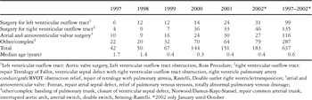
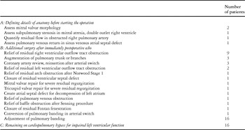
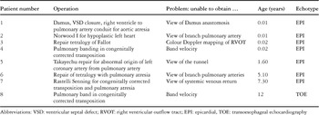
We evaluated our experience with 639 intraoperative ultrasound studies over a five year time period. During this period, from January 1997 until October 2002, a total number of 2737 patients underwent cardiac surgery at the Birmingham Children's Hospital National Health Service Trust, including 454 closed heart operations. The number of intraoperative ultrasound studies and demographic data of all patients are summarized in Table 1. During the period, there was a constant rise in the use of intraoperative ultrasound studies. Table 2 gives an overview of the numbers of examinations in each year, and for each type of surgery. Maintaining a relatively constant number of operations, there was a steady rise in the number of intraoperative ultrasound studies, while the age of the patients decreased over the years (Fig. 2). The intraoperative ultrasound assessment had influence on surgical management in 63 of 639 examinations (9.9%) (Table 3). Review of the data to the end of 2001 revealed 35 of 456 instances (7.7%) of significant influence on management. More detailed analysis for cohort of patients examined during the year 2002 revealed alteration of management in 28 of 183 patients (15.3%). Of these, 8 changes (4.4%) were primarily based on the ultrasound findings. During the same period, there were 8 of 183 (4.4%) cases where intraoperative ultrasound studies failed to assess important aspects of the repair, and thus were considered incomplete (Table 4).
Table 1. Demographic details of patients undergoing intraoperative echocardiography 1997–2002.

Table 2. Number of patients for each type of surgery in which intraoperative echocardiography was performed.


Figure 2. Evolution of the intraoperative echocardiographies over time: Number of intraoperative echocardiographies (light tined) compared to the number of operations (dark tinted) and to the median age of the patients at the time of the operation (open circles).
Table 3. Consequences of intraoperative echocardiographies performed in the years 1997 to 2002. From a total number of 639 examinations, 63 had obvious consequences.

Table 4. Limitations of intraoperative echo in 2002; total number of examinations: 183.

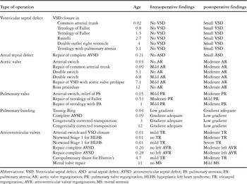
The postoperative transthoracic ultrasound findings were significantly different from the intraoperative findings in 27 of 183 examinations (14.7%) in 2002 (Table 5). In 62 patients undergoing closure of ventricular septal defects, we carried out intraoperative assessment for residual shunting. In 6 patients, postoperative findings differed from the intraoperative evaluation. In 4 out of 10 patients undergoing banding of the pulmonary trunk, there was a significant difference between the measured intra- and postoperative Doppler gradient (Table 5).
Table 5. Patients in whom transthoracic postoperative echocardiographic findings were different from intraoperative echocardiographic findings in 2002. Number of patients: 27.

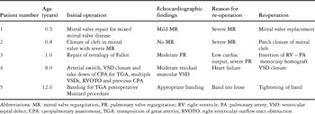
Of the total number of 639 patients who underwent intraoperative assessment, 18 (2.8%) required surgical reintervention within 2 weeks postoperatively. In 13 of these, review of the data did not document that early reintervention could have been anticipated at the time of the original intraoperative study. The remaining 5 patients, in whom the need for early reinterventions could have been predicted, are listed in detail in Table 6. In the remaining 2098 patients who did not undergo intraoperative ultrasound evaluation, there were 43 early reoperations (2.0%).
Table 6. Surgical reinterventions within 14 days; patients in whom intraoperative echocardiography might have predicted significant residual defects 1997–2002.

Discussion
The possibility of assessing both the morphology of the heart and residual haemodynamic lesions upon completion of the surgical repair has resulted in intraoperative ultrasound becoming an integral part of both paediatric1, 2, 4 and adult cardiac surgery.5 The technique has been shown to improve outcome and technique of surgical repair,6, 7 and to be cost effective.8, 9 The primary role of intraoperative ultrasound is the assurance of a good result of surgery before closing the chest and transferring the patient to the intensive care unit. In light of the advances of transthoracic ultrasound in the preoperative assessment of congenital cardiac malformations, the routine practice of pre-bypass studies in our, and others'7, 10, 11 experience is no longer indicated.
During the five-year period of observation included in our study, there has been a dramatic increase in the use of intraoperative ultrasound studies, from one-tenth of cases in 1997 to almost half in 2002 (Table 2 and Fig. 2). This can be explained by three factors. Firstly, the appreciation of the detailed insights that can be gained by the technique immediately after bypass. Secondly, the introduction of dedicated 10 megaherz short focus transducers, which allow for high quality studies in neonates. Thirdly, the appointment of a new consultant surgeon, keen to assess the quality of surgery upon completion of the operative procedures. The increasing number of young patients over the years (Table 2 and Fig. 2) reflects the more complex cardiac anatomy encountered in these patients needing early cardiac surgery. It is this population who may benefit most greatly from intraoperative echocardiographic evaluation.
Although our own service has not received appropriate funding, we feel obliged to provide it when performing surgery for complex congenital cardiac malformations, despite the significant demands both on manpower and equipment. At present, we do not provide this service for more straightforward surgical interventions, such as closure of isolated septal defects or creation of cavopulmonary anastomoses, including the Fontan procedure.
Looking at the rate of consequences arising from the intraoperative ultrasound studies (Table 3), and the rate of reintervention, our current strategy in using this technique of monitoring has been very successful. The incidence of detecting unsuspected residual defects by intraoperative ultrasound has been reported to range from 7 to 43%.6, 10, 12, 13 Such lesions are frequently of little significance. The immediate evaluation at the termination of bypass has been reported to support the need for surgical reintervention during the same anaesthesia in from one-twentieth to one-tenth of cases.2, 7, 13, 14 Those numbers are comparable with our experience.
Epicardial imaging yields good views of virtually any part of the heart and great vessels. Some questions, however, remained unanswered in our patients (Table 4). Limitations of imaging may reflect the large distances between the region of interest and the transducer (patient 6 in Table 4), or the interposition of air and the movement of the transducer, together with the movements of the underlying heart (patient 3 in Table 4). While the quality of the cross-sectional images is almost always adequate, colour Doppler imaging can at times be limited, especially in sites of low velocity of the flow of blood (patients 5 and 7 in Table 4). The size of the operating field, as well as the position of the bypass cannulas, may limit adequate positioning of the tranducer, especially in small patients (patient 1 in Table 4). In transoesophageal echocardiography, the position of the transducer is very limited within the oesophagus. The main disadvantage is the inability of a perfect Doppler alignment to a vessel to obtain the maximal velocity of flow of blood to calculate stenotic gradients (patient 8 in Table 4).
Although in recent years transoesophageal echocardiography has largely been favoured as the optimal modality for intraoperative imaging, with a very low rate of complications even in small children,3, 7, 8, 15, 16 in our institution we elect to use epicardial ultrasound in the majority of cases. We are concerned about the potential of significant oesophageal damage caused by the transoesophageal probe during long bypass procedures in neonates and infants. In our experience, the epicardial approach has provided very adequate information in almost all patients studied. The studies are routinely performed by the surgeon, with the cardiologist attending and providing advice when needed. Interpretation of the findings in the context of the overall haemodynamic situation, and details of the surgical repair performed, are conducted as a group discussion between surgeons, cardiologists and anaesthetists. This approach, in our experience, is also highly beneficial in training new members of the team, and obviates the discussion of who is competent to conduct such studies.4, 17–21
Postoperative transthoracic echocardiography showed additional findings in a number of patients (Table 5). Small residual ventricular septal defects, leaking valves, and Doppler gradients across the banded pulmonary trunk, accounted for the most frequent discrepancies between intra- and postoperative echocardiographies. This is likely to be due to an altered overall haemodynamic status influenced by large changes in circulating volumes of blood, and vascular resistance under general anaesthesia with mechanical ventilation, and in the setting of recent cardiopulmonary bypass.13 In two of our patients, both with very mild atrioventricular valvar regurgitation documented immediately after bypass, severe regurgitation was documented on later postoperative studies. We feel that disintegration of the surgical repair is the most likely explanation for this finding. Other lesions, such as small residual ventricular septal shunts around the suture lines securing the patch, are common findings immediately after bypass. Most of these small communications will have closed spontaneously by the time of discharge from hospital.22 We would suggest that the provision of routine intraoperative ultrasound studies for the assessment of atrial or ventricular septal defect closure is not indicated. Intraoperative ultrasound studies have become an integral part in the monitoring of the banded pulmonary trunk for retraining the left ventricle prior to double switch procedure, or in patients with a failing Mustard procedure.23 The size of the band is adjusted on the basis of observed changes in ventricular size and function. In future we may expand the use of routine intraoperative ultrasound studies to the immediate assessment of cavopulmonary shunts, in particular when there has been concomitant reconstruction of the pulmonary arteries during completion of the Fontan procedure.14, 24–26
We conclude that intraoperative ultrasound studies should be employed as a part of the routine monitoring during the majority of complex cardiac surgical procedures for congenital cardiac disease. The technique contributes significantly to management immediately after the completion of bypass, and identifies the majority of patients who benefit from early revision of the surgical repair. It is essential that this important role is recognised, and that the technique is fully implemented and funded in congenital cardiac units.
Acknowledgements
We gratefully acknowledge the assistance of John Stickley, Database Manager, who assisted in acquisition of the data and its statistical analysis. We also thank the staff of the cardiac theatres, and the cardiac anaesthetists at Birmingham Children's Hospital, for their enthusiastic support