Aneurysms of one of the aortic sinuses of Valsalva are rare lesions, which may be congenital or acquired. Aneurysms arise mostly from the right coronary or the non-coronary sinus, only 5% arising from the left coronary sinus. The dilated sinus may bulge into one of the cardiac chambers; the most feared complication is a sudden catastrophic rupture of the aneurysm. Owing to their clinical silence, unruptured aneurysms remain either undiagnosed or become corrected before rupture. Here we present a unique case of an adolescent with Down syndrome and sudden rupture of sinus of Valsalva, with documented normal aortic sinuses in infancy.
Case report
A 16-year-old boy with Down syndrome was referred to us because of chest pain after intensive swimming. A loud heart murmur, which had never appreciated before, was noted by his general practitioner. There was no preceding history of an infection, trauma, or chest pain. Echocardiographic screening for congenital heart defects was performed at the age of 2 weeks because of trisomy 21. Normal sinuses of Valsalva were documented at that time.
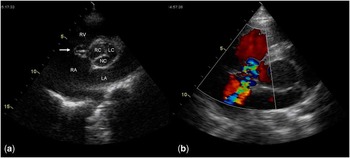
On examination, a healthy looking young man with Down syndrome without heart failure was seen. Auscultation revealed a grade III/VI continuous murmur at the lower left sternal border. A 12-lead electrocardiogram showed normal sinus rhythm and a right bundle branch block, without any signs of myocardial ischaemia. Transthoracic echocardiography demonstrated that the right coronary aortic sinus was dilated and protruded into the right ventricle. A fistulous communication with a windsock appearance, between the right sinus of Valsalva and right atrium, was seen through the anteroseptal commissure of the tricuspid valve (Fig 1). The right atrium and right ventricle were dilated. Mild aortic regurgitation through the valve itself was also present. The aortic valve was normally developed. Doppler features of significant diastolic run-off were recorded in the descending and abdominal aorta. No other intracardiac abnormalities were demonstrated.
Transthoracic echocardiogram. ( a ) Parasternal short-axis view demonstrating a ruptured aneurysm of the right coronary sinus (arrow) protruding as a windsock into the RA. ( b ) Colour flow Doppler demonstrating that the blood stream diverts from the ruptured aneurysm predominantly to the RA. LA = left atrium; LC = left coronary sinus; NC = non-coronary sinus; RA = right atrium; RC = right coronary sinus; RV = right ventricle.

Surgical repair involved resection of the membranous “windsock”, leaving a large central defect in the right coronary sinus and a defect in the anteroseptal commissure of the tricuspid valve. Both defects were closed with a xenopercardial patch, respectively, through the aortotomy and the right atrium, thereby restoring normal aortic root and tricuspid valve geometry. Histological examination of the resected aneurysm ruled out infective endocarditis. Both the aortic and tricuspid valves appeared normal 7 years after surgery. All three aortic sinuses have a normal appearance and are not dilated.
Discussion
Cardiovascular anomalies are found in 40–50% of children with Down syndrome, the most common being septal defects and tetralogy of Fallot.Reference Irving and Chaudhari 1 Reports of sinus of Valsalva aneurysm in adult patients with Down syndrome are scarce.Reference Lee, Joseph, Lambert, Mazer and Hutchison 2 This is only the second report in a child.Reference Benatar, Decraene and Feenstra 3
Aneurysm of the sinus of Valsalva is a rare cardiac anomaly with a marked male preponderance; their incidence was observed to be highest among Asians.Reference Zhao, Seng and Yan 4 Congenital aneurysms may be associated with structural defects such as doubly committed juxta-arterial ventricular septal or occasionally perimembranous ventricular septal defect, coronary anomalies, bicuspid aortic valve, aortic regurgitation, pulmonary stenosis, tetralogy of Fallot, coarctation, or atrial septal defect. Occasionally, it may be difficult to define whether the aneurysm was acquired or congenital. Congenital aneurysms are more common, caused by weakness at the juncture of the aortic media and the annulus fibrosus. A finger-like aneurysmal sac develops as a result of the weakened tissue and prolonged exposure to high aortic pressures. Endocarditis, chest trauma, or strenuous physical activity may result in further weakening of the thinned wall, eventually leading to rupture of the aneurysm. Rupture results most often in the right sinus aneurysm protruding into the right ventricle. The next most common site of rupture is into the right atrium from an aneurysm of the non-coronary sinus.
Unruptured aneurysms are often found incidentally during diagnostic studies. They may, however, present with myocardial ischaemia due to obstruction of the coronary blood flow or with right or left ventricular outflow tract obstruction. When the aneurysm ruptures into one of the cardiac chambers, the size of the fistula, rapidity of rupture, and the chamber into which the rupture occurs determines the clinical presentation. An acute, large rupture may present with dramatic onset of marked precordial chest pain along with severe dyspnoea with sudden haemodynamic collapse. Symptoms often follow physical stress, with acute dyspnoea and chest pain developing after strenuous physical activity.Reference Zhao, Seng and Yan 4 Small perforations of aneurysms may present with an asymptomatic continuous murmur. The long-term results of surgical repair are excellent.Reference Takach, Reul and Duncan 5 Percutaneous treatment of an aneurysm of sinus of Valsalva is in certain cases feasible in children as well.Reference Srivastava and Radha 6
Rupture of an aneurysm of sinus of Valsalva has been observed in Noonan syndrome, 22q11 microdeletion syndrome, and in tetralogy of Fallot.Reference Abuchaibe, Dobrolet, Peicher, Ventura and Welch 7 , Reference Mithani, Polimenakos and Santucci 8 The occurrence of a ruptured aortic sinus aneurysm in tetralogy of Fallot, 22q11 microdeletion syndrome, and Down syndrome may be a reflection of a joint underlying connective tissue abnormality. Progressive aortic root and ascending aortic dilatation is typical for syndromes such as Marfan, Loeys–Dietz, Ehlers–Danlos, and Turner syndrome,Reference Yetman and Graham 9 but may be found also in Noonan syndrome or chromosome 22q11 deletion syndrome.Reference Abuchaibe, Dobrolet, Peicher, Ventura and Welch 7
The patient reported by Benatar was found to have a continuous cardiac murmur on routine school examination; echocardiography revealed a fistula from an aneurysmally dilated non-coronary sinus into the right atrium.Reference Benatar, Decraene and Feenstra 3 Our patient was screened for congenital heart defects at the age of 2 weeks because of trisomy 21. The sinuses were documented to be normal. Most likely, an acquired aneurysm developed during childhood and ruptured after an episode of intensive swimming.
Late development of valve dysfunction in adolescents and young adults with Down syndrome has been reported and may be associated with collagen abnormalities documented in this syndrome.Reference Geggel, O'Brien and Feingold 10 Although the need for a second cardiovascular evaluation of Down syndrome patients before reaching adult age is still discussed, current evidence does not prescribe a rigid schedule for routine echocardiography in adults with Down syndrome in the absence of a known congenital heart defect.Reference Lin, Basson and Goldmuntz 11 The prevalence of rupture of Valsalva sinuses in Down syndrome is very rare and does not probably differ from normal population.
Conclusion
We report the case of an adolescent with Down syndrome and acute rupture of the aneurysm of sinus of Valsalva in the face of previously documented normal aortic sinuses. This case shows that paediatricians should have a low threshold for cardiac re-evaluation of any patient with Down syndrome with the new onset complaints or murmurs.
Financial support
None.
Conflict of interest
None.