




No CrossRef data available.
Article contents
The clinical profile of Ebstein's malformation as seen from the fetus to the adult in 52 patients
Published online by Cambridge University Press: 20 January 2005
Abstract
Ebstein's malformation of the tricuspid valve is a rare but complex congenital cardiac lesion characterised by a variable degree of dysplasia and displacement of the proximal attachments of its inferior and septal leaflets from the true atrioventricular junction. The aim of our retrospective study is to report the risk factors for mortality, and to determine the clinical profile as seen in 52 cases diagnosed in our service between 1978 and 2002, concentrating in particular on the outcome for the neonatal patient. There were 26 females and 26 males, and the age at presentation ranged from 30 weeks gestational age to 46 years. We found 23 associated cardiac anomalies in 20 cases. Of the patients, 11 patients (21%) died. Actuarial survival at 30 years was 65%. Predictors of death included fetal or neonatal presentation, presence of associated defects, a grade within the Celermajer index of 3 or 4, and a cardiothoracic ratio equal to or greater than 65%. The diagnosis was made in the neonatal period in 24 patients, nine of whom died. Of the 15 survivors, only 4 are free of symptoms. Surgical treatment was undertaken in 9 patients, with a mortality rate of 33% without late deaths, with all the survivors being in good condition. The mean period of follow-up for the 41 living patients was 16.5 years. An arrhythmia of variable severity appeared during the evolution of 27 patients (66%). At present, only 7 cases are in the functional class III or IV of the grading system of the New York Heart Association, but 25 patients (61%) needed some medical treatment, meanly for arrhythmic events. Thus, fetal and neonatal presentation of Ebstein's malformation is associated with a poor outcome. Moreover, the echocardiographic appearance, marked cardiomegaly, and the presence of associated lesions are all risk factors for mortality. Arrhythmia and need of medical treatment are common in older children and adults. Survival after surgical treatment is associated with a good outcome.
Information
- Type
- Original Article
- Information
- Copyright
- © 2004 Cambridge University Press
Ebstein's malformation is a rare complex congenital cardiac lesion of the tricuspid valve and right ventricle, characterised by a variable degree of dysplasia and displacement of the proximal attachments of the inferior and septal leaflets from the true atrioventricular junction. The abnormally positioned valve then divides the right ventricle into two components, the proximal one between the atrioventricular junction and the displaced valvar orifice, the so-called atrialised portion, and the distal one that constitutes the functional part of the right ventricular chamber.1–4 Histologic alterations have been reported involving both the left and right ventricular myocardium, but especially thinning of the right ventricular wall, with an increase of the content of fibrous tissue in both left and right free ventricular walls, this last finding being associated with poor right ventricular compliance and early mortality.5–7 Ebstein's malformation is estimated to occur in between one in 10,000 and 50,000 live births, and it represents from 0.03 to 1% of all congenital cardiac malformations, and accounts for two-fifths of all congenital malformations afflicting the tricuspid valve.8 The defect is frequently associated with an atrial septal defect or pulmonary stenosis, and less frequently with others cardiopathies.6, 9
Its natural history is extremely variable, depending on the severity of dysplasia and displacement of the attachment of the valvar leaflets and the degree of myocardial involvement, with a corresponding broad clinical spectrum ranging from severe disease causing fetal or neonatal death to mild disease with survival to very old age. The mortality rate for neonatal presentation ranges between one-quarter and one-half,10–14 and it is currently expected that adults with mild forms will have a better prognosis.15–18
In the last two decades, cross-sectional echocardiography has permitted diagnosis during fetal and neonatal life,19–21 and also allowed different parameters to be established for classification of the defect.22–24 In this respect, the index established by Celermajer and his colleagues6 is considered to be good predictor of the morbidity and mortality of the lesion.6, 13, 14, 25
It is the highly variable clinical features of the condition, together with the initially poor results of surgery, which accounts for the fact that, for many years, Ebstein's malformation was regarded as a condition preferably treated medically. More recently, the improvement in surgical techniques has made surgery a valuable alternative at an early stage in the natural history of the disease.26, 30
The aim of this study is to report risk factors for mortality, and to determine the clinical profile, on the basis of our experience with 52 patients diagnosed with Ebstein's malformation.
Methods
We have carried out a retrospective review of the medical files, presence or absence of cardiomegaly on chest X-rays films, echocardiographic studies, surgical reports, electrophysiologic studies, functional grading within the system of the New York Heart Association, and autopsy findings in 52 patients with Ebstein's malformation presenting in our Department between March 1978 and March 2002. Cardiac catheterisation was only performed in the cases with associated anomalies, ventricular dysfunction, and as preoperative studies. We have excluded patients with variants such as an imperforate tricuspid valve, an imperforate pulmonary valve, or combined atrioventricular and ventriculo-arterial discordant connections (“congenitally corrected transposition”). Early mortality has been defined as death within the first month of life, and late mortality as death thereafter, with or without surgical treatment.
Survival and risk factor analysis
Values were compared using the chi-square test or Fisher's exact test, and statistical significance was inferred at a p value of less than 0.05.
We generated Kaplan-Meier survival curves for the overall group, and for the different clinical subgroups. Variables were evaluated using the Cox regression model to estimate relative risk factors associated with death.31, 32 Univariate analysis was performed when variables were available in equal or more than half of the subjects, specifically for neonatal diagnosis, other presenting features such as cyanosis, congestive heart failure, murmur, and arrhythmia, associated cardiac defects, a cardiothoracic ratio equal or greater than 0.65, a grading in the Celermajer index of III or IV, arrhythmia as symptom during evolution, and the need for surgical treatment. The multivariate regression model could not be used to predict late deaths because of the small number of events. Relative risk factors are indicated with 95% confidence limits. Statistical analysis was done by SPSS for Windows 10.0 computer program.
Patients were censored at death, whether “natural” or postoperative, and survival beyond the last contact with our hospital was not presumed.
Results
Population data
There were 26 females and 26 males. The age at presentation ranged from 30 weeks of gestational age to 46 years, with a median of 5.6 years. There were 3 cases identified during fetal life, 21 neonates presenting up to 1 month of age, 8 infants presenting from 1 month to 2 years, 13 children presenting from the age of 2 to 10 years, 4 adolescents first seen at ages from 10 to 18 years, and 3 adults first seen over the age of 18 years.
Presenting features
The mode of presentation differed for each age group. Table 1 shows the major presenting clinical features used in the evaluation of the 52 patients. In some patients, we encountered more than one presenting feature. In neonates, cyanosis was more common (90%) than in older patients, whereas a murmur was more frequent (84%) in subjects younger than 10 years. In adolescents and adults, these patients accounting for 13% of our total group, there was no specific mode of presentation.
Table 1. Presenting feature.

Neonatal diagnosis
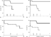
In 24 cases (42% of total), the diagnosis was made either during the fetal or neonatal period. In fifteen of them (62%), other cardiac anomalies were present, and nine needed drug treatment in the neonatal care unit. Moreover, nine of these patients died, with 4 early and 5 late deaths. Of the 15 survivors, 11 are now symptomatic, and only 4 (16.6%) are free of symptoms and do not require medical treatment. The actuarial survival rate at 1 year of follow-up was 71%, reducing to 66% at 10 years, and 44% at 30 years (Fig. 1A).

Figure 1. Kaplan-Meier survival curves. A: Cases with fetal or neonatal diagnosis (broken line) and the rest of patients (continuous line). B: Cases with associated cardiac defects (broken line) and cases without association (continuous line). C: Cases with Celermajer index of Grade 1 or 2 (continuous line), and cases with Grade 3 or 4 (broken line). D: Cases with Cardiothoracic index less than 65% (continuous line) and cases with index equal or greater than 65% (broken line).
Associated cardiac defects
We found 23 associated cardiac anomalies, other than a patent oval foramen or atrial septal defect, in 20 patients, this representing 38% of the total group. The distribution of lesions in the different age groups is shown in Figure 2. Valvar pulmonary stenosis is the more frequent lesion, accounting for 39% of the total associated defects. Three cases of left ventricular cardiomyopathy were suspected after an echocardiographic scan. Actuarial analysis of cases with and without associated cardiac anomalies is shown in Figure 1B.

Figure 2. Associated cardiac defects presenting at different age groups. RVOTO: Right ventricular outflow tract obstruction; PAPVR: partially anomalous pulmonary venous return; PDA: patent ductus arteriosus.
The Celermajer index
At the time of diagnosis, or during the period of follow up, we were able to use our cross-sectional echocardiographic findings to calculate the Celermajer index in 42 patients (80%). Of these, 23 cases fell in Grade I or II (54%), and all of these patients are still alive at 30 years of follow-up, with only 2 needing surgery, and only 1 being in class III or IV of the system of the New York Heart Association. Of the 19 patients falling into Grade 3 or 4 of the index, 7 died, 6 needed surgery, and 6 are in Grade III or IV of the system of the New York Heart Association. Actuarial analysis for the patients in whom we were able to calculate the Celermajer index are shown in Figure 3.

Figure 3. Evolution of 52 cases of Ebstein's malformation. In the fourth file, number of neonates included in the four mean evolution forms: late/early death, and symptomatic/asymptomatic survivors.
Cardiothoracic ratio
We were able to document the cardiothoracic ratio from chest X-ray films taken at presentation in all the patients. A value equal or greater than 0.65 was found in 15 patients (29%), 10 of whom were neonates, with 7 of these patients dying, 5 early. In addition, 4 of these patients needed surgery, 3 patients are on medical treatment and in Grade III or IV of the system of the New York Heart Association, and only one is free of symptoms and without any treatment. Actuarial analysis of the cases with a cardiothoracic ratio equal or greater than 0.65, and those with a ratio of less than 0.65, is shown in Figure 1D.
Surgery
Surgical treatment was undertaken in 9 cases, these patients having a mean age of 13.5 years, with a range from 1 month to 31 years. The indications for operation were persistent cyanosis in 6 cases, and heart failure without an adequate response to medical treatment in 3. Of the cases, 5 were diagnosed during the first year of life. The mean Celermajer index was 1, with a range from 0.66 to 1.41, and the average cardiothoracic ratio was 0.61, with a range from 0.52 to 0.67. The surgical techniques used are shown in Table 2. Perioperative mortality was 33%. Late deaths were not observed. The mean period of follow-up is now 8.5 years, with a range from 2 to 16 years. Only one patient is now in class III of the grades of the New York Heart Association. This patient has atrial flutter, and responds poorly to medical treatment. None of the 6 survivors required a new operation, and they are still in good clinical condition.
Table 2. Operative procedures.

Outcome
Figure 3 shows the evolution of our 52 patients with Ebstein's malformation, with special reference to those first seen during the neonatal period.
- Mortality. There was a global mortality of 21%, with 11 patients dying. The actuarial survival rate was 86% at 1 year, 84% at 10 years, 74% at 20 years, and 65% at 30 years (Fig. 4). Table 3 shows the data of these cases.
- Early deaths. In the neonatal period, four patients (7.7%) died, all of them with heart failure, and one with additional sepsis. Associated cardiac anomalies, and hemodynamic deterioration, as presenting features were present in all. In two cases, the Celermajer index was IV, and the cardiothoracic ratio had a mean of 0.65, with a range from 0.60 to 0.68. Only one neonate required surgery, specifically a pulmonary valvotomy, but died within the first 24 postoperative hours. None of the patients presented with arrhythmias.
- Late deaths. Seven patients died late (14.6%), at a mean age of 14 years, and with a range from 2 months to 31 years. Of these, 4 had cardiac failure, 2 with severe arrhythmias, and 1 in the operating room. Associated cardiac abnormalities were present in 5 patients, all of them with haemodynamic deterioration, except in one with a severe arrhythmia. The Celermajer index was 3 in 5 cases, 4 in one, and had not been registered in the last one. The average cardiothoracic ratio was 0.69, with a range from 0.59 to 0.83. Surgical treatment was needed in two patients, both of whom died, one during replacement of the tricuspid valve and closure of atrial septal defect, and the other 7 days after a one-stage Fontan operation with plication of the right ventricle and closure of the atrial septal defect. A supraventricular arrhythmia appeared during the evolution of 3 patients, 2 having atrial fibrillation, and one developing supraventricular tachycardia that needed multiple ablations.
- Survivors. The 41 patients who survived were followed up for a mean period of 16.5 years, with a range from 2.5 to 28 years. Associated cardiac anomalies were found in 11 cases (27%). The Celermajer index was calculated in 35 cases, and was Grade 3 or 4 in 12 (30%). The cardiothoracic ratio was measured in all, having a mean of 0.58, and a range from 0.49 to 0.68. Surgical treatment was needed in 6 patients (see Table 2). In 27, a disturbance of electrical conduction of variable severity appeared during the follow-up, the majority with isolated premature atrial contractions, but also 6 patients with paroxysmal supraventricular tachycardia, 3 with second degree atrioventricular block, and 1 with atrial fibrillation. Only 7 patients (17%) are in functional class III or IV of the grades of the New York Heart Association, but 61% (25 cases) have needed some treatment.

Figure 4. Kaplan-Meier survival curve indicating percent survival with follow-up.
Table 3. Early and late mortality.

Analysis of risk factors
We analysed six variables as possible predictors for death (Table 4). Four of them were associated with death in a univariate analysis model, namely fetal or neonatal diagnosis, presence of associated anomalies, grade 3 or 4 in the Celermajer index, and a cardiothoracic ratio equal or greater than 65%.
Table 4. Risk factor identification.

Discussion
Ebstein's malformation is an uncommon heart defect that accounts for less than 1% of all cases of congenital cardiac disease, but represents two-fifths of all congenital malformations of the tricuspid valve. There is an equal distribution between sexes, and in most of cases occur sporadically. Some familial cases have been reported.33 Its association with chromosomal abnormalities and multiple malformation syndromes is rare. Its incidence is reputedly higher than expected in infants born to women who have ingested lithium during early pregnancy. More recent evaluation, nonetheless, suggests that perhaps this fact has been overemphasized.34
Natural history. Ebstein's malformation has an extremely variable natural history, depending of the degree of displacement of the proximal attachments of the valvar leaflets and the myocardial involvement. Nowadays, the defect is usually diagnosed prenatally or in early life. Patients with a severe form may demonstrate profound congestive heart failure and cyanosis, and die early in infancy, or even during fetal life. In contrast, patients with a mildly deformed tricuspid valve may reach adulthood without symptoms. Typically, symptoms present in infancy, improve as the pulmonary vascular resistance decreases, but then return later in life. The symptoms may include dyspnea on exertion, arrhythmia, or cyanosis. In the majority of patients, the malformation lies between these extremes, but sudden death is not uncommon, even in cases believed to be asymptomatic.12–14
Limitations of study. Our study includes 52 patients referred to our hospital throughout the last 24 years. Over this long period, diagnostic and therapeutic techniques have evolved, and the number of patients with undiagnosed Ebstein's malformation has probably changed. Thus, some patients will be alive with as yet unrecognised defects. The net effect of such patients on the survival curves presented in this report is uncertain.
The malformation in the fetus. The malformation is one of few congenital cardiac defects that frequently causes fetal cardiac dysfunction, with cardiomegaly, hydrops, and tachyarrhythmias. Because these abnormalities are easily identified by echocardiography, Ebstein's malformation is one of the most common lesions referred by the obstetrician for fetal cardiac study. Prenatal diagnosis is easy, and is associated with poor prognosis. Massive cardiomegaly can impair normal development of the lungs, such that many of these babies die in the immediate postnatal period because they cannot be adequately ventilated.21, 35 In our series, three patients were diagnosed prenatally. All presented cardiac failure and severe cyanosis at birth, and needed vasoactive drugs and mechanical ventilation. One patient died before the first month of life, but presently the other two are well with treatment after 6 years of follow-up.
Diagnosis. Echocardiography is the most important diagnostic test for Ebstein's malformation, permitting accurate assessment of the anatomy of the tricuspid valve, the size and contractility of the functional right ventricle, and the associated cardiac defects. Nowadays, we use the easily measurable Celermajer index for determining prognosis. To calculate this index, we compare the area of the right atrium and atrialised right ventricle with the total area of the right ventricle plus the left atrium and left ventricle as measured in a four-chamber view at end-diastole.12 This ratio reflects both the anatomic displacement of the tricuspid valvar leaflets and the physiologic consequences of tricuspid stenosis, regurgitation, or both.6, 13 Our findings confirm that this single echocardiographic measure gives important prognostic information. Thus, 8 of our 11 patients who died were in Grade 3 or 4 of the index.
In many institutions, cardiac catheterisation is avoided, due to the great risk of severe tachyarrhythmia during the procedure.24 In our experience, only 10 patients of the 52 were submitted to haemodynamic study, albeit without major complications.
Associated lesions. Some authors have noted an increased mortality in patients with associated cardiovascular lesions.12–14 We found that the 6 of our 11 fatal cases had an associated cardiac defect, 4 of them pulmonary stenosis. Only 17% of those surviving had a cardiovascular defect associated with the Ebstein's malformation.
Arrhythmias. Conduction disturbances are common particularly in the older patients, and are usually related to accessory atrioventricular connections and atrial flutter or fibrillation. These arrhythmias tend to be recurrent and resistant to drug treatment, and have been related with late sudden death. Paroxysmal supraventricular tachycardia, by far the most frequent rhythm disorder, is thought to occur in approximately one-sixth of patients. It is often the result of an accessory conduction pathway producing Wolff-Parkinson-White syndrome. In such instances, successful medical control may be difficult to achieve.27, 36 Ablation of the accessory connections has been performed infrequently in Ebstein's malformation.37, 38 In our study, despite no correlation being found between the presence of arrhythmia and mortality, occurring in only 3 of the 11 deaths, we have found a high incidence of arrhythmias and conduction anomalies in the subsequent evolution of the disease, occurring in 27 of the 42 survivors.
Treatment. The management of Ebstein's malformation depends on the age at presentation, the anatomic severity of the lesion, the presence of associated defects, and the clinical features.
Neonates and infants falling within Grade 1 or 2 of the Celermajer index have a good outcome, and palliation should be undertaken for any associated lesions. Those falling in Grade 3 or 4, however, have a poor prognosis. In these cases, surgery is also associated with a very poor outcome. Possible strategies for these children include the creation of functional tricuspid atresia by closing the valve and excising the atrial septum, with insertion of a systemic-to-pulmonary arterial shunt with a view to later Fontan-type surgery or cardiac transplantation.14, 29, 30 In agreement with other authors, our experience confirms the bad prognosis of neonatal diagnosis, this feature accounting for over four-fifths of our total deaths. Moreover, almost a half of the cases needing drugs or mechanical ventilation during the neonatal period had a fatal evolution. We have not had the choice of planning a functionally univentricular strategy for treatment of any of our neonates, with only one of our 24 neonates undergoing surgical treatment, this patient dying after a pulmonary valvotomy. Moreover, it is significant that the seven-tenths of the surviving neonates have needed medical treatment in the period of follow-up.
Older children and adults can be managed medically until symptoms appear, when good results may be obtained by surgery. Many authors have reported different surgical techniques for Ebstein's malformation.39–43 It seems that the most important component of repair is the creation of a competent tricuspid valve, coupled with restoration of the shape and compliance of the right ventricle. Despite modifications in the plication procedure, there is no evidence of benefit to right ventricular performance, particularly in view of long-term results.40 In general, whenever possible, valvar repair is preferred to replacement, because it avoids anticoagulation, and in children also avoids replacement of the prothesis because of growth. A functionally univentricular strategy is possible in older children, but inadequate size of the pulmonary arteries, or left ventricular dysfunction, are often seen, limiting the postoperative survival. Since 1986, we have performed a variation of the Fontan procedure in selected patients.44 The Pérez de León-technique consists in anastomosing the right atrium and the pulmonary trunk with a prosthetic conduit, and closing the atrial septal defect. We have had the opportunity to use this technique in two patients, one adolescent and one adult, with good results. At all ages, however, there is an increasing risk of arrhythmias, and persistent risk of late hemodynamic deterioration and sudden death.36
Risk factors. The fetus or neonate with unfavourable morphologic features is at high risk of dying at less than 3 months of age, whereas the older child with unfavourable anatomy who has survived the neonatal period may be more likely to have poor cardiac function and require plication, valvar replacement, or a univentricular approach. Associated cardiovascular lesions, when they obstruct pulmonary blood flow, may be associated with a poor outcome. Cardiomegaly, and Grade 3 or 4 of the Celermajer index, were consistently associated with a poor prognosis in our series. Neonates often may have congestive heart failure, cyanosis and impressive cardiomegaly. If intensive therapy, including mechanical ventilation, sympatho-mimetic agents and infusion of prostaglandins, is required the prognosis is poor. In our experience, however, surgical treatment, when the patient survives, is associated with a good outcome.
Conclusion
We found that fetal and neonatal diagnosis of Ebstein's malformation, presence of associated defects, a high grade of cardiomegaly, and an increased echocardiographic index, were all risk factors for poor prognosis. Survivors of surgical treatment had a good outcome, with the optimal therapeutic strategy depending on age and the anatomic features of the disease.
References
Ebstein W. Uber einen sehr seltenen Fall von Insufizienz der Valvula tricuspidalis, bedingt durch eine angeborene hochgradige misbildung derselben. Arch Anat Physiol Wiss Med 1866; 33: 238–254.Google Scholar
Becker AE, Becker MJ, Edwards JE. Pathologic spectrum of dysplasia of the tricuspid valve. Arch Path 1971; 91: 167–178.Google Scholar
Watson H. Natural history of Ebstein's malformation of the tricuspid valve in childhood and adolescence: international co-operative study. Br Heart J 1974; 36: 412–427.Google Scholar
Anderson KR, Lie JT. Pathologic anatomy of Ebstein's malformation of the heart revisited. Am J Cardiol 1978; 41: 739–745.Google Scholar
Armengol Rofes AJ, Serrano Durán M, Albert Brotons DC, Sánchez López C, Casaldáliga Ferrer J, Girona Comas JM. Ebstein's malformation of the tricuspid valve. A review of 35 cases. An Esp Pediatr 1996; 44: 139–144.Google Scholar
Celermajer DS, Dodd SM, Greenwald SE, Wyse RKH, Deanfield JE. Morbid Anatomy in Neonates with Ebstein's malformation of the tricuspid valve: pathophysiologic and clinical implications. J Am Coll Cardiol 1992; 19: 1049–1053.Google Scholar
Lev M, Liberthson RR, Joseph RH, et al. The pathologic anatomy of Ebstein's disease. Arch Path 1970; 90: 334–343.Google Scholar
Ammash NM, Warnes CA, Connolly HM, Danielson GK, Seward JB. Mimics of Ebstein's malformation. Am Heart J 1997; 134: 508–513.Google Scholar
De Lonlay-Debeney P, De Blois MC, Bonnet D, et al. Ebstein malformation associated with rearrangements of chromosomal region 11q. Am J Med Genet 1998; 80: 157–159.Google Scholar
Zuberbuhler JR, Allwork SP, Anderson RH. The spectrum of Ebstein's malformation of the tricuspid valve. J Thorac Cardiovasc Surg 1979; 77: 202–211.Google Scholar
Anderson KR, Zuberbuhler JR, Anderson RH, Becker EA, Lie JT. Morphologic spectrum of Ebstein's malformation of the heart: a review. Mayo Clin Proc 1979; 54: 174–180.Google Scholar
Roberson DA, Silverman NH. Ebstein's malformation: echocardiographic and clinical features in the fetus and neonate. J Am Coll Cardiol 1989; 14: 1300–1307.Google Scholar
Celermajer DS, Cullen S, Sullivan ID, Spiegelhalter DJ, Wyse RKH, Deanfield JE. Outcome in neonates with Ebstein's malformation. J Am Coll Cardiol 1992; 19: 1041–1046.Google Scholar
Celermajer DS, Bull C, Till JA, et al. Ebstein's malformation: presentation and outcome from fetus to adult. J Am Coll Cardiol 1994; 23: 170–176.Google Scholar
Adams JCL, Hudson R. A case of Ebstein's malformation surviving to age 79. Br Heart J 1956; 18: 129–131.Google Scholar
Attie F, Casanova JM, Zabal C, et al. Ebstein's malformation. Clinical profile in 174 patients. Arch Inst Cardiol Mex 1999; 69: 17–25.Google Scholar
Garrido Martín A, Oliver Ruiz JM, Domínguez Melcon FJ, González AE, Sobrino Daza JA. Congenital dysplasia of tricuspid valve (Ebstein like) in a 73-year-old male with severe tricuspid insufficiency. Rev Esp Cardiol 2000; 52: 1008–1010.Google Scholar
Attie F, Rosas M, Rijlaarsdam M, et al. The adult patient with Ebstein malformation. Outcome in 72 unoperated patients. Medicine 2000; 79: 27–36.Google Scholar
Sharland GK, Chita SK, Allan LD. Tricuspid valve dysplasia or displacement in intrauterine life. J Am Coll Cardiol 1991; 17: 944–949.Google Scholar
Hornberger LK, Sahn DJ, Kleinman CS, Copel JA, Reed KL. Tricuspid valve disease with significant tricuspid insufficiency in the fetus: diagnosis and outcome. J Am Coll Cardiol 1991; 17: 167–173.Google Scholar
Sharland G. In: Allan L, Hornberger G, Sharland G (eds). Textbook of Fetal Cardiology. London, Greenwich Medical Media 1990: pp 133–147.
Shiina A, Seward JB, Tajik AJ, Hagler DJ, Danielson GK. Two-dimensional echocardiographic-surgical correlation in Ebstein's malformation: preoperative determination of patients requiring tricuspid valve plication vs replacement. Circulation 1983; 68: 534–544.Google Scholar
Shiina A, Seward JB, Edwards WD, Hagler DJ, Tajik AJ. Two dimensional echocardiographic spectrum of Ebstein's malformation: detailed anatomic assessment. J Am Coll Cardiol 1984; 3: 356–370.Google Scholar
Seward JB. Ebstein's malformation ultrasound imaging and hemodynamic evaluation. Echocardiography 1993; 10: 641–664.Google Scholar
Yetman AT, Freedom RM, McCrindle BW. Outcome in cyanotic neonates with Ebstein's malformation. Am J Cardiol 1998; 81: 749–754.Google Scholar
Dearani JA, Danielson GK. Congenital Heart Surgery Nomenclature and Database Project: Ebstein's malformation and tricuspid valve disease. Ann Thorac Surg 2000; 69: S106–S117.Google Scholar
Mair DD. Ebstein's malformation: natural history and management. J Am Coll Cardiol 1992; 19: 1047–1048.Google Scholar
Knott-Craig CJ, Overholt ED, Ward KE, Ringewald JM, Baker SS, Razook JD. Repair of Ebstein's malformation in the symptomatic neonate: an evolution of technique with 7-year follow-up. Ann Thorac Surg 2002; 73: 1786–1792.Google Scholar
Chavaud S, Fuzellier JF, Berrebi A, et al. Bi-directional cavopulmonary shunt associated with ventriculo and valvuloplasty in Ebstein's malformation: benefits in high risk patients. Eur J Cardiothorac Surg 1998; 13: 514–519.Google Scholar
Starnes VA, Pitlick PT, Bernstein D, Griffin ML, Choy M, Shumway NE. Ebstein's malformation appearing in the neonate. A new surgical approach. J Thorac Cardiovasc Surg 1991; 101: 1082–1087.Google Scholar
Kaplan EL, Meier P. Nonparametric estimations from incomplete observations. J Am Stat Assoc 1958; 53: 457–481.Google Scholar
Cox DR, Oakes D. Analysis of Survival Data. London: Chapman and Hall, 1984; 91: 110.
Balaji S, Dennis NR, Keeton BR. Familial Ebstein's malformation: a report of six cases in two generations associated with mild skeletal abnormalities. Br Heart J 1991; 66: 26–28.Google Scholar
Cohen LS, Friedman JM, Jefferson JW, Johnson EM, Weiner ML. A revaluation of risk of in utero exposure to lithium. JAMA 1994; 271: 146–150.Google Scholar
Pavlova M, Fouron JC, Drblik SP, et al. Factors affecting the prognosis of Ebstein's malformation during fetal life. Am Heart J 1998; 135: 1081–1085.Google Scholar
Till J, Celermajer DS, Deanfield JE. The natural history of arrhythmia in Ebstein's malformation [abstract]. J Am Coll Cardiol 1992; 19 (Suppl A): 273A.Google Scholar
Lazorishinets VV, Glagola MD, Stychinsky AS, Rudenko MN, Knyshov GV. Surgical treatment of Wolf-Parkinson-White syndrome during plastic operations in patients with Ebstein's malformation. Eur Cardiothorac Surg 2000; 18: 487–490.Google Scholar
Pass RH, Williams MR, Quaegebeur JM, Liberman L, Hordof AJ. Intraoperative linear catheter ablation of accessory pathways in children with Ebstein's malformation undergoing tricuspid annuloplasty. Am J Cardiol 2002; 90: 817–819.Google Scholar
Chauvaud SM, Mihaileanu SA, Gaer JAR, Carpentier AC. Surgical treatment of Ebstein's malformation – the “Hôpital Broussais” experience. Cardiol Young 1996; 6: 4–11.Google Scholar
Augustin N, Schmidt-Habelmann P, Wottke M, Meisner H, Sebening F. Results after surgical repair of Ebstein's malformation. Ann Thorac Surg 1997; 63: 1650–1656.Google Scholar
Chavaud S. Ebstein's malformation. Surgical treatment and results. Thorac Cardiovasc Surg 2000; 48: 220–223.Google Scholar
Marianeschi SM, McElhinney DB, Reddy VM, Silverman NH, Hanley FL. Alternative approach to the repair of Ebstein's malformation: intracardiac repair with ventricular unloading. Ann Thorac Surg 1998; 66: 1546–1550.Google Scholar
Kreutzer C, Mayorquim RC, Kreutzer GO, et al. Experience with one and a half repair. J Thorac Cardiovasc Surg 1999; 117: 662–668.Google Scholar
Maître Azcárate MJ, Quero Jiménez M, Amirati A. Bidimensional and Doppler echocardiographic evaluation of the atrio-pulmonary anastomosis. XXII Annual Meeting of the AEPC. Dubrovnik, May 1986.Google Scholar
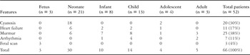
Table 1.
Kaplan-Meier survival curves. A: Cases with fetal or neonatal diagnosis (broken line) and the rest of patients (continuous line). B: Cases with associated cardiac defects (broken line) and cases without association (continuous line). C: Cases with Celermajer index of Grade 1 or 2 (continuous line), and cases with Grade 3 or 4 (broken line). D: Cases with Cardiothoracic index less than 65% (continuous line) and cases with index equal or greater than 65% (broken line).

Associated cardiac defects presenting at different age groups. RVOTO: Right ventricular outflow tract obstruction; PAPVR: partially anomalous pulmonary venous return; PDA: patent ductus arteriosus.

Evolution of 52 cases of Ebstein's malformation. In the fourth file, number of neonates included in the four mean evolution forms: late/early death, and symptomatic/asymptomatic survivors.
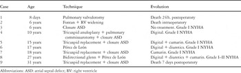
Table 2.

Kaplan-Meier survival curve indicating percent survival with follow-up.
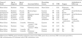
Table 3.
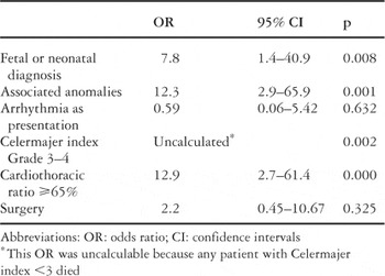
Table 4.
 You have
Access
- 32
- Cited by