The improving care and increased long-term survival rate among children with different types of cardiomyopathy has increased the number of children with heart failure.Reference Patel and Shaddy 1 Heart failure is associated with impairment of cardiac electrical conducting system in about 30% of patients causing intra-ventricular and inter-ventricular conduction delay and results in cardiac dyssynchrony. This dyssynchrony greatly reduces the pumping efficiency of the ventricles in patients with heart failure owing to delayed activation of the left lateral wall, mitral regurgitation, worsened ventricular dilation, impaired left ventricular filling, and decreased parameters of cardiac contractility.Reference Burkhardt and Wilkoff 2 – Reference Baldasseroni, Opasich and Gorini 4 Wide QRS complex on the electrocardiogram is evidence of dyssynchrony and is associated with the increased risk of mortality in patients with congestive heart failure and is recognised as an important predictor of poor outcome if left untreated.Reference Shamim, Francis and Yousufuddin 5
Adequate identification of left ventricular dyssynchrony is important in management patients with heart failure. Several recent imaging modalities were proved to be effective in diagnosing left ventricular dyssynchrony. These techniques vary from simple conventional M-mode and Doppler echocardiography to the more advanced tissue Doppler imaging, three-dimensional echocardiography, two-dimensional speckle tracking imaging, and, most recently, three-dimensional speckle tracking imaging as well as the conductance catheter techniques.Reference Bleeker, Bax, Steendijk, Schalij and van der Wall 6 , Reference Zhang and Yu 7
Experience with mechanical dyssynchrony and data in children are still limited because of the relative lack of paediatric studies. We hypothesised that dyssynchrony can be reliably measured in children with congestive cardiac failure and is associated with reduced systolic ventricular function. The aim of this study was to evaluate cardiac dyssynchrony in children with congestive heart failure due to dilated cardiomyopathy.
Materials and methods
This was a prospective case–control analysis of 34 consecutive Egyptian children presented to a tertiary paediatric cardiology centre with newly diagnosed congestive heart failures due to dilated cardiomyopathy. The study was conducted between June, 2011 and November, 2012. Heart failure was classified according to the Modified Ross Heart Failure Classification for Children.Reference Hsu and Pearson 8 An age- and sex-matched cohort of 30 patients was studied as a control group. They had an echocardiographic examination as a part of their routine assessment and before starting active treatment. Inclusion criteria included recently diagnosed cases of dilated cardiomyopathy with congestive heart failure and age between 2 and 5 years. Exclusion criteria included other cardiac causes of heart failure, other systemic conditions that could affect the cardiac conditions, previous use of cardiac or non-cardiac medications that could affect the heart functions, including anti-failure medications, age outside the range of 2–5 years, and difficult acquisition and analysis of time-strain curves.
All children were evaluated by a paediatric cardiologist. A complete history taking, thorough clinical examination, complete blood pictures, chest X-ray, electrocardiogram, oxygen saturation, and echocardiographic examination by conventional two-dimensional echocardiography, tissue Doppler, and speckle tracking strain analysis were carried out by a sonographer blinded to the patients’ studies. To avoid intra-observer variability, two examinations were conducted by the same sonographer for each patient within 1 week and we considered the average results. The data obtained from the normal group were used as normal values that were comparable to normal published data.Reference Kaku, Takeuchi and Tsang 9 , Reference Al-Biltagi, Serag, Hefidah and Mabrouk 10 The sonographer used the same techniques to obtain the systolic and diastolic indices. The study was approved by the Institutional Ethical and Research Review Board. All the parents of the included children signed a written informed consent before enrolment into the study.
Conventional echo-Doppler examination
Echocardiographic examination was conducted using Vingmed Vivid-7 (General Electric Vingmed, Milwaukee, Wisconsin, United States of America). Data acquisition was performed with a 3.5-MHz transducer at a depth of 16 cm in the parasternal and apical views. M-mode and two-dimensional images obtained and stored in cine loop format from three consecutive beats according to the recommendation of the American Society of Echocardiography.Reference Picard, Adams and Bierig 11 All children were examined in a semi-supine, left lateral position. The left ventricle volumetric study and ejection fraction were carried out using unique three-dimensional echocardiography cardiac ultrasound imaging transducers combined with enhanced four-dimensional software applications (TomTec v1.2; TomTec Imaging Systems, Fulda, Germany) in apical four- and two-chamber cut planes, which were optimised to avoid foreshortening. The echocardiographic quantification techniques were used according to the American Society of Echocardiography guidelines for children.Reference Lopez, Colan and Frommelt 12
Left ventricular dyssynchrony by tissue Doppler imaging
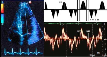
Tissue Doppler imaging was performed using the same machine and probe at a depth of 16 cm in the parasternal and apical views – standard long-axis and two- and four-chamber images. Using pulsed-wave angle-corrected colour-coded tissue Doppler imaging filters, the baseline was adjusted to a low-velocity range (−20 to 20 cm/second), and Doppler frame rates were varied between 80 and 115 frames/second depending on the sector width of the range of interest, with minimal gain setting to minimise background noise and to obtain the highest quality images. The sample volume was placed within the myocardium equidistant from the endocardial and epicardial borders. From the apical four-chamber planes, the myocardial velocity curves of septal mitral valve annulus, lateral mitral valve annulus, and lateral tricuspid valve annulus were recorded. The electrocardiogram was connected and traced simultaneously. The beginning of QRS complex was used as a reference point.Reference Al-Biltagi, Serag, Hefidah and Mabrouk 10 Figure 1 showed waves and times obtained by tissue Doppler imaging.
Waves and times derived by pulsed wave tissue Doppler lateral mitral annular tissue Doppler tracing. s: peak velocity during ventricular systole; e: peak velocity during early ventricular diastole; a: peak velocity during atrial contraction. IVCT=isometric (isovolumic) contraction time; IVRT=isometric (isovolumic) relaxation time.

Intra-ventricular left ventricular dyssynchrony was studied from the colour Doppler images by offline analysis using commercial software (Echopac 6.1; General Electric Vingmed). The tissue Doppler imaging data were analysed by an experienced observer blinded to the clinical data. Sample volumes were placed at the basal level in the septum and lateral wall, using the four chamber images, to derive velocity graphs. The systolic wave (S) reflects the systolic function of either the right or left ventricle. The early/atrial (E'/A') ratio of tricuspid and mitral valve annulus reflects the diastolic function of the right and left ventricle, respectively. Isometric contraction time was defined as the time duration between the beginnings of QRS complex in the electrocardiogram to the beginning of tissue Doppler imaging S-wave. The isometric relaxation time was defined as the interval between the end of S-wave and the beginning of the early wave. Both isovolumic contraction time and isovolumic relaxation time were corrected for heart rate. At least 10 cardiac cycles were recorded at a speed of 100 mm/second and the images were stored electronically. Tissue Doppler imaging measurements were reported according to reported against published Z-scores.Reference Eidem, McMahon and Cohen 13 From these data, the time from the beginning of the QRS complex (on electrocardiogram) to peak systolic velocities in the septum and lateral wall were assessed, and the difference between time-to-peak systolic velocity of the basal septal and basal lateral segments was calculated as a measure of intra-ventricular dyssynchrony, referred to as the septal-to-lateral delay.Reference Delgado, Ypenburg and van Bommel 14
Speckle tracking strain analysis
For speckle tracking analysis, standard greyscale two-dimensional images were acquired in the apical four-chamber view as well as the parasternal short-axis views at the level of the papillary muscles. Special care was taken to avoid oblique views from the mid-level short-axis images and to obtain images with the most circular geometry possible. All of the images were recorded with a frame rate of 30 frames/second using EchoPac 6.1 software.Reference Suffoletto, Dohi, Cannesson, Saba and Gorcsan 15 From an end-systolic single frame, a region of interest was traced on the endocardial cavity interface by a point-and-click approach. Then an automated tracking algorithm followed the endocardium from this single frame throughout the cardiac cycle. Further adjustment of the region of interest was done to ensure that all of the myocardial regions were included. Next, acoustic markers, the so-called speckles, equally distributed in the region of interest, could be followed throughout the entire cardiac cycle. The distance between the speckles was measured as a function of time, and parameters of myocardial deformation could be calculated. Finally, the myocardium was divided into six segments that are colour coded and displayed into six segmental time-strain curves for radial strain, circumferential strain, and longitudinal strain. For each type of strain analysed, two different parameters for dyssynchrony were obtained: maximal time delay between peak systolic strain of two segments, between the antero-septal and postero-lateral wall. For radial strain and circumferential strain, difference between times to peak-systolic strain of the antero-septal and posterior segments – antero-septal and posterior segments delay. For longitudinal strain, four-chamber view was used to calculate the difference between time-to-peak systolic strain of the basal septal and basal lateral left ventricular segment. Standard deviation of time-to-peak strain from multiple segments was calculated to evaluate left ventricular global synchrony.Reference Perk, Tunick and Kronzon 16
Statistical analysis
The statistical power of the study was more than 90% (using a computerised program: Power and Precision V3; www.PowerAnalysis.com). Data are presented as mean (±standard deviation) values. The two-way analysis of variance with repeated measures and χ2-test by SPSS V.16 were used to identify statistically significant differences in the different parameters among the groups. For all analyses, a statistical significance of p-value <0.05 was used. Wilcoxon’s signed-rank test was used to assess the normality of distributions of the data. The Bonferroni correction/adjustment procedure was performed to avoid “significance” because of chance only, in multiple comparisons with echocardiographic parameters. Correlation between variables was evaluated using Pearson’s correlation coefficient.
Results
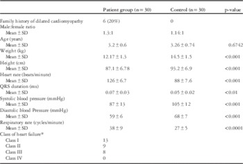
The study included 30 children with congestive heart failure due to dilated cardiomyopathy – after exclusion of four children because of difficulty in acquisition and analysis of time-strain curves – and 30 normal children as a control group. Their demographic data were shown in Table 1. There were no significant differences in age (p=0.6) and sex between these patients and the control group. The age of the patient group ranged from 2 to 4 years with a mean of 3.2±0.6 years, whereas that of the control group ranged from 2.5 to 5 years with a mean of 3.26±0.74 years. Of the congestive heart failure children, 52% (17 children) were male and 43% (13 children) were female, whereas 16 (53%) children in the control group were male and 14 (47%) were female. Children with congestive heart failure had significantly lower weight and height than children in the control group (p<0.001). Both the heart rate and respiratory rate were significantly higher in the patient group than in control group (p<0.001). On the other hand, the patient group had significantly lower systolic blood pressure and diastolic blood pressure than the control group (p<0.001). The table also showed significant statistical but clinically insignificant increase in QRS complex duration in the patient group than in the control group but with normal morphology (p<0.01). None of our patients had wide QRS complex more than the upper limit of normal or had abnormal morphology.
Demographic data and clinical features of both the patient and control groups.

* According to Modified Ross Heart Failure Classification for Children
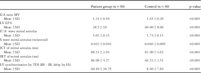
Tables 2 and 3 illustrated comparison of echocardiographic and tissue Doppler imaging features in the patient and control groups. The left ventricular ejection fraction was significantly lower in the patient group than in the control (p<0.001) group that denoted the presence of systolic dysfunction in the patient group. At the same time, conventional trans-mitral Doppler echocardiography showed left ventricular diastolic dysfunction as indicated by the lower E/A ratio of trans-mitral valve flow (p<0.001). Tissue Doppler showed the presence of both systolic, as indicated by decreased S-wave and prolonged isovolumic contraction time, and diastolic, as indicated by decreased E'/A' wave at mitral valve annulus and prolonged isovolumic relaxation time, dysfunction in the patient group than in the control group. It also showed more significant prolongation of the mean of antero-septal and posterior segments delay, circumferential strain parameters (63.26±35.68 ms) in children with congestive heart failure than in the control group (26.60±16.72 ms) (p<0.005).
Comparison of conventional and tissue Doppler echocardiographic features in the patient and control groups.

BS−BL delay is the difference between time to peak systolic velocity of the basal septal and basal lateral segments; E/A ratio MV=ratio of the early (E)-to-late (A) ventricular filling velocities across mitral valve; E'/A' is the ratio between early diastolic myocardial relaxation velocity below the baseline as the annulus ascends away from the apex; and myocardial velocity associated with atrial contraction; ICT=isometric contraction time; IRT=isometric relaxation time; LS=longitudinal strain; LV=left ventricular; LV EF=LV ejection fraction; S=LV systolic tissue Doppler velocity; TDI=tissue Doppler imaging
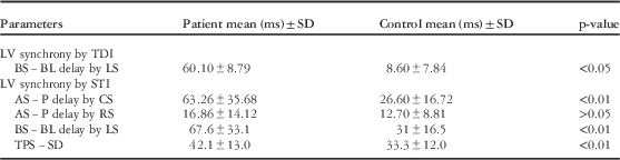
LV synchrony by TDI and STI in the studied groups.

AS−P delay=difference between time to peak systolic strain of the anteroseptal and posterior segment; BS−BL delay=difference between time to peak systolic strain of the basal septal and basal lateral segments; CS=circumferential strain; LV=left ventricular; LS=longitudinal strain; RS=radial strain; STI=speckle tracking imaging; TDI=tissue Doppler imaging; TPS−SD=standard deviation of time to peak strain
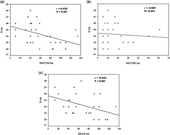
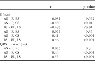
Table 3 showed the comparison of left ventricular synchronisation by speckle tracking imaging findings in the patient and the control group. It showed significant increase in the mean of antero-septal and posterior segments delay, circumferential strain parameters of patients with congestive heart failure (63.26±35.68 ms) than that of the control group (26.60±16.72 ms) (p=0.003). A significant increase in the mean basal septal and basal lateral segments delay parameters was present in the patient group with congestive heart failure (67.6±22 ms) than that of the control group (31±16.5 ms) (p=0.002). On the other hand, there was no significant increase in the mean antero-septal and posterior segments delay, and radial strain parameters was present in patients with congestive heart failure (16.86±14.12 ms) compared with the control group (12.70±8.8 ms) (p>0.05). It should be mentioned that no obvious specific clustering pattern was present for early and late contracting segments. At the same time, a significant left ventricular global dyssynchrony as indicated by standard deviation of time-to-peak strain was present in the patient group (42.1±13) when compared with the control group (33.3±12) (p=0.008). Intra-observer agreement of all echocardiographic measurements was present in both the patient and control groups (k between 70 and 82). Table 4 and Figure 2a–c showed a significant negative correlation between left ventricular S'-wave and left ventricular ejection fraction with left ventricular dyssynchrony derived from circumferential strain and longitudinal strain, whereas no significant correlation was present between the S' and left ventricular ejection fraction with left ventricular dyssynchrony from radial strain. At the same time, a significant positive correlation was present between QRS complex duration and left ventricular dyssynchrony derived from circumferential strain and longitudinal strain.
Correlation between LV S' and LV dyssynchrony derived from ( a ) circumferential dyssynchrony (CS), ( b ) radial dyssynchrony, and ( c ) longitudinal dyssynchrony (LS). ( a – c ) show the presence of a significant negative correlation between LV S' and LV dyssynchrony derived from circumferential strain and longitudinal strain, whereas there is no significant correlation between S' and LV dyssynchrony from radial strain. CS=circumferential strain; LV=left ventricular; LS=longitudinal strain.

Correlation between QRS duration, LV S' and LV dyssnchrony.

AS−P=difference between time to peak systolic strain of the anteroseptal and posterior segment; BS−BL=difference between time to peak systolic strain of the basal septal and basal lateral segments; CS=circumferential strain; LS=longitudinal strain; LV=left ventricular; RS=radial strain
There is a significant negative correlation between LV S' and LV dyssynchronisation derived from CS and LS, whereas there is no significant correlation between S' and LV dyssynchronisation from RS
Discussion
Cardiac dyssynchrony is present in a considerable number of patients with congestive heart failure and is an important factor that might worsen heart failure, impair cardiac output, and may be a major cause of morbidity and mortality.Reference Bleeker, Bax and Fung 17 Because of few studies concerned with cardiac dyssynchrony in children, we conducted this study to evaluate left ventricular dyssynchrony using tissue Doppler imaging and speckle tracking imaging in 30 children with congestive heart failure due to dilated cardiomyopathy. In the current study, tissue Doppler imaging showed a significant reduction in mitral valve annulus systolic wave velocity (S) in children with congestive heart failure when compared with the control group that denoted systolic dysfunction. This was previously confirmed in another study where the systolic myocardial velocity (S) at the lateral mitral annulus was a valid measure of longitudinal systolic function and correlated well with the measurements of left ventricular ejection fraction and peak dP/dt.Reference Marwick, Case and Leano 18 At the same time, a significant decrease in mitral annulus E'/A' ratio was present; indicating the presence of diastolic dysfunction using the advantage that assessment of diastolic function by tissue Doppler imaging is less load dependent than that provided by standard Doppler techniques. E' reflects the velocity of early myocardial relaxation as the mitral annulus ascends during early rapid left ventricular filling. Peak E' velocity can be measured from any aspect of the mitral annulus from the apical views, with the lateral annulus most commonly used. Because of intrinsic differences in myocardial fibre orientation, septal E' velocities are slightly lower than lateral E' velocities.Reference Ho, Sweitzer and McDonough 19
In this study, the maximum time delay between basal septal and basal lateral systolic velocities as measured by tissue Doppler imaging was significantly higher in children with congestive heart failure than the control group, indicating left ventricular dyssynchrony. This agreed with the work of Bax et alReference Bax, Ansalone and Breithardt 20 who found that the tissue Doppler-derived systolic velocity is sensitive in detecting left ventricular reverse remodelling and strain.
Two-dimensional speckle tracking imaging is a relatively new echocardiographic technique that could allow the study of all three types of deformation. Measurements of radial strain, circumferential strain, and longitudinal strain have also recently been validated by cardiac MRI. More importantly, two-dimensional speckle tracking imaging is an angle independent and a strain imaging technique; it enables differentiating those myocardial segments with active movement from those with passive movement, that is, scarred tissue tethered by the non-scarred segments.Reference Delgado, Ypenburg and van Bommel 21 Our results demonstrated the usefulness of two-dimensional speckle tracking imaging in the diagnosis of left ventricular dyssynchrony in all three deformation types: radial, circumferential, and longitudinal. It assessed the presence of left ventricular dyssynchrony parameters with circumferential strain – antero-septal and posterior segments delay – and longitudinal strain – basal septal and basal lateral segments delay; no changes in the left ventricular dyssynchrony were observed with radial strain – antero-septal and posterior segments delay. Sutherland et alReference Sutherland, Di Salvo, Claus, D’hooge and Bijnens 22 showed that the evaluation of left ventricular dyssynchrony using speckle tracking strain analysis was feasible and that substantial left ventricular dyssynchrony was present in all three deformation types – radial, circumferential, and longitudinal – in depressed left ventricular function and dilated cardiomyopathy. Strain myocardial imaging parameters and strain imaging may represent useful techniques to diagnose left ventricular dyssynchrony. In particular, myocardial strain is a dimensionless index of change in myocardial length in response to an applied force, and is expressed as fractional or per cent change. This technique has a theoretic advantage that it is relatively immune to cardiac translational motion and tethering, allowing differentiation between active systolic contraction and passive motion.Reference Bleeker, Bax and Fung 17
In the current study, a significant negative correlation was present between both left ventricular S-wave velocity and left ventricular ejection fraction and left ventricular dyssynchrony derived from circumferential strain and longitudinal strain, whereas no significant correlation was present between S and left ventricular ejection fraction and left ventricular dyssynchrony from radial strain. This denotes that the severity of dyssynchrony may adversely affect systolic function of the left ventricle and may lead to impaired cardiac output. However, as the correlation coefficient between tissue Doppler imaging S-wave velocity versus longitudinal strain-derived basal septal and basal lateral segments delay was fairly small (r=−0.4), such conclusion is not quite robust. Studying cardiac dyssynchrony in children with dilated cardiomyopathy and congestive heart failure may help in understanding the pathophysiology of this condition and may help to guide and monitor the proposed treatment. The study also showed significant positive correlation between prolongation of QRS complex duration and left ventricular dyssynchrony. This agreed with the work of Andrikopoulos et al who demonstrated that the duration of QRS was correlated with inter-ventricular but not intra-ventricular dyssynchrony in heart failure patients.Reference Andrikopoulos, Tzeis and Kolb 23 Andrikopoulos et al showed that narrowing of the QRS duration was correlated with improved cardiac function in paediatric population.Reference Gokhale, Husain, Nicholson, Texter, Zaidi and Cua 24 The presence of narrow QRS complex in our cases may be related to the recent onset of dilated cardiomyopathy in our cases. In our study, no obvious specific clustering pattern for early and late contracting segments was found in the presence of a narrow QRS complex. The absence of clustering is considered as a sign of random contractile pattern and dyssynchrony. This may indicate that the ventricular dyssynchrony associated with dilated cardiomyopathy is not only caused by electromechanical dyssynchrony but may be related to other intrinsic factors related to the cardiomyocyte as a result of myocyte injury and necrosis associated with myocardial fibrosis, which results in impaired mechanical function.Reference Leong, Chakrabarty and Shipp 25
The limitations of the current study were the relatively few numbers of patients. The study also concerned with studying the left ventricle only, ignoring the right ventricle, which is also of paramount importance specially related to the inter-ventricular dyssynchrony. We did not differentiate between those patients who had significant dyssynchrony from those who had insignificant dyssynchrony as well. At the same time, we did not have long-term follow-up to evaluate the course and the progression of those who had significant dyssynchrony. However, as there are relatively few studies concerned with studying inter-ventricular cardiac dyssynchrony in children with heart failure due to dilated cardiomyopathy, this study may be helpful to add some highlights about cardiac dyssynchrony in paediatric age group.
Conclusion
The study concluded that children with congestive heart failure due to dilated cardiomyopathy usually suffer from significant intra-ventricular dyssynchrony that negatively correlated with left ventricular systolic function. Tissue Doppler study and speckle tracking strain analysis are helpful tools to detect the presence of cardiac dyssynchrony in those children. Studying cardiac dyssynchrony in children with dilated cardiomyopathy and congestive heart failure may help in understanding the pathophysiology of this condition and could help to guide and monitor the proposed treatment.
Acknowledgement
The authors would like to thank all the members of Pediatric Department, Tanta University, Egypt, for their support.
Conflicts of Interest
None.
Financial Support
This research received no specific grant from any funding agency, commercial, or not-for-profit sectors.
Ethical Standards
The authors assert that all procedures contributing to this work comply with the ethical standards of the Helsinki Declaration of 1975, as revised in 2008, and has been approved by the Tanta Faculty of Medicine Research Ethics Board.