Introduction
Human alveolar echinococcosis (AE) is a severe parasitic zoonosis caused by infection with the metacestode Echinococcus multilocularis. In Japan, E. multilocularis infection is prevalent in Hokkaido where local red foxes serve as the definitive host and small rodents serve as the intermediate host. In order to detect AE in Hokkaido, mass screening of residents for this disease has been undertaken over the past 20 years as a part of administrative countermeasures (Kimura et al., Reference Kimura, Furuya, Kawase, Sato, Yamano, Takahashi, Uraguchi, Ito, Yagi and Sato1999).
The initial screening process utilized to detect AE is divided into two steps. The first step involves the use of an enzyme-linked immunosorbent assay (ELISA) (Sato et al., Reference Sato, Mitamura, Arai and Kumagai1983a, Reference Sato, Mitamura, Arai and Kumagaib), and the second step consists of both a Western blotting (WB) (Furuya et al., Reference Furuya, Sato and Kumagai1987, Reference Furuya, Sasaki, Honma, Kumagai, Sato, Takahashi and Uchino1989, Reference Furuya, Nishizuka, Honma, Kumagai, Sato, Takahashi and Uchino1990; Nagano et al., Reference Nagano, Sato and Furuya1995) and ultrasound-based tests. These tests are often used in provisional confirmation prior to surgical treatment. The final diagnosis for most AE patients is based on histopathological findings following surgery (Condon et al., Reference Condon, Rausch and Wilson1988; Furuya et al., Reference Furuya, Sato, Kawanaka, Takahashi, Saeada, Honma and Tamura2001). The Hokkaido Institute of Public Health (HIPH) has played an important role in the serological aspect of the mass screening process. Furthermore, the procedures utilized in the detection of AE have also brought about numerous improvements in the testing of serological samples at a more general level.
In the original WB method undertaken between 1987 and 2000, diagnosis of AE was based on the presence of 66, 55 and 30–35 kDa bands (Furuya et al., Reference Furuya, Sasaki, Honma, Kumagai, Sato, Takahashi and Uchino1989). Subsequent to this time and due to a higher sensitivity and specificity, the presence of 26–28, 18 and 7–8 kDa bands is now used for the provisional confirmation of the disease (Furuya et al., Reference Furuya, Kawanaka, Yamano, Sato and Honma2004; Yamano et al., Reference Yamano, Yagi, Furuya, Sawada, Honma and Sato2005).
On the basis of diagnosis using these low molecular weight antigens, numerous serum samples from people previously screened and from patients suspected to have AE in hospitals were examined between 2001 and 2006. As a result, we were able to identify 81 new AE cases in this 6-year period. However, 5 of the 81 cases demonstrated no reactivity with the 18 kDa antigen, although reactivity with one or both of the 26–28 and 7–8 kDa antigens was evident. In this paper, we report on the five registered AE patients with interesting serological characteristics and discuss the importance of making diagnoses based on the overall reactivity against plural antigens.
Materials and methods
Serum samples
A total of 278,696 serum samples were collected for the primary screening (ELISA) process undertaken between 2001 and 2006. From these samples, 848 individuals were recalled for a secondary examination involving WB, ultrasound, computed tomography and/or magnetic resonance imaging. In addition, 897 sera were also tested as part of a routine examination of hospital-suspected cases.
Antigen production
ELISA and WB tests were undertaken employing a crude antigen extracted from E. multilocularis cysts. To prepare the antigen, protoscoleces were collected from cysts in cotton rat liver which had been experimentally inoculated with metacestodes of E. multilocularis. The protoscoleces were repeatedly washed with phosphate-buffered saline (PBS, pH 7.4) and treated with 0.2% Triton X-100 in PBS. This solution was then centrifuged for 15 min at 500 rpm and the supernatant dialysed against PBS for 24 h at 4°C to remove detergent. The total protein concentration of the antigen solution was set to 12 μg ml− 1.
ELISA protocol
ELISAs were performed as described previously by Sato et al. (Reference Sato, Mitamura, Arai and Kumagai1983a, Reference Sato, Mitamura, Arai and Kumagaib) with some minor modifications. Flat-bottomed microplates (EIA/RIA 8-well strip, high-bind type, Corning, New York, USA) were coated with crude antigen (100 μl/well) for 4 h at 37°C. Serum samples diluted at 1:250 in 0.05% Tween–PBS (100 μl/well) were then added to the wells of the microplates and incubated overnight at 4°C. After washing, 100 μl of anti-human IgG alkaline phosphatase conjugated antibody (Sigma, St Louis, Missouri, USA; 1:2500 in 0.05% Tween–PBS) was added and incubated for 4 h at 37°C. After further washing, bound antibodies were detected by the presence of a reaction for alkaline phosphatase on p-nitrophenyl-phosphate (0.1 mg/well) in 10% diethanolamine buffer (pH 9.8) following an incubation period of 8 min at 37°C (100 μl/well). The reaction was stopped with the addition of 50 μl of 3N NaOH and the optical density (OD) values read at 405 nm on a microplate reader (Model 680, BIORAD, Hercules, California, USA). Between each incubation step, plates were washed three times with 0.05% Tween-PBS (300 μl/well).
Preliminary experiments undertaken in 1983 gave a mean OD value of 0.94 ± 0.21 for 46 confirmed AE patients. Therefore, the diagnostic criteria for the current study were defined based on the mean OD and mean OD minus 2 standard deviations as follows: OD 0–0.50 = negative; 0.51–0.99 = quasi-positive; over 1.00 = positive (Sato et al., Reference Sato, Mitamura, Arai and Kumagai1983a). A negative mean OD value (n = 471) was defined as 0.29 under these conditions.
Western blot protocol
Sodium dodecyl sulphate–polyacrylamide gel electrophoresis (SDS–PAGE) was undertaken as described by Laemmli (Reference Laemmli1970), utilizing a crude E. multilocularis antigen preparation. Antigens were separated electrophoretically on SDS–15% polyacrylamide gels and the separated antigens were electroblotted on to a nitrocellulose membrane. The membrane was subsequently blocked in 5% skimmed milk in PBS for 1 h at room temperature (RT) and then incubated with patient sera at 1:100 in PBS containing 0.05% Tween 20 and 5% skimmed milk for 1.5 h at RT. After washing, the membrane was treated with an anti-human IgG alkaline phosphatase conjugated antibody (Sigma), diluted at 1:2500 in 0.05% Tween–PBS, for 1 h at RT. After further washing, the membrane was exposed to BCIP/NBT substrate solution (PerkinElmer Life Sciences, Gaithersburg, Maryland, USA) for colour development. We adopted multiple markers including 26–28, 18 (the main marker utilized) and 7–8 kDa bands in our WB testing. The criteria for judgement are presented in table 1.
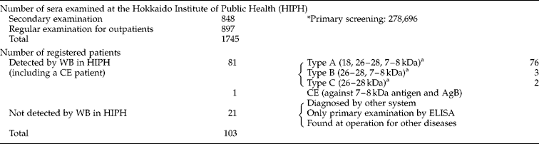
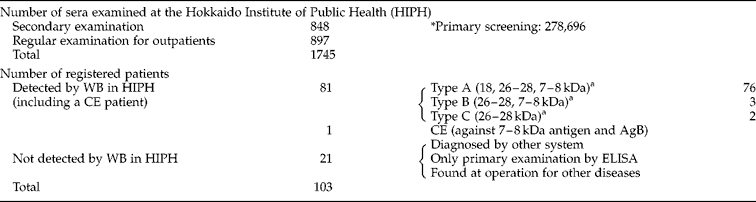
Results of Western blot (WB) testing and a summary of echinococcosis detection between 2001 and 2006.
CE, cystic echinococcosis.
a Type A and B are positive and Type C is quasi-positive.
* The first step of the screening process by ELISA.
In addition, the commercially available immunoblot assay kit, ECHINOCOCCUS Western Blot IgG (Ldbio Diagnostics, Lyon, France) was also utilized in this study. The operation of this kit and the interpretation of subsequent results were undertaken in accordance with the manufacturer's recommendations.
Results
An outline of examination results and echinococcosis detection
Table 1 presents an outline of the examination results and echinococcosis detection. Confirmative WB tests were carried out on a total number of 1745 sera between 2001 and 2006, including both secondary examinations undertaken as part of the Hokkaido serological mass screening system and the regular examination of suspected outpatients in hospitals. Under the ‘law concerning prevention of infectious diseases and medical care for patients of infections’, 103 patients from Hokkaido were newly reported to the Infectious Disease Surveillance Center as having echinococcosis in these 6 years. From our review study, it was identified that HIPH directly participated in the detection of at least 82 cases (79.6%), including one cystic echinococcosis (CE) patient. Among these 81 AE patients, 79 cases were found to be positive and two quasi-positive in our WB testing procedure. In addition, 21 of the newly registered 103 patients included cases that were diagnosed by other systems undertaken by some facilities, and were detected only via primary screening using ELISA, or were identified during surgery associated with other diseases.
As outlined in table 1, 81 AE patients detected by our WB testing were classified into three groups. Type A comprised 76 positive cases that demonstrated reactivity against the 18, 26–28 and 7–8 kDa antigens, Type B consisted of three positive cases showing reactivity against the 26–28 and 7–8 kDa antigens, and Type C included two quasi-positive cases that reacted only to the 26–28 kDa antigen. The Type A cases, that demonstrated reactivity to the 18 kDa antigen, are thought to represent typical positive AE patterns. In contrast, Types B and C (n = 5) represented patients with active AE and with characteristic sero-negativity for antibody directed to the 18 kDa antigen.
Five atypical cases
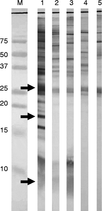
Five atypical patients of Types B (n = 3) and C (n = 2) were comprehensively diagnosed based on serological examination, diagnostic imaging and histopathological analysis. These patients characteristically lacked the presence of an 18 kDa band on their immunoblots (fig. 1), and similar results were also obtained using a commercially available immunoblot kit.
Immunoblot analysis of patients showing sero-negativity against the 18 kDa antigen. Lanes: M, the molecular size marker; 1, a positive control (Type A); 2 and 3 are sera from Type B patients (Cases 2 and 3); 4 and 5 are sera from Type C patients (Cases 4 and 5). The diagnostic bands are indicated by arrows.

Case 1 was a 70-year-old Japanese male of Type B, the details of whom have already been reported (Yamano et al., Reference Yamano, Yagi, Furuya, Sawada, Honma and Sato2005).
Case 2 was a 49-year-old Japanese male of Type B, who was detected via the Hokkaido screening system in 2003. His ELISA result was quasi-positive (OD 0.658) in the primary screening process, and a WB test undertaken as part of a secondary confirmation step was judged to be positive for the 26–28 and 7–8 kDa bands. When he consulted a physician for a thorough examination, several cysts were identified in S2/3, S5 and S7 of the liver using ultrasonography and computed tomography. He subsequently underwent surgery in September 2003, and histopathological confirmation was carried out.
Case 3 was a 43-year-old Japanese male of Type B. In this patient, several cysts with calcification were identified in S6/7 and S8 of the liver using ultrasonography and computed tomography following a medical examination. A serum sample for serological testing was collected in September 2004. The subsequent ELISA test demonstrated an OD value within the negative range (OD 0.417). Further WB tests identified the presence of 26–28 and 7–8 kDa protein bands and were judged to be positive. The cysts were surgically removed in December 2004 and histopathological confirmation was carried out.
Case 4 was a 71-year-old Japanese female of Type C. Upon consultation with a physician for ureteral calculus, a phyma of 5 cm in diameter was identified in S8 of the liver using computed tomography. A subsequent serological test was undertaken on a serum sample collected in February 2005. Both ELISA and WB analysis gave a quasi-positive result, with an OD value of 0.731 and the presence of a 26–28 kDa band, respectively. This patient received a resection of phyma in March 2005, and a diagnosis of AE was confirmed following histopathogical analysis.
Case 5 was a 19-year-old Japanese male of Type C. Following a medical examination for stomach ache, a phyma with calcification was identified in the left lobe of the liver using ultrasonography, computed tomography and magnetic resonance imaging. A serological test was undertaken on a serum sample collected in February 2005. An ELISA test demonstrated an OD value within the positive range (OD 1.51); however, a WB test identified the presence of only the 26–28 kDa band and was thus judged to be quasi-positive. The patient underwent surgery in May 2006 and a diagnosis of AE was confirmed following histopathological analysis.
Discussion
AE is prevalent in the forest and farming areas of the northern hemisphere and is endemic to the Hokkaido region of Japan. CE, which is caused by infection with E. granulosus, occurs most often around areas of the world involved in the raising of livestock and is generally regarded to be of minor importance in imported cases in Japan (Furuya et al., Reference Furuya, Kawanaka, Sato, Honma and Tamura2000). Therefore, the accurate diagnosis of AE is a major priority in Japan, especially in Hokkaido.
A serological screening system using ELISA (Sato et al., Reference Sato, Mitamura, Arai and Kumagai1983a, Reference Sato, Mitamura, Arai and Kumagaib) and WB (Furuya et al., Reference Furuya, Sato and Kumagai1987, Reference Furuya, Sasaki, Honma, Kumagai, Sato, Takahashi and Uchino1989, Reference Furuya, Nishizuka, Honma, Kumagai, Sato, Takahashi and Uchino1990; Nagano et al., Reference Nagano, Sato and Furuya1995) tests has been established previously to detect patients with AE residing in Hokkaido. These methods employ a crude antigen extracted from E. multilocularis cysts which detects numerous antibodies directed specifically against E. multilocularis antigens.
In our original WB testing procedures, protein bands of 55–66 kDa and a broad smear band including polysaccharides ranging from 30–35 kDa were identified as markers for AE when separated by SDS–PAGE (8% polyacrylamide gel). More recently, Em18, which is located in the 18 kDa region of immunoblots, has been reported to be a good marker for AE detection (Ito et al., Reference Ito, Nakao, Kutsumi, Lightowlers, Itoh and Sato1993, Reference Ito, Schantz and Wilson1995, Reference Ito, Sako, Yamasaki, Mamuti, Nakaya, Nakao and Ishikawa2003). It was subsequently demonstrated that the Em18 antigen is the breakdown product of EM10 (55–66 kDa antigen) following proteolysis by cysteine proteinases (Sako et al., Reference Sako, Nakao, Nakaya, Yamasaki, Gottstein, Lightowlers, Schantz and Ito2002). These results confirmed that Em18 and 55–66 kDa antigens were basically equal in the diagnosis of AE with WB testing. In addition, a commercially available immunoblot kit that detects bands ranging from 7 to 28 kDa has been developed and manufactured in France (Liance et al., Reference Liance, Janin, Bresson-Hadni, Vuitton, Houin and Piarroux2000) for the detection of echinococcosis. Thus, we began to investigate the use of low molecular weight antigens in the identification of AE (Yamano et al., Reference Yamano, Yagi, Furuya, Sawada, Honma and Sato2005). Our improved WB method monitors the presence of three marker bands ranging from 7 to 28 kDa. This concept is similar to that utilized by the above-mentioned commercially available kit. However, in the utilization of crude antigen, the 7–8 kDa band is usually accompanied by an approximately 12 kDa band which appears to be related to antigen B (AgB) (Leggatt et al., Reference Leggatt, Yang and McManus1992). Moreover, some CE cases may be detected due to cross-reaction with the 26–28 and/or 7–8 kDa antigens, a result that may also occur with the commercially available kits (see table 1).
In the previously utilized WB testing procedures, we identified patients demonstrating sero-negativity for the 55/66 kDa antigens. Similarly, we detected five patients showing sero-negativity for the 18 kDa antigen over a period of 6 years, using the application of low molecular weight markers. The prognosis for these five patients is good as a result of surgical treatment, suggesting that precise serodiagnosis aids in the improvement of curative effects.
It has recently become popular to use low molecular weight antigens as a diagnostic marker for AE. However, 6.2% (5 of 81) of patients showed sero-negativity against the 18 kDa antigen, a main marker for AE. Unfortunately this frequency is quite significant. We have also reported previously that AE patients demonstrating sero-negativity against the 18 kDa antigen were detected at a frequency of 21.7%, due to the presence of the 26–28 kDa and/or 7–8 kDa bands when using the commercially available immunoblot kits (Furuya et al., Reference Furuya, Kawanaka, Yamano, Sato and Honma2004). Similar findings regarding sero-negativity against the 18 kDa antigen were also reported for Chinese and French AE patients (Nirmalan & Craig, Reference Nirmalan and Craig1997; Liance et al., Reference Liance, Janin, Bresson-Hadni, Vuitton, Houin and Piarroux2000). These findings may indicate that the positive rate against the 18 kDa antigen in sera from AE patients is not sufficiently high and that the detection of multiple bands on an immunoblot test may be required for the correct diagnosis of AE.
The recent development of recombinant antigens has also contributed to an improvement in serodiagnostic examinations. These antigens include EmII/3 (Vogel et al., Reference Vogel, Gottstein, Muller and Seeback1988; Felleisen & Gottstein, Reference Felleisen and Gottstein1994; Kouguchi et al., Reference Kouguchi, Suzuki, Yamano, Honma and Sawada2005), EM10 (Frosch et al., Reference Frosch, Frosch, Pfister, Schaad and Bitter-Suermann1991; Helbig et al., Reference Helbig, Frosch, Kern and Frosch1993; Hubert et al., Reference Hubert, Cordero, Frosch and Solomon1999) and Em18 (Ito et al., Reference Ito, Xiao, Liance, Sato, Sako, Mamuti, Ishikawa, Nakao, Yamasaki, Nakaya, Bardonnet, Bresson-Hadni and Vuitton2002). Unfortunately, no single antigen identified to date has proven successful in detecting all AE patients. Therefore, it is concluded that we need to pay particular attention to the specific reactions to plural different diagnostic antigens such as the 26–28 and 7–8 kDa proteins to minimize the risk of inaccurate diagnosis of AE.