
Introduction
Parasitism is a close association in which parasites may influence their hosts at several organizational levels (Barber and Wright, Reference Barber and Wright2005). Often these effects of parasites on the hosts increase the parasite's likelihood of transmission (Poulin, Reference Poulin1998). The alimentary canal of fish has been shown to be a favourite place for the growth, establishment and reproduction of enteric parasites (Sayyaf Dezfuli et al., Reference Sayyaf Dezfuli, Giari and Bosi2021a). Intestinal parasites often induce changes in the morphology of the host tissue that might affect organ physiology (Fairweather, Reference Fairweather and Beckage1997). Inflammation due to fish enteric helminths is well documented (Dezfuli et al., Reference Dezfuli, Arrighi, Domeneghini and Bosi2000, Reference Dezfuli, Castaldelli, Bo, Lorenzoni and Giari2011a, Reference Dezfuli, Giari, Squerzanti, Lui, Lorenzoni, Sakalli and Shinn2011b; Taraschewski, Reference Taraschewski2000; Sayyaf Dezfuli et al., Reference Sayyaf Dezfuli, Giari and Bosi2021a, Reference Sayyaf Dezfuli, Pironi, Maynard, Simoni and Bosi2022) and consists of a complex series of homoeostatic mechanisms involving circulatory, immune, nervous and endocrine systems (Serna-Ducque and Esteban, Reference Serna-Duque and Esteban2020; Sayyaf Dezfuli et al., Reference Sayyaf Dezfuli, Giari and Bosi2021a). In at least some host–parasite systems, changes in host physiology are caused by parasite action on the fish neuroendocrine system (NES) (Øverli et al., Reference Øverli, Páll, Borg, Jobling and Winberg2001; Herbison, Reference Herbison2017; Helland-Riise et al., Reference Helland-Riise, Vindas, Johansen, Nadler, Weinersmith, Hechinger and Øverli2020).
Several amines, peptides and other molecules permit communication between nervous, endocrine and immune cells, which often produce neuromodulators (O'Dorisio and Panerai, Reference O'Dorisio and Panerai1990). Some publications have recorded the essential role of the NES in inflammatory processes caused by intestinal parasites in mammals (see Fairweather, Reference Fairweather and Beckage1997). Interestingly, most mammalian enteric neuropeptides also are present in fish and probably act in the same way (Holmgren, Reference Holmgren1985). The number of studies concerning the physiological role of neuromodulators of the fish enteric NES is growing (Bosi et al., Reference Bosi, Bermùdez and Domeneghini2007; Holmgren and Olsson, Reference Holmgren, Olsson, Bernier, Van Der Kraak, Farrell and Brauner2009; Takei and Loretz, Reference Takei, Loretz, Grosell, Farrel and Brauner2011; Blanco et al., Reference Blanco, Bertucci, Valenciano, Delgado and Unniappan2017; Ceccotti et al., Reference Ceccotti, Giaroni, Bistoletti, Viola, Crema and Terova2018). The NES controls and regulates intestinal physiology and in fish with enteric parasites this system is challenged with an overload. A recent review by Serna-Duque and Esteban (Reference Serna-Duque and Esteban2020) suggested 3 types of neuroendocrine responses against parasites: fight, protect and escape.
In wild fish, parasitic infections are frequent (Feist and Longshaw, Reference Feist and Longshaw2008) and in cultured fish parasites and diseases reduce reproductive performance and have a negative impact on feed conversion efficiency, which influences animal welfare (Shinn et al., Reference Shinn, Pratoomyot, Bron, Paladini, Brooker and Brooker2015). Indeed, fish parasites decrease host growth (Hoai, Reference Hoai2020), in some cases provoke mortality (Tavares-Dias and Martins, Reference Tavares-Dias and Martins2017), some parasites are zoonotic and may challenge food safety (Levsen et al., Reference Levsen, Svanevik, Cipriani, Mattiucci, Gay, Hastie, Bušelić, Mladineo, Karl, Ostermeyer, Buchmann, Højgaard, González, Pascual and Pierce2018). The purpose of this investigation was to detect and then compare the occurrence and distribution of putative neuromodulators in the intestine of several fish species infected with parasites.
The gut NES
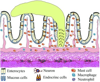
During digestion, in the lumen of the alimentary canal food is reduced to small nutritive molecules absorbed for the body energy balance and waste is ejected. According to Takei and Loretz (Reference Takei, Loretz, Grosell, Farrel and Brauner2011) the lumen represents a continuum with the environment, the ‘outside world’, which makes the lumen a gateway for the entrance of pathogens. Fish living in water are continuously challenged with a plethora of micro-organisms, pollutants and parasites. The activities involved in the physiological control of vertebrate gut function, including defence against pathogens, are mediated by the NES (Palmer and Greenwood-Van Meerveld, Reference Palmer and Greenwood-Van Meerveld2001; Sayyaf Dezfuli et al., Reference Sayyaf Dezfuli, Giari and Bosi2021a) formed by the enteric nervous system (ENS) and the enteric endocrine system (EES) (Takei and Loretz, Reference Takei, Loretz, Grosell, Farrel and Brauner2011) (Fig. 1).
Schematic view of the intestinal wall of fish with attached acanthocephalan. The intestinal mucosa is lined by a simple columnar epithelium consisting of typical epithelial cells with a sparse intermingling of mucus cells and ECs. Note the presence of the myenteric plexus with neurons between 2 muscle layers.

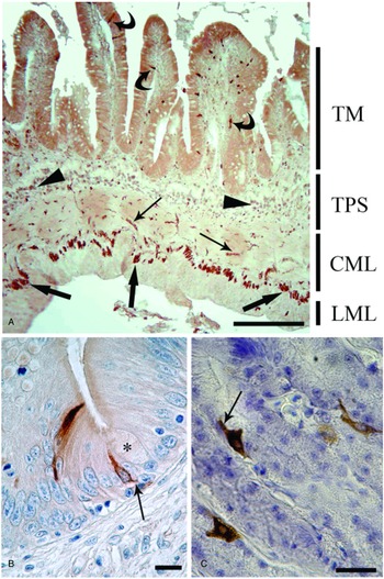
In fish, the ENS consists of neurons scattered in the gut wall or aggregated in small groups interconnected with nerve fibres and nerve bundles, named myenteric plexus, running between the inner circular and the outer longitudinal muscle layers (Furness, Reference Furness and Furness2006a; Ceccotti et al., Reference Ceccotti, Giaroni, Bistoletti, Viola, Crema and Terova2018) (Figs 1 and 2A). Fish lack a well-organized submucosal plexus to innervate the mucosa; nevertheless, in some instances the nerve fibres were reported in the tunica propria-submucosa (Ceccotti et al., Reference Ceccotti, Giaroni, Bistoletti, Viola, Crema and Terova2018). The extrinsic autonomic nervous system controls the ENS through the cranial vagus nerve and the spinal splanchnic nerves (Holmgren and Olsson, Reference Holmgren, Olsson, Bernier, Van Der Kraak, Farrell and Brauner2009; Blanco et al., Reference Blanco, Calo and Soengas2021). The neurons of the ENS innervate each component of the gut wall (muscle, epithelium, blood vessels and innate immune cells located in the connective layer beneath the epithelium) (Furness, Reference Furness and Furness2006a).
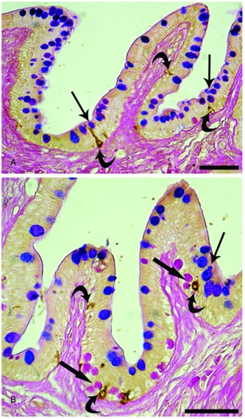
The EES is considered the largest endocrine organ in the body (Ahlman and Nilsson, Reference Ahlman and Nilsson2001; Blanco et al., Reference Blanco, Calo and Soengas2021). The main components of the EES are the endocrine cells (ECs, Figs 1 and 2B, C), many of which are randomly scattered in the epithelium of the gastrointestinal tract and sometimes are in close proximity to nerve fibres and immune cells (Holmgren and Olsson, Reference Holmgren, Olsson, Bernier, Van Der Kraak, Farrell and Brauner2009; Stakenborg et al., Reference Stakenborg, Viola and Boeckxstaens2020). There are 2 morphologically distinct types of ECs, ‘open’ and ‘closed’. The first type is a spindle-shaped cell, with its basal region near the basement membrane of the epithelium and the subtle apex exposed to the intestinal lumen (Takei and Loretz, Reference Takei, Loretz, Grosell, Farrel and Brauner2011; Kulkarni et al., Reference Kulkarni, Ganz, Bayrer, Becker, Bogunovic and Rao2018; Blanco et al., Reference Blanco, Calo and Soengas2021) (Fig. 2B). The closed type EC is also situated near the basal membrane, but does not reach the epithelial apex (Takei and Loretz, Reference Takei, Loretz, Grosell, Farrel and Brauner2011; Kulkarni et al., Reference Kulkarni, Ganz, Bayrer, Becker, Bogunovic and Rao2018; Blanco et al., Reference Blanco, Calo and Soengas2021) (Fig. 2C). The ECs, also called ‘paraneurons’ (Fujita and Kobayashi, Reference Fujita and Kobayashi1977; Day and Salzet, Reference Day and Salzet2002; Takei and Loretz, Reference Takei, Loretz, Grosell, Farrel and Brauner2011), pick up luminal/tissue stimuli with their membrane receptors and release specific chemical messengers from an axonal-like process at their basolateral membrane, called the ‘neuropod’ (Takei and Loretz, Reference Takei, Loretz, Grosell, Farrel and Brauner2011; Latorre et al., Reference Latorre, Sternini, De Giorgio and Greenwood-Van Meerveld2016; Kulkarni et al., Reference Kulkarni, Ganz, Bayrer, Becker, Bogunovic and Rao2018; Bosi et al., Reference Bosi, Lorenzoni, Carosi and Sayyaf Dezfuli2020) (Fig. 2B and C). The proximity of the ECs to mucous cells and mast cells might suggest their involvement in fish immune responses to the pathogens (Bosi et al., Reference Bosi, Shinn, Giari and Sayyaf Dezfuli2015; Sayyaf Dezfuli et al., Reference Sayyaf Dezfuli, DePasquale, Castaldelli, Giari and Bosi2017, Reference Sayyaf Dezfuli, Giari and Bosi2021a) (Fig. 3) and their connection with sensory neurons of the gut plexuses are consistent with the role of the ECs in direct transmission of information to the brain (Kaelberer et al., Reference Kaelberer, Buchanan, Klein, Barth, Montoya, Shen and Bohórquez2018; Ye et al., Reference Ye, Bae, Cassilly, Jabba, Thorpe, Martin, Lu, Wang, Thompson, Lickwar, Poss, Keating, Jordt, Clardy, Liddle and Rawls2021).
Components of the enteric nervous system and EES in several fish–helminth systems. (A) Salmo trutta infected with Pomphorhynchus laevis (out of microscope field), showing neurons of the myenteric plexus (thick arrows) and nerve fibres (thin arrows) immunoreactive to the anti-SP between the inner and outer intestinal muscle layers. The curved arrows indicate ECs in the epithelium. In the tunica propria-submucosa, numerous SP-positive mast cells are evident (arrowheads). CML, LML, circular and longitudinal muscle layer of tunica muscularis; TM, tunica mucosa; TPS, tunica propria-submucosa; scale bar: 100 μm. (B) Oncorhynchus mykiss parasitized with Eubothrium crassum (out of microscope field) showing 2 open-type ECs immunoreactive to anti-CCK8 in the rainbow trout intestinal epithelium; 1 of them is near a mucus cell (asterisk). Note the neuropod (thin arrow) of the cell on the right; scale bar: 10 μm. (C) Esox lucius harbouring Acanthocephalus lucii (out of microscope field), showing 2 closed-type ECs immunoreactive to anti-somatostatin-14 in the gastric epithelium of the pike. The neuropod is evident in the top left EC (thin arrow); scale bar: 20 μm.

Close relationship between ECs and epithelial components of the innate defence against parasites. Both images are from Silurus glanis intestine infected with the cestode Glanitaenia osculata (not shown). (A) ECs (curved arrows) immunoreactive to anti-serotonin close to the epithelial mucus cells (thin arrows). Immunohistochemistry counterstained with the Alcian blue/periodic acid Schiff method (AB/PAS). The AB/PAS stain reveals mucus cells in blue, magenta or violet depending on their content of acidic, neutral or mixed (acidic + neutral) mucins; scale bar: 50 μm. (B) ECs (curved arrows) immunoreactive to anti-met-enkephalin near intraepithelial mast cells (thick arrows). Mast cells are magenta due to periodic acid Schiff. Immunohistochemistry counterstained with the AB/PAS stain; scale bar: 50 μm.

The ENS–EES can function independently from the brain and spinal cord and are able to direct several physiological functions of the gastrointestinal tract without receiving commands from the central nervous system (Furness, Reference Furness and Furness2006a). The ECs detect several substances, each of them inducing an intracellular signal that leads to the release of a specific signal molecule, mainly a peptide (Sternini et al., Reference Sternini, Anselmi and Rozengurt2008; Blanco et al., Reference Blanco, Calo and Soengas2021). The released signal molecule can act locally on the neighbouring tissue components, or enter the circulatory system to reach more distant cells (Blanco et al., Reference Blanco, Calo and Soengas2021).
The interactions between components of the ENS–EES are mediated by several signal molecules produced by them, and some other molecules are secreted by cells of the innate immune system (Holmgren and Olsson, Reference Holmgren, Olsson, Bernier, Van Der Kraak, Farrell and Brauner2009; Hernández-Bello et al., Reference Hernández-Bello, Escobedo, Guzmán, Ibarra-Coronado, López-Griego and Morales-Montor2010; Sayyaf Dezfuli et al., Reference Sayyaf Dezfuli, Giari and Bosi2021a). In the past, ECs were classified based on the nature of the molecular signal they produce; recently molecular biology assays revealed they are more complex than previously recognized and are able to express and secrete different hormones and neurotransmitters in relation to the gut regions where they are located (Ye and Rawls, Reference Ye and Rawls2021).
The primary stimulus for the physiological activity of the gut is the ingestion of food, and its progression along the alimentary canal stimulates the ECs (Holmgren and Olsson, Reference Holmgren, Olsson, Bernier, Van Der Kraak, Farrell and Brauner2009; Takei and Loretz, Reference Takei, Loretz, Grosell, Farrel and Brauner2011). The sequential contraction and relaxation of the smooth muscle layer ensures mixture and the progressive movement of the digesta along the alimentary canal; the secretive/absorptive activity of the gut mucosa, pancreas and liver reduces the size of the food particles and macromolecules to small nutritional molecules that can be absorbed (Holmgren and Olsson, Reference Holmgren, Olsson, Bernier, Van Der Kraak, Farrell and Brauner2009). Several authors have reviewed the molecules involved in fish physiological activity (Holmgren and Olsson, Reference Holmgren, Olsson, Bernier, Van Der Kraak, Farrell and Brauner2009; Volkoff et al., Reference Volkoff, Unniappan, Kelly, Bernier, Van der Kraak, Farrel and Brauner2009; Takei and Loretz, Reference Takei, Loretz, Grosell, Farrel and Brauner2011; Volkoff, Reference Volkoff2016; Serna-Duque and Esteban, Reference Serna-Duque and Esteban2020; Blanco et al., Reference Blanco, Calo and Soengas2021), thus, in the following sections only a brief description of signal molecules and their main functions will be provided.
The NES control of gut motility
The main excitatory neurotransmitter in the fish gut is acetylcholine (Szabó et al., Reference Szabó, Nemcsók, Kása and Budai1991; Holmgren and Olsson, Reference Holmgren, Olsson, Bernier, Van Der Kraak, Farrell and Brauner2009; Ceccotti et al., Reference Ceccotti, Giaroni, Bistoletti, Viola, Crema and Terova2018). Several other peptides like tachykinin substance P (SP) and galanin function as co-transmitters in cholinergic gut neurons (Furness, Reference Furness2000; Bosi et al., Reference Bosi, Di Giancamillo, Arrighi and Domeneghini2004, Reference Bosi, Shinn, Simoni, Arrighi and Domeneghini2006, Reference Bosi, Bermùdez and Domeneghini2007; Holmgren and Olsson, Reference Holmgren, Olsson, Bernier, Van Der Kraak, Farrell and Brauner2009; Ceccotti et al., Reference Ceccotti, Giaroni, Bistoletti, Viola, Crema and Terova2018). Seemingly, the contraction of the smooth muscle cells is regulated by the calcitonin gene-related peptide (CGRP) and the amine serotonin (Shahbazi et al., Reference Shahbazi, Karila, Olsson, Holmgren, Conlon and Jensen1998; Holmgren and Olsson, Reference Holmgren, Olsson, Bernier, Van Der Kraak, Farrell and Brauner2009; Ceccotti et al., Reference Ceccotti, Giaroni, Bistoletti, Viola, Crema and Terova2018). It has been mentioned that the largest neuronal population in the myenteric plexus of the fish gut is formed by serotonergic neurons (Olsson et al., Reference Olsson, Holmberg and Holmgren2008b; Ceccotti et al., Reference Ceccotti, Giaroni, Bistoletti, Viola, Crema and Terova2018). Other molecules that function as excitatory neurotransmitters in the gut neurons of fish are well known. For example, the peptide ghrelin, which signals satiety, is secreted by endocrine epithelial cells of the stomach and modulates the contraction of smooth muscle cells (Olsson et al., Reference Olsson, Holbrook, Bompadre, Jönsson, Hoyle, Sanger, Holmgren and Andrews2008a; Holmgren and Olsson, Reference Holmgren, Olsson, Bernier, Van Der Kraak, Farrell and Brauner2009).
In the fish gut, relaxation of smooth muscle cells is mainly induced by vasoactive intestinal peptide (VIP), pituitary adenylate-cyclase-activating polypeptide (PACAP) and by nitric oxide gas (Holmgren and Olsson, Reference Holmgren, Olsson, Bernier, Van Der Kraak, Farrell and Brauner2009; Takei and Loretz, Reference Takei, Loretz, Grosell, Farrel and Brauner2011; Ceccotti et al., Reference Ceccotti, Giaroni, Bistoletti, Viola, Crema and Terova2018). Nitric oxide is not stored in the cell but is synthesized on demand by activation of a specific enzyme, neuronal-nitric oxide synthase (n-NOS) (Furness, Reference Furness and Furness2006b).
Some signal molecules modulate gut muscle peristalsis: their release alone or in combination with other substances can induce contraction or relaxation of the organ. In addition to its excitatory function, the peptide CGRP can inhibit gut muscle motility (Shahbazi et al., Reference Shahbazi, Karila, Olsson, Holmgren, Conlon and Jensen1998; Holmgren and Olsson, Reference Holmgren, Olsson, Bernier, Van Der Kraak, Farrell and Brauner2009; Ceccotti et al., Reference Ceccotti, Giaroni, Bistoletti, Viola, Crema and Terova2018). The peptide bombesin can stimulate stomach smooth muscle cells, although it was considered as a weak inhibitor of muscle contraction in some fish species (Holmgren and Jönsson, Reference Holmgren and Jönsson1988; Holmgren and Olsson, Reference Holmgren, Olsson, Bernier, Van Der Kraak, Farrell and Brauner2009). The main function of somatostatin is the inhibition of gastric acid secretion (Chu and Schubert, Reference Chu and Schubert2013; Bohórquez and Liddle, Reference Bohórquez, Liddle, Feldman, Friedman and Brandt2015). Notwithstanding, the inhibition of cholinergic neurons of the smooth muscle cells by somatostatin has also been documented in fish stomach (Grove and Holmgren, Reference Grove and Holmgren1992a, Reference Grove and Holmgren1992b; Holmgren and Olsson, Reference Holmgren, Olsson, Bernier, Van Der Kraak, Farrell and Brauner2009).
The NES control of alimentary physiology
In vertebrates, specific brain regions regulate feeding behaviour under the influence of molecular signals, produced by the brain itself or by the gut and its annexes (Volkoff, Reference Volkoff2016). The feeding activity is inhibited or stimulated in the presence of molecular signals released by neurons of the hypothalamus as well as neurons of ENS, and/or ECs of EES (Volkoff, Reference Volkoff2016).
Several signal molecules are involved in the control and regulation of the gastrointestinal status and consequently of the fish feeding behaviour (Le Bail and Boeuf, Reference Le Bail and Boeuf1997; Takei and Loretz, Reference Takei, Loretz, Grosell, Farrel and Brauner2011). The signal molecules with orexigenic action are ghrelin, galanin, neuropeptide Y (NPY) and β-endorphin. Conversely, gastrin/cholecystokinin8 (CCK8), gastrin releasing peptide (GRP)/bombesin, PACAP, VIP, CGRP, tachykinins and serotonin are considered anorectic signal molecules (Holmgren and Olsson, Reference Holmgren, Olsson, Bernier, Van Der Kraak, Farrell and Brauner2009; Takei and Loretz, Reference Takei, Loretz, Grosell, Farrel and Brauner2011; Volkoff, Reference Volkoff2016).
When digesta enters the fish proximal intestine, the local release of glucagon inhibits gastric acid secretion and acts as a short-term signal for the ingestion of food (Olsson and Holmgren, Reference Olsson and Holmgren2001; Takei and Loretz, Reference Takei, Loretz, Grosell, Farrel and Brauner2011). The ECs secrete CCK8, which induces gall bladder contraction and the entrance of bile in the intestinal lumen (Volkoff, Reference Volkoff2016). Moreover, CCK8 stimulates the synthesis and secretion of pancreatic enzymes (Rønnestad et al., Reference Rønnestad, Kamisaka, Conceição, Morais and Tonheim2007; Takei and Loretz, Reference Takei, Loretz, Grosell, Farrel and Brauner2011; Volkoff, Reference Volkoff2016; Blanco et al., Reference Blanco, Calo and Soengas2021).
Intra-cerebroventricular or -peritoneal injections of bombesin, CCK8, PACAP and VIP inhibit food intake in fish (Himick and Peter, Reference Himick and Peter1994a, Reference Himick and Peter1994b; Matsuda et al., Reference Matsuda, Maruyama, Nakamachi, Miura, Uchiyama and Shioda2005; Volkoff et al., Reference Volkoff, Unniappan, Kelly, Bernier, Van der Kraak, Farrel and Brauner2009). The effects of the different neuroendocrine molecules on fish feeding behaviour are several and variegated; many molecules are involved in food intake or prohibit food intake. For instance, CCK8 stimulates cholinergic neurons to reduce gastric emptying (Olsson et al., Reference Olsson, Aldman, Larsson and Holmgren1999) and simultaneously stimulates the gallbladder to release bile (Aldman and Holmgren, Reference Aldman and Holmgren1995). Both CCK8 and CGRP are synthesized by neurons and ECs and are involved in several functions like control of peristalsis, vasodilation and regulation of food intake concurrently with other signal molecules (Shahbazi et al., Reference Shahbazi, Karila, Olsson, Holmgren, Conlon and Jensen1998; Martínez-Álvarez et al., Reference Martínez-Álvarez, Volkoff, Muñoz-Cueto and Delgado2009).
With reference to orexigenic signal molecules, galanin injected in the fish brain influenced the feeding behaviour of tench, Tinca tinca, and goldfish, Carassius auratus (Guijarro et al., Reference Guijarro, Delgado, Pinillos, López-Patiño, Alonso-Bedate and De Pedro1999; Volkoff and Peter, Reference Volkoff and Peter2001; Volkoff, Reference Volkoff2016). Another orexigenic peptide, NPY is expressed in the fish brain and intestine and its increase induces feeding activity (Volkoff, Reference Volkoff2016). Ghrelin treatment can negatively affect food uptake in the rainbow trout, Oncorhynchus mykiss, and the Atlantic salmon, Salmo salar. Conversely, the same molecule increased foraging activity in brown trout, Salmo trutta. Thus, the function of ghrelin in fish feeding behaviour seems to be species-specific (Volkoff, Reference Volkoff2016). All of the studies on molecules involved in fish feeding activity were conducted on uninfected fish, or did not specify whether the fish were infected. To better understand host–parasite interactions, more studies on both uninfected and infected fish are needed.
The NES control of intestinal immunity
In fish, the amine serotonin is synthesized by neurons, ECs and immune cells, primarily by mast cells (Bosi et al., Reference Bosi, Shinn, Giari, Simoni, Pironi and Dezfuli2005a, Reference Bosi, Domeneghini, Arrighi, Giari, Simoni and Dezfuli2005b, Reference Bosi, Shinn, Giari and Sayyaf Dezfuli2015; Dezfuli et al., Reference Dezfuli, Castaldelli, Bo, Lorenzoni and Giari2011a; Ceccotti et al., Reference Ceccotti, Giaroni, Bistoletti, Viola, Crema and Terova2018). The tachykinin SP is a pro-inflammatory peptide, secreted by mast cells (Powell et al., Reference Powell, Wright and Burka1991; Serna-Duque and Esteban, Reference Serna-Duque and Esteban2020). Moreover, the endogenous opioids leu- and met-enkephalin are reported in the intestinal mast cells of diverse fish species (Bosi et al., Reference Bosi, Domeneghini, Arrighi, Giari, Simoni and Dezfuli2005b; Dezfuli et al., Reference Dezfuli, Lui, Giovinazzo, Boldrini and Giari2009; Sayyaf Dezfuli et al., Reference Sayyaf Dezfuli, Giari, Lorenzoni, Carosi, Manera and Bosi2018b). In the gut as in other organs, mast cells are strategically located at perivascular sites and very close to enteric nervous fibres (Dezfuli et al., Reference Dezfuli, Bo, Lorenzoni, Shinn and Giari2015; Sayyaf Dezfuli et al., Reference Sayyaf Dezfuli, Fernandes, Galindo, Castaldelli, Manera, DePasquale, Lorenzoni, Bertin and Giari2016; Serna-Duque and Esteban, Reference Serna-Duque and Esteban2020).
Serotonin, SP and enkephalins are common signal molecules of nervous, endocrine and immune systems, respectively (Bosi et al., Reference Bosi, Domeneghini, Arrighi, Giari, Simoni and Dezfuli2005b; Holmgren and Olsson, Reference Holmgren, Olsson, Bernier, Van Der Kraak, Farrell and Brauner2009; Dezfuli et al., Reference Dezfuli, Lui, Giari, Castaldelli, Shinn and Lorenzoni2012; Gabanyi et al., Reference Gabanyi, Muller, Feighery, Oliveira, Costa-Pinto and Mucida2016; Sayyaf Dezfuli et al., Reference Sayyaf Dezfuli, Giari, Lorenzoni, Carosi, Manera and Bosi2018b). The immunomodulatory role of serotonin and other pro-inflammatory molecules in inflammation mechanisms was reported in Wang et al. (Reference Wang, Sharkey and McKay2018) and Serna-Duque and Esteban (Reference Serna-Duque and Esteban2020). An update on the involvement of the NES in the fish immune response to parasites is presented in the recent review by Sayyaf Dezfuli et al. (Reference Sayyaf Dezfuli, Giari and Bosi2021a).
Effects of parasites on the gut NES of fish
Effects of intracellular parasites
Very limited investigations have been done on the effects of intracellular parasites on the NES of fish gut. Pioneering studies were done on the turbot Scophthalmus maximus harbouring the myxozoan Enteromyxum scophthalmi (Bermúdez et al., Reference Bermúdez, Vigliano, Quiroga, Nieto, Bosi and Domeneghini2007; Losada et al., Reference Losada, Bermúdez, Faílde, Di Giancamillo, Domeneghini and Quiroga2014; Robledo et al., Reference Robledo, Ronza and Harrison2014) and on the channel catfish, Ictalurus punctatus, infected with the bacterium Edwardsiella ictaluri (Li et al., Reference Li, Zhang, Wang, Lu, Nandi, Mohanty, Terhune, Liu and Peatman2012). In the fish gut, E. scophthalmi penetrates into the epithelial cells where it grows and proliferates, damaging cells and invading the epithelium of other digestive canal regions (Redondo et al., Reference Redondo, Palenzuela and Álvarez-Pellitero2004; Bermudez et al., Reference Bermúdez, Vigliano, Quiroga, Nieto, Bosi and Domeneghini2007). This myxozoan provokes a severe enteritis, epithelial cell desquamation and high fish mortality (Branson et al., Reference Branson, Riaza and Alvarez-Pellitero1999).
The effects of E. scophthalmi on the ENS–EES of the turbot are summarized in Table 1. In the parasitized turbot, a high number of ECs immunoreactive to the antibodies anti-CCK8, somatostatin, serotonin, SP, leu- and met-enkephalin were noticed in comparison with the uninfected fish (Bermúdez et al., Reference Bermúdez, Vigliano, Quiroga, Nieto, Bosi and Domeneghini2007; Losada et al., Reference Losada, Bermúdez, Faílde, Di Giancamillo, Domeneghini and Quiroga2014). In the same host–parasite system, Robledo et al. (Reference Robledo, Ronza and Harrison2014) found that the gene expressions for gastrin/CCK-like peptide, CCK2-receptor, gastric inhibitory peptide (GIP) and galanin receptor 1 are all downregulated in turbot harbouring the myxozoan. Concerning bombesin, glucagon and VIP, a reduced number of neuroendocrine components was observed in the turbot infected with the myxozoan (Bermúdez et al., Reference Bermúdez, Vigliano, Quiroga, Nieto, Bosi and Domeneghini2007; Losada et al., Reference Losada, Bermúdez, Faílde, Di Giancamillo, Domeneghini and Quiroga2014). Moreover, Bermúdez et al. (Reference Bermúdez, Vigliano, Quiroga, Nieto, Bosi and Domeneghini2007) showed an increase in the number of macrophages immunoreactive to the antibodies anti-SP and -leu-enkephalin in the gut regions that are heavily parasitized with E. scophthalmi.
Effect of intestinal intracellular parasites’ occurrence on the NES of fish

Source: Bermúdez et al. (Reference Bermúdez, Vigliano, Quiroga, Nieto, Bosi and Domeneghini2007); Li et al. (Reference Li, Zhang, Wang, Lu, Nandi, Mohanty, Terhune, Liu and Peatman2012); Nam et al. (Reference Nam, Moon, Kim, Kong, Kim, Kim, Jee and Lee2013); Losada et al. (Reference Losada, Bermúdez, Faílde, Di Giancamillo, Domeneghini and Quiroga2014); Robledo et al. (Reference Robledo, Ronza and Harrison2014).
Ns, neurons; ECs, endocrine cells; MAs, macrophages; MCs, mast cells.
The normal-reversal acronym indicates an increase-decrease of immune-positive cells or gene expression, respectively.
The Gram-negative bacterium E. ictaluri is an aetiologic agent of enteric septicaemia of the channel catfish; the pathogen is considered responsible for high fish mortality (Li et al., Reference Li, Zhang, Wang, Lu, Nandi, Mohanty, Terhune, Liu and Peatman2012). Catfish experimentally infected with E. ictaluri showed a changed transcriptional response, which increased from 693 genes at 3 h to 1035 genes at 3 days post-infection (Li et al., Reference Li, Zhang, Wang, Lu, Nandi, Mohanty, Terhune, Liu and Peatman2012). Among the detected genes, only 3 were related to peptides of the NES, namely ghrelin, peptide YY (PYY) and somatostatin; they were downregulated in the catfish harbouring bacteria (Li et al., Reference Li, Zhang, Wang, Lu, Nandi, Mohanty, Terhune, Liu and Peatman2012). Nonetheless, high expression of a species-specific variant of PACAP (PACAP-related peptide, PRP) was detected in the intestine and pyloric caeca of the olive flounder Paralichthys olivaceus infected with Edwardsiella tarda (Nam et al., Reference Nam, Moon, Kim, Kong, Kim, Kim, Jee and Lee2013). The experimental evidences suggested a regulatory role of PRP in the intestinal immune system of the olive flounder infected with the bacterium (Nam et al., Reference Nam, Moon, Kim, Kong, Kim, Kim, Jee and Lee2013).
Effects of trematodes
Recent insight into histopathology due to different species of fish trematodes (flukes) was presented in Sayyaf Dezfuli et al. (Reference Sayyaf Dezfuli, Giari and Bosi2021a). Most trematodes are not a serious threat to fish health; however, some species that occur in non-gut sites are more pathogenic. Mass mortality in cultured marine species leading to significant socio-economic losses was attributed to flukes (Power et al., Reference Power, Nowak, Cribb and Bott2020). Most intestinal adult flukes attach to the mucosa by sucker(s) (Fig. 4A) and feed mainly on epithelial cells (Jennings, Reference Jennings, Florkin and Scheer1968). The parasites destroy the mucosal epithelium covering the intestinal folds, causing local inflammation and cell necrosis (Sayyaf Dezfuli et al., Reference Sayyaf Dezfuli, Giari and Bosi2021a).
Fish infected with intestinal digeneans. (A) The digenean Helicometra fasciata (arrow) attached with its sucker (arrowheads) to an intestinal fold (curved arrow) of the European eel. Azan Mallory stained; scale bar: 100 μm. (B) ECs (curved arrows) immunoreactive to anti-met-enkephalin in the intestinal epithelium of the Squalius cephalus infected with a digenean (d); scale bar: 100 μm. (C) Chelon saliens parasitized with Dicrogaster contractus (out of microscope field). Pituitary adenylate cyclase-activating polypeptide-immunoreactive ECs (curved arrows), and neurons and nerve fibres (thin arrows) in the infected mullet intestine; scale bar: 100 μm.

There are several studies on the histopathological effects of trematode infection in fish intestine (Sayyaf Dezfuli et al., Reference Sayyaf Dezfuli, Castaldelli and Giari2018a, Reference Sayyaf Dezfuli, Giari and Bosi2021a; Dumbo and Avenant-Oldewage, Reference Dumbo and Avenant-Oldewage2019). In comparison with other helminth taxa, such as Acanthocephala, the shallow attachment of trematodes to the intestinal mucosa (Fig. 4B) did not seem to induce massive engagement of the host EES. Similarly, neurons and nerve fibres of the myenteric plexus immunoreactive to the anti-PACAP did not show changes in number between uninfected mullet, Chelon saliens, and conspecifics infected with the intestinal trematode Dicrogaster contractus (this study) (Fig. 4C).
Effects of Cestoda
The class Cestoda includes less than 5000 species of intestinal parasites of vertebrates. The alteration of fish feeding behaviour by cestodes has been documented in the stickleback, Gasterosteus aculeatus, the second intermediate host for the tapeworm Schistocephalus solidus (Barber and Huntingford, Reference Barber and Huntingford1995; Ranta, Reference Ranta1995). The presence of S. solidus reduces the level of monoamines in the brain, making the fish more active in feeding (Øverli et al., Reference Øverli, Páll, Borg, Jobling and Winberg2001). Furthermore, this cestode manipulates host behaviour via the immune and neuroendocrine axis, which aids transmission to the definitive host, fish-eating birds (Scharsack et al., Reference Scharsack, Gossens, Franke and Kurtz2013).
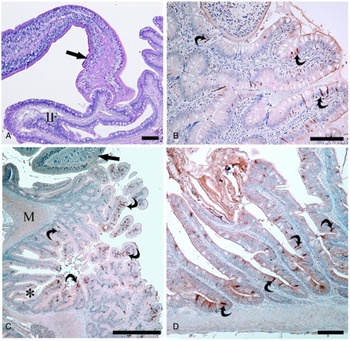
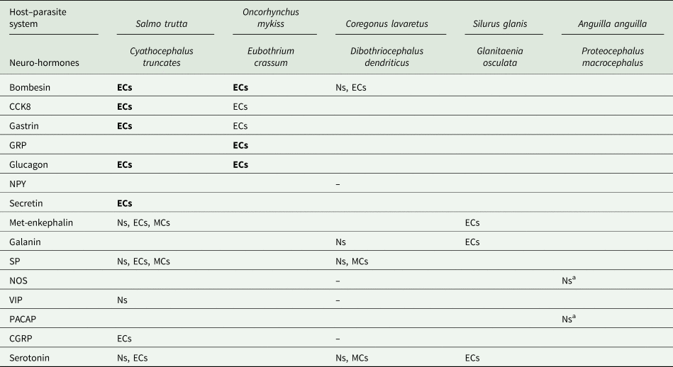
The effects of cestodes on the ENS–EES of the fish gut have been studied in diverse host–parasite systems (Table 2). The damage to the gut mucosa is mainly due to specialized structures of the scolex including the bothria and suckers. In the brown trout infected with Cyathocephalus truncatus, the most infected gut region was the tract where the bile duct opens (Dezfuli et al., Reference Dezfuli, Arrighi, Domeneghini and Bosi2000), probably because worm development and growth are stimulated by the bile components (Smyth, Reference Smyth1969). Most cestode species do not cause severe damage to the intestinal wall because they do not penetrate deeply into the gut wall (Dezfuli et al., Reference Dezfuli, Pironi, Campisi, Shinn and Giari2010) (Fig. 5A). Nevertheless, in some exceptions (e.g. Monobothrium wageneri in T. tinca) worms reach the muscle layer, destroying the intestinal wall architecture and provoking an intense inflammatory response (see Dezfuli et al., Reference Dezfuli, Giari, Squerzanti, Lui, Lorenzoni, Sakalli and Shinn2011b).
EES in fish infected with intestinal cestodes. (A) Silurus glanis parasitized with G. osculata (arrow). IF, intestinal fold. Haematoxylin–eosin stain; scale bar: 50 μm. (B) Salmo trutta infected with Cyathocephalus truncatus (arrow). ECs (curved arrows) immunoreactive to anti-glucagon in the intestinal epithelium are evident; scale bar: 100 μm. (C) Numerous ECs (curved arrows) immunoreactive to anti-CCK8 in the intestine of O. mykiss infected with the cestode E. crassum (arrow). The asterisk indicates a pyloric caecum. M, muscle layer; scale bar: 500 μm. (D) High number of ECs (curved arrows) in the intestinal epithelium of the stickleback Gasterosteus aculeatus parasitized with cestodes (not shown); scale bar: 50 μm.

Results of the effects of intestinal cestodes on the NES of fish

Source: Dezfuli et al. (Reference Dezfuli, Arrighi, Domeneghini and Bosi2000, Reference Dezfuli, Giari, Arrighi, Domeneghini and Bosi2003, Reference Dezfuli, Pironi, Shinn, Manera and Giari2007a, Reference Dezfuli, Pironi, Simoni, Shinn and Giari2007b); Sayyaf Dezfuli et al., (Reference Sayyaf Dezfuli, DePasquale, Castaldelli, Giari and Bosi2017); Bosi et al. (Reference Bosi, Shinn, Giari, Simoni, Pironi and Dezfuli2005a); unpublished data of this study.
Ns, neurons; ECs, endocrine cells; MCs, mast cells.
–, not detected.
The normal-reversal acronym indicates an increase-decrease of immune-positive cells, respectively.
a This study.
In the brown trout–C. truncatus system, the presence of cestodes was associated with a low number of ECs secreting some of the main digestive neuro-hormones in comparison with non-parasitized fish (Dezfuli et al., Reference Dezfuli, Arrighi, Domeneghini and Bosi2000) (Table 2, Fig. 5B). In contrast, in the stickleback– and rainbow trout–cestode systems, more CCK8-secreting ECs were documented (Bosi et al., Reference Bosi, Shinn, Giari, Simoni, Pironi and Dezfuli2005a) (Table 2, Fig. 5C and D). In Dibothricephalus dendriticus-infected internal organs of the powan, Coregonus lavaretus, the larvae become encapsulated and numerous mast cells containing SP and serotonin were seen inside the cyst (Dezfuli et al., Reference Dezfuli, Pironi, Simoni, Shinn and Giari2007b). Moreover, within the C. lavaretus stomach wall, an increase in nervous elements containing bombesin, SP and galanin was reported around the encysted larvae (Dezfuli et al., Reference Dezfuli, Pironi, Simoni, Shinn and Giari2007b) (Table 2).
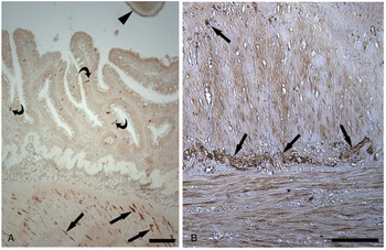
In the myenteric plexus of the intestine of parasitized Salmoniformes, Dezfuli et al. (Reference Dezfuli, Arrighi, Domeneghini and Bosi2000, Reference Dezfuli, Pironi, Simoni, Shinn and Giari2007b) reported an increase in the number of nervous components secreting modulators such as met-enkephalin (Fig. 6A), galanin, SP, VIP and serotonin. Herein, we observed a greater number of nerve fibres immunoreactive to anti-PACAP in the intestine of the European eel, Anguilla anguilla harbouring Proteocephalus macrocephalus (Fig. 6B) than were seen in uninfected eel. Moreover, in some fish–parasite systems, met-enkephalin and serotonin were observed in a high number of mast cells (see Table 2).
Enteric nervous system and EES in fish infected with intestinal cestodes. (A) Salmo trutta infected with C. truncatus (arrowhead). Epithelial ECs (curved arrows) and nerve fibres (thick arrows) in the inner circular muscle layer, both immunoreactive to anti-met-enkephalin; scale bar: 100 μm. (B) Anguilla anguilla harbouring Proteocephalus macrocephalus (not shown). High magnification of neurons and nerve fibres (thick arrows) immunoreactive to pituitary adenylate cyclase-activating polypeptide in the myenteric plexus; scale bar: 100 μm.

Effects of Acanthocephala
Taxon Acanthocephala is a relatively small group of helminths of about 1200 species (Smales, Reference Smales and Schmidt-Rhaesa2015) that infects the digestive tract of a wide range of vertebrates, particularly fish (]Sayyaf Dezfuli et al., Reference Sayyaf Dezfuli, Giari and Bosi2021a). Acanthocephalans are easily recognizable by their attachment organ, a retractable proboscis covered with hooks (Fig. 7A) that enables them to firmly attach to the host's gut wall (Sayyaf Dezfuli and Giari, Reference Sayyaf Dezfuli, Giari, Schierwater and DeSalle2022). Within this taxon, members of several genera, e.g. Pomphorhynchus and Acanthocephalus, cause extensive damage to their host intestine and visceral serosa by deep penetration of the proboscis; damage was heavier in fish intestines with the highest intensity of infection (Taraschewski, Reference Taraschewski2000; Dezfuli et al., Reference Dezfuli, Pironi, Giari, Domeneghini and Bosi2002, Reference Dezfuli, Castaldelli, Bo, Lorenzoni and Giari2011a). Recently the first papers reporting severe effects of an acanthocephalan in a farmed fish have been published. Several records dealt with outbreaks of Neoechinorhynchus buttnerae on farmed tambaqui, Colossoma macropomum, in Brazil. The parasite infects 100% of the tambaqui population, impairing host food uptake with impacts on host growth and great economic losses (Silva-Gomes et al., Reference Silva-Gomes, Gomes Coelho-Filho, Viana-Silva, Braga-Oliveira, Bernardino and Costa2017). Tambaqui is the most produced fish species in Amazonian and Brazilian aquaculture and N. buttnerae is the main obstacle to success for tambaqui farming. Considering the severity of economic losses in Brazilian aquaculture caused by N. buttnerae, the perception that acanthocephalans are harmless parasites of fish should be revised.
Fish intestine infected with acanthocephalans. (A) Salmo trutta parasitized with Dentitruncus truttae, parasite proboscis (arrow) reached tunica propria-submucosa (asterisk). AB/PAS; scale bar: 100 μm. (B) Squalius cephalus harbouring P. laevis, showing anti-galanin immunoreactive neurons and nerve fibres (thick arrows) in the myenteric plexus of the parasitized chub. The thin arrows show subtle nerve fibres in the connective capsule (asterisk) around the bulb of the acanthocephalan (not shown); scale bar: 50 μm. (C) Salmo trutta infected with P. laevis. High number of nerve endings (arrows) immunoreactive to anti-VIP in inner circular muscle layer of fish intestine. The asterisk indicates the acanthocephalan proboscis; scale bar: 50 μm. (D) Salmo trutta–P. laevis system, showing neurons and nerve fibres (thick arrows) of the myenteric plexus immunoreactive to anti-VIP. Note the occurrence of numerous nerve endings in longitudinal muscle layers (thin arrows). The asterisk shows the bulb of worm surrounded by a connective capsule (cc); scale bar: 50 μm. (E) Coregonus lavaretus infected with D. truttae (not shown), several ECs (curved arrows) immunoreactive to anti-SP in the intestinal epithelium are evident. The ECs are close to mucus cells secreting mainly mixed mucins (acid + neutral, deep violet stain, AB/PAS); scale bar: 50 μm. (F) Squalius cephalus–P. laevis system. In the intestinal epithelium of the infected chub, a high number of ECs (curved arrows) immunoreactive to anti-met-enkephalin are visible. The asterisk indicates the acanthocephalan; scale bar: 100 μm.

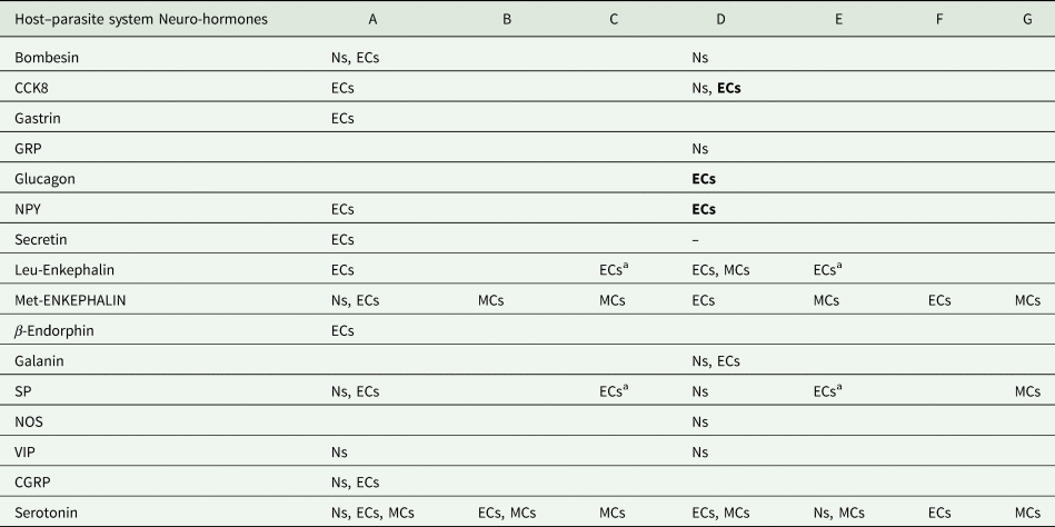
The acanthocephalan Pomphorhynchus laevis parasitizes freshwater fishes like brown trout (Dezfuli et al., Reference Dezfuli, Pironi, Giari, Domeneghini and Bosi2002), chub, Squalius cephalus (Bosi et al., Reference Bosi, Domeneghini, Arrighi, Giari, Simoni and Dezfuli2005b, Reference Bosi, Shinn, Giari and Sayyaf Dezfuli2015), common barbel, Barbus barbus and the Welsh catfish, Silurus glanis (Dezfuli et al., Reference Dezfuli, Castaldelli, Bo, Lorenzoni and Giari2011a) (Table 3). In brown trout, P. laevis occupied the posterior part of the middle intestine, behind the pyloric caeca region, especially when it co-occurred with the cestode C. truncatus (Dezfuli et al., Reference Dezfuli, Pironi, Giari, Domeneghini and Bosi2002). The effects of P. laevis on the NES of different hosts are summarized in Table 3, which shows the increase/decrease of cell densities detected with immunohistochemical protocols. Changes were evident in the neurons that secrete putative modulators of smooth muscle cell activity (galanin, CGRP, SP, VIP, n-NOS) (Fig. 7B–D), and in the ECs that produce anorexi-orexigenic hormones (CCK8, glucagon, NPY) (Dezfuli et al., Reference Dezfuli, Pironi, Giari, Domeneghini and Bosi2002; Bosi et al., Reference Bosi, Domeneghini, Arrighi, Giari, Simoni and Dezfuli2005b, Reference Bosi, Shinn, Giari and Sayyaf Dezfuli2015) (Table 3). In this study in the intestine of the common barbel, we observed an increase in the number of ECs secreting leu-enkephalin and SP when P. laevis occurred (Table 3, Fig. 7E). Likewise, we noticed a high number of ECs producing leu-enkephalin and SP in the intestine of C. lavaretus infected with Dentitruncus truttae (Table 3, Fig. 7F). In the intestinal connective capsule around the P. laevis bulb and proboscis, we observed nervous fibre network immunoreactive to bombesin, SP and galanin (Fig. 7B) (Dezfuli et al., Reference Dezfuli, Pironi, Giari, Domeneghini and Bosi2002; Bosi et al., Reference Bosi, Domeneghini, Arrighi, Giari, Simoni and Dezfuli2005b). Moreover, a high number of cells secreting endogenous opioids were found in parasitized intestine of some fish species. These results support the important role of these modulators in the physiological mechanisms of the fish digestive tract (Dezfuli et al., Reference Dezfuli, Pironi, Giari, Domeneghini and Bosi2002, Reference Dezfuli, Castaldelli, Bo, Lorenzoni and Giari2011a; Bosi et al., Reference Bosi, Domeneghini, Arrighi, Giari, Simoni and Dezfuli2005b, Reference Bosi, Shinn, Giari and Sayyaf Dezfuli2015). Indeed, in the intestine of common barbel and Welsh catfish harbouring P. laevis, the occurrence of mast cells at the site of infection and their positive immunoreactivity to enkephalins and serotonin antibodies were reported by Dezfuli et al. (Reference Dezfuli, Castaldelli, Bo, Lorenzoni and Giari2011a).
Results of the effects of acanthocephalan infection on the NES of fish

Source: Dezfuli et al. (Reference Dezfuli, Pironi, Giari, Domeneghini and Bosi2002, Reference Dezfuli, Giari, Arrighi, Domeneghini and Bosi2003, Reference Dezfuli, Giovinazzo, Lui and Giari2008, Reference Dezfuli, Lui, Giovinazzo, Boldrini and Giari2009, Reference Dezfuli, Castaldelli, Bo, Lorenzoni and Giari2011a); Sayyaf Dezfuli et al. (Reference Sayyaf Dezfuli, Giari, Lorenzoni, Carosi, Manera and Bosi2018b); Bosi et al. (Reference Bosi, Domeneghini, Arrighi, Giari, Simoni and Dezfuli2005b, Reference Bosi, Shinn, Giari and Sayyaf Dezfuli2015); unpublished data of this study.
Ns, neurons; ECs, endocrine cells; MCs, mast cells.
–, not detected.
The normal-reversal acronym indicates an increase-decrease of immune-positive cells, respectively.
Host–parasite system: A: Salmo trutta–Pomphorhynchus laevis; B: Salmo trutta–Dentitruncus truttae; C: Coregonus lavaretus–Dentitruncus truttae; D: Squalius cephalus–Pomphorhynchus laevis; E: Barbus barbus–Pomphorhynchus laevis; F: Silurus glanis–Pomphorhynchus laevis; G: Esox lucius–Acanthocephalus lucii.
a This study.
Effects of Nematoda
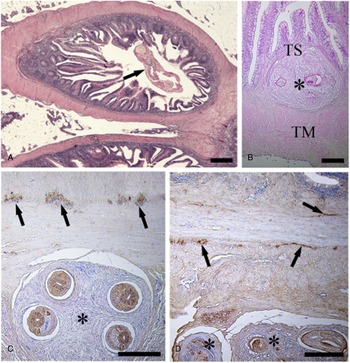
Nematoda or roundworms are estimated to be more than 40 000 species, inhabiting a broad range of environments (Anderson, Reference Anderson2000). About 7000 species are estimated to be parasites of animals and plants. Several nematode species infect fish as definitive or paratenic hosts, occurring free in the gut lumen or encysted in internal organs and filet (Sayyaf Dezfuli et al., Reference Sayyaf Dezfuli, Giari and Bosi2021a, Reference Sayyaf Dezfuli, Simoni, Bosi, Palomba, Mattiucci, Giulietti, Bao, Levsen and Cipriani2021c). Nematodes' affinity for a specific organ site depends on their nutritional and/or environmental requirements, and on the fish species (Bahlool et al., Reference Bahlool, Skovgaard, Kania, Haarder and Buchmann2012; Moravec and Justine, Reference Moravec and Justine2020). The host tissue responses to invading nematodes range from acute to chronic inflammation and encapsulation of the parasite (Fig. 8A and B) (Bahlool et al., Reference Bahlool, Skovgaard, Kania, Haarder and Buchmann2012; Sayyaf Dezfuli et al., Reference Sayyaf Dezfuli, Giari and Bosi2021a, Reference Sayyaf Dezfuli, Maestri, Lorenzoni, Carosi, Maynard and Bosi2021b, Reference Sayyaf Dezfuli, Simoni, Bosi, Palomba, Mattiucci, Giulietti, Bao, Levsen and Cipriani2021c).
Fish intestine parasitized with nematodes. (A) Nematode (arrow) inside the C. lavaretus pyloric caecum is evident. Haematoxylin–eosin stain; scale bar: 200 μm. (B) Anguilla anguilla intestine is infected with Contracaecum rudolphii larva (asterisk) between the tunica propria-submucosa (TS) and tunica muscularis (TM). Haematoxylin–eosin stain; scale bar: 200 μm. (C) Microphotograph shows C. rudolphii larvae inside a cyst (asterisk) on the outer surface of the eel gut. In the myenteric plexus, neurons and nerve fibres (arrows) are immunoreactive to the anti-met-enkephalin; scale bar: 200 μm. (D) Some C. rudolphii larvae in 2 cysts on external surface of the eel gut, neurons and nerve fibres (arrows) of the myenteric plexus immunoreactive to anti-leu-enkephalin are visible; scale bar: 200 μm.

Liver seems to be one of the most preferred organs for nematodes, often with a high intensity of infection and the entire parenchyma occupied by parasites (Sayyaf Dezfuli et al., Reference Sayyaf Dezfuli, Fernandes, Galindo, Castaldelli, Manera, DePasquale, Lorenzoni, Bertin and Giari2016, Reference Sayyaf Dezfuli, Simoni, Bosi, Palomba, Mattiucci, Giulietti, Bao, Levsen and Cipriani2021c). Similar findings were noticed in the liver of Gymnotus inaequilabiatus and Micromesistius poutassou infected respectively with the nematodes Brevimulticaecum sp. and Anisakis simplex. In fish liver, nerve fibres of sympathetic and parasympathetic innervation run along the portal triad, which includes branches of the hepatic artery, the portal vein and the biliary duct, although no intraparenchymal nerve fibres were observed in the trout liver (Esteban et al., Reference Esteban, Jiménez, Barroso, Pedrosa, del Moral, Rodrigo and Peinado1998; Peinado et al., Reference Peinado, del Moral, Jiménez, Rodrigo and Esteban2002).
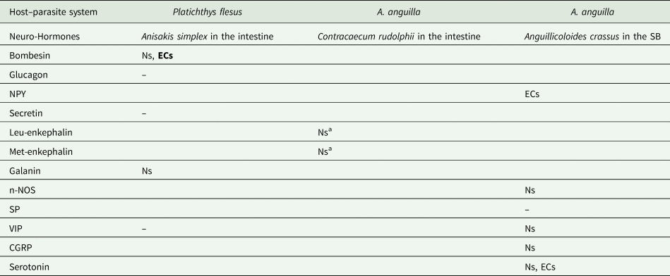
An increase in the number of ECs secreting some neuromodulators was reported in the intestine of the flounder, Platichthys flesus parasitized with A. simplex (Table 4) (Dezfuli et al., Reference Dezfuli, Pironi, Shinn, Manera and Giari2007a). Moreover, in the same host–parasite system, bombesin- and galanin-positive nerve fibres showed a higher density in the myenteric plexus of infected vs uninfected P. flesus intestine (Table 4). Similarly, in fish parasitized with acanthocephalans (Fig. 7B), bombesin nerve fibres formed a fine net among connective cells of the capsule surrounding the parasite (Dezfuli et al., Reference Dezfuli, Pironi, Giari, Domeneghini and Bosi2002, Reference Dezfuli, Pironi, Shinn, Manera and Giari2007a). In recent immunohistochemical tests on A. anguilla parasitized with the extraintestinal larvae of the nematode Contracaecum rudolphii, we observed an increased density of nerve fibres containing endogenous opioids, n-NOS and PACAP (Table 4, Fig. 8C and D).
Results of the effects of nematode on the NES of fish

Source: Dezfuli et al. (Reference Dezfuli, Pironi, Shinn, Manera and Giari2007a), Sayyaf Dezfuli et al. (Reference Sayyaf Dezfuli, Maestri, Lorenzoni, Carosi, Maynard and Bosi2021b); unpublished data of this study.
Ns, neurons; ECs, endocrine cells; MCs, mast cells.
–, not detected.
The normal-reversal acronym indicates an increase-decrease of immune-positive cells, respectively.
a This study.
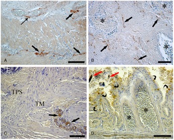
The nematode Anguillicoloides crassus infects the swim bladder (SB) of at least 6 eel species (Lefebvre et al., Reference Lefebvre, Wielgoss, Nagasawa and Moravec2012). Several authors have suggested that the parasite impairs organ functions and significantly compromises the success of the eels' spawning migration (Pelster et al., Reference Pelster, Schneebauer and Dirks2016). In the SB of the European eel parasitized with A. crassus, Sayyaf Dezfuli et al. (Reference Sayyaf Dezfuli, Maestri, Lorenzoni, Carosi, Maynard and Bosi2021b) noticed an increase in intramural neurons and nerve fibres immunoreactive to CGRP, n-NOS, VIP and serotonin antibodies (Table 4, Fig. 9A–C). Furthermore, in the same host–parasite system, ECs positive to NPY and serotonin antibodies were scattered among the gas gland cells of the epithelium of the SB (Table 4, Fig. 9D) (Sayyaf Dezfuli et al., Reference Sayyaf Dezfuli, Maestri, Lorenzoni, Carosi, Maynard and Bosi2021b).
SB of A. anguilla infected with the nematode Anguillicoloides crassus (not shown). (A) Nerve fibres (thick arrows) immunoreactive to the anti-CGRP are numerous in the infected SB. Many subtle nerve fibres (thin arrow) are near blood vessels; scale bar: 50 μm. (B) The arrows indicate several nerve fibres immunoreactive to anti-VIP in a region rich in blood vessels (asterisks); scale bar: 50 μm. (C) Large ganglia with numerous neurons immunoreactive to anti-n-NOS in the tunica muscularis (TM) of the infected eel SB. E, epithelium; TPS, tunica propria-submucosa; scale bar: 100 μm. (D) Numerous anti-NPY immunoreactive ECs (curved arrows) in the epithelium of the infected eel SB. The blood vessels are ectatic and filled with erythrocytes (asterisks). The red thick arrows indicate 2 parasite larvae; scale bar: 100 μm.

Effects of parasites at ultrastructural levels
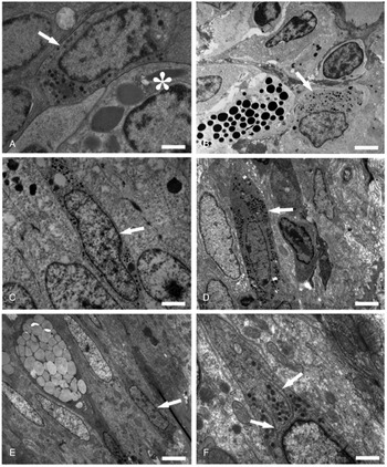
By transmission electron microscope occurrence of enteric ECs in different fish–parasite systems was documented. Figure 10A shows the contact between an EC and mast cell in the epithelium of a mullet harboured an acanthocephalan. ECs were present also in tunica-propria-submucosa of the intestine of some fish species (e.g. S. trutta infected with acanthocephalan, Fig. 10B) (Dezfuli et al., Reference Dezfuli, Arrighi, Domeneghini and Bosi2000, Reference Dezfuli, Giari, Arrighi, Domeneghini and Bosi2003). In all fish, the ECs were readily characterized by the shape and electron density of their secretory granules (Fig 10A, C and D). The ECs with euchromatic nucleus (Fig. 10A–F) were approximately elongated in shape. Some ECs though were seen lodged between the basal portion of some epithelial cells without a free border towards the gut lumen (Fig. 10A, C, D, E and F). The cytoplasms of the ECs were darker than those of the surrounding cells and were observed to contain numerous round-to-oval-shaped secretory granules (Fig. 10A–F). The secretory granules were of approximately similar sizes and were filled with a finely electron-dense material (Fig 10A–F). Free ribosomes and rough endoplasmic reticulum with long cisternae were frequently seen next to secretory granules (Fig. 10F). In all fish species, ECs contained few mitochondria and did not possess well-developed Golgi complexes.
Transmission electron micrographs of neuroendocrine cells in the intestine of different fish species infected with helminths. In all neuroendocrine cells, numerous small electron-dense granules in cytoplasm are evident. (A) Intestine of Chelon ramada infected with digeneans; within the epithelium a neuroendocrine cell (arrow) is attached to a mast cell (asterisk); scale bar: 0.80 μm. (B) A neuroendocrine cell (arrow) and a mast cell within the tunica propria-submucosa of the intestine of C. lavaretus harbouring a nematode; scale bar: 3 μm. (C) A neuroendocrine cell (arrow) between nuclei of 2 intestinal epithelial cells of E. lucius parasitized with an acanthocephalan; scale bar: 1.4 μm. (D) Micrograph shows a neuroendocrine cell (arrow) within the epithelium of A. anguilla intestine infected with digeneans; scale bar: 2 μm. (E) A neuroendocrine cell (arrow) within the intestinal epithelium of S. glanis harbouring a cestode; scale bar: 3 μm. (F) In a higher magnification of Fig. 2, several small electron-dense granules are visible near the rough endoplasmic reticulum (arrows) in the cytoplasm; scale bar: 0.6 μm.

Effects of parasites on gut physiology
Intestinal parasites induce a regional inflammation altering the morphological structure of the gut wall, which affects biochemical and physiological homoeostasis (Fairweather, Reference Fairweather and Beckage1997; Palmer and Greenwood-Van Meerveld, Reference Palmer and Greenwood-Van Meerveld2001). The inflammation induces changes in the ENS with neuronal loss and/or hyper-innervation with consequences for nervous functions (Kulkarni et al., Reference Kulkarni, Ganz, Bayrer, Becker, Bogunovic and Rao2018). In addition, the structural changes induced by intestinal parasites can affect the EES with alterations in the gastrointestinal tract's normal functions (Fairweather, Reference Fairweather and Beckage1997; Palmer and Greenwood-Van Meerveld, Reference Palmer and Greenwood-Van Meerveld2001). Several host–parasite systems were investigated to clarify the mechanisms underlying changes in host physiology and behaviour (Klein, Reference Klein2003; Thomas et al., Reference Thomas, Adamo and Moore2005), nevertheless, the reasons for most changes remain unknown (Shaw and Øverli, Reference Shaw and Øverli2012).
Alterations induced by parasites depend on parasite density (Weinersmith et al., Reference Weinersmith, Hanninen, Sih, Mcelreath and Earley2016). In California killifish Fundulus parvipinnis experimentally infected with the brain-encysting trematode Euhaplorchis californiensis, parasite density determines the level at which the pathogen influences serotonin neural transmission (Helland-Riise et al., Reference Helland-Riise, Vindas, Johansen, Nadler, Weinersmith, Hechinger and Øverli2020). Little is known about the effects of intestinal parasites on the NES of the alimentary canal; conversely, many papers have been published on this topic in healthy/uninfected fish.
Often, the effects on fish tissue depend on the pathogen's intensity of infection. Accordingly, in the E. scophthalmi–turbot and E. ictaluri–channel catfish systems, high intensity of infection and its detrimental effects on the intestinal epithelium were responsible for severe enteritis and fish high mortality (Branson et al., Reference Branson, Riaza and Alvarez-Pellitero1999; Redondo et al., Reference Redondo, Palenzuela and Álvarez-Pellitero2004; Li et al., Reference Li, Zhang, Wang, Lu, Nandi, Mohanty, Terhune, Liu and Peatman2012). Many digenean and cestode species, with their shallow attachment to the intestinal mucosa, do not cause appreciable tissue damage. In contrast, numerous acanthocephalan species, with the hook-covered holdfast on their proboscis, cause severe changes to normal gut architecture (Taraschewski, Reference Taraschewski2000; Dezfuli et al., Reference Dezfuli, Pironi, Giari, Domeneghini and Bosi2002, Reference Dezfuli, Giari, Arrighi, Domeneghini and Bosi2003, Reference Dezfuli, Pironi, Campisi, Shinn and Giari2010, Reference Dezfuli, Castaldelli, Bo, Lorenzoni and Giari2011a). In the following sections, we discuss the effects of fish intestinal parasites on the main physiological mechanisms.
Neuroendocrine and immune system interactions in infected fish
Several studies have pointed out the essential role of the ENS–EES in inflammatory processes caused by intestinal parasites (Fairweather, Reference Fairweather and Beckage1997; Bosi et al., Reference Bosi, Shinn, Giari, Simoni, Pironi and Dezfuli2005a, Reference Bosi, Domeneghini, Arrighi, Giari, Simoni and Dezfuli2005b, Reference Bosi, Shinn, Giari and Sayyaf Dezfuli2015; Halliez and Buret, Reference Halliez and Buret2015; Sayyaf Dezfuli et al., Reference Sayyaf Dezfuli, DePasquale, Castaldelli, Giari and Bosi2017, Reference Sayyaf Dezfuli, Giari and Bosi2021a). The correct release of cytokines, hormones and neuropeptides relies on interaction between the immune, nervous and endocrine systems (Lomax et al., Reference Lomax, Linden, Mawe and Sharkey2006; Di Giovangiulio et al., Reference Di Giovangiulio, Verheijden, Bosmans, Stakenborg, Boeckxstaens and Matteoli2015). One of the most important effects of parasites on their hosts is the activation of the innate and adaptive immune system, thanks to release of signal molecules, like the opioids leu- and met-enkephalin, by host cells (Mola et al., Reference Mola, Bertacchi, Gambarelli and Pederzoli2004; Nardocci et al., Reference Nardocci, Navarro, Cortés, Imarai, Montoya, Valenzuela, Jara, Acuña-Castillo and Fernández2014; Sayyaf Dezfuli et al., Reference Sayyaf Dezfuli, Giari, Lorenzoni, Carosi, Manera and Bosi2018b). In host–fish systems investigated so far, an increase in the number of ECs and/or nervous components containing enkephalins has been observed in parasitized vs uninfected fish (Bosi et al., Reference Bosi, Domeneghini, Arrighi, Giari, Simoni and Dezfuli2005b, Reference Bosi, Shinn, Giari and Sayyaf Dezfuli2015; Dezfuli et al., Reference Dezfuli, Castaldelli, Bo, Lorenzoni and Giari2011a; Losada et al., Reference Losada, Bermúdez, Faílde, Di Giancamillo, Domeneghini and Quiroga2014; Sayyaf Dezfuli et al., Reference Sayyaf Dezfuli, DePasquale, Castaldelli, Giari and Bosi2017). The opioids are signal molecules involved in the communication between neuroendocrine and immune systems (Stefano et al., Reference Stefano, Scharrer, Smith, Hughes, Magazine, Bilfinger, Hartman, Fricchione, Liu and Makman2017). Indeed, enkephalins were often found in mast cells (Bosi et al., Reference Bosi, Domeneghini, Arrighi, Giari, Simoni and Dezfuli2005b; Dezfuli et al., Reference Dezfuli, Lui, Giovinazzo, Boldrini and Giari2009, Reference Dezfuli, Castaldelli, Bo, Lorenzoni and Giari2011a; Sayyaf Dezfuli et al., Reference Sayyaf Dezfuli, Giari, Lorenzoni, Carosi, Manera and Bosi2018b) and macrophage aggregates (Losada et al., Reference Losada, Bermúdez, Faílde, Di Giancamillo, Domeneghini and Quiroga2014). The occurrence of intestinal parasites is associated with mucous hypersecretion and enkephalins are involved in mucous discharge (Zoghbi et al., Reference Zoghbi, Trompette, Claustre, Homsi, Garzón, Jourdan, Scoazec and Plaisancié2006; Bosi et al., Reference Bosi, Shinn, Giari and Sayyaf Dezfuli2015; Sayyaf Dezfuli et al., Reference Sayyaf Dezfuli, DePasquale, Castaldelli, Giari and Bosi2017). In the case of enteric helminth infection, the overproduction and release of mucous at the luminal surface has been considered a defence against parasites (Dezfuli et al., Reference Dezfuli, Pironi, Campisi, Shinn and Giari2010; Bosi et al., Reference Bosi, Giari, DePasquale, Carosi, Lorenzoni and Dezfuli2017).
In fish gut, serotonergic neurons are the largest neuronal population of the myenteric plexus (Anderson and Campbell, Reference Anderson and Campbell1988; Olsson et al., Reference Olsson, Holmberg and Holmgren2008b; Ceccotti et al., Reference Ceccotti, Giaroni, Bistoletti, Viola, Crema and Terova2018) and serotonin is stored and secreted by macrophages, mast cells and T lymphocytes (Margolis and Gershon, Reference Margolis and Gershon2016; Herr et al., Reference Herr, Bode and Duerschmied2017; Serna-Duque and Esteban, Reference Serna-Duque and Esteban2020). Moreover, serotonin is synthesized by ECs as a paracrine hormone (Bermúdez et al., Reference Bermúdez, Vigliano, Quiroga, Nieto, Bosi and Domeneghini2007; Dezfuli et al., Reference Dezfuli, Giovinazzo, Lui and Giari2008, Reference Dezfuli, Castaldelli, Bo, Lorenzoni and Giari2011a; Bosi et al., Reference Bosi, Shinn, Giari and Sayyaf Dezfuli2015; Sayyaf Dezfuli et al., Reference Sayyaf Dezfuli, DePasquale, Castaldelli, Giari and Bosi2017, Reference Sayyaf Dezfuli, Maestri, Lorenzoni, Carosi, Maynard and Bosi2021b). In almost all known host–parasite systems, infected fish have more ECs containing serotonin than do uninfected conspecifics. In parasitized fish, high amounts of serotonin are consistent with a pro-inflammatory activity, whereas the higher number of neurons immunoreactive to serotonin indicates neuroprotective and anti-inflammatory functions (Serna-Duque and Esteban, Reference Serna-Duque and Esteban2020). In the gut epithelium of Welsh catfish harbouring the tapeworm Glanitaenia osculata, serotonin-containing ECs were observed close to the mucous and mast cells, suggesting an endocrine–immune interaction (Sayyaf Dezfuli et al., Reference Sayyaf Dezfuli, DePasquale, Castaldelli, Giari and Bosi2017). In the intestine of S. cephalus parasitized with an acanthocephalan, opioid peptides and the cholinergic co-mediator galanin were documented in ECs very near to the mucous cells (Bosi et al., Reference Bosi, Shinn, Giari and Sayyaf Dezfuli2015). Intestinal mucous cells are considered a component of the mucosal immune system with a key role in regulating inflammation (Johansson and Hansson, Reference Johansson and Hansson2014; Bosi et al., Reference Bosi, Shinn, Giari and Sayyaf Dezfuli2015, Reference Bosi, Giari, DePasquale, Carosi, Lorenzoni and Dezfuli2017).
The expression of SP in different immunocompetent cells (Stoyanova and Gulubova, Reference Stoyanova and Gulubova2002; Bermudez et al., Reference Bermúdez, Vigliano, Quiroga, Nieto, Bosi and Domeneghini2007) and its pro-inflammatory activity (Maggi, Reference Maggi1997) are well known. The tachykinin SP was noticed in the neuroendocrine and immune components of turbot infected with myxozoans (Bermudez et al., Reference Bermúdez, Vigliano, Quiroga, Nieto, Bosi and Domeneghini2007) and in fish parasitized with tapeworms and acanthocephalans (Dezfuli et al., Reference Dezfuli, Pironi, Giari, Domeneghini and Bosi2002, Reference Dezfuli, Pironi, Simoni, Shinn and Giari2007b; Bosi et al., Reference Bosi, Domeneghini, Arrighi, Giari, Simoni and Dezfuli2005b; Sayyaf Dezfuli et al., Reference Sayyaf Dezfuli, Giari, Lorenzoni, Carosi, Manera and Bosi2018b; this study). Moreover, Nam et al. (Reference Nam, Moon, Kim, Kong, Kim, Kim, Jee and Lee2013) found a high expression of a PRP in the intestine and pyloric caeca of P. olivaceus infected with E. tarda. The administration of this peptide in vivo to olive flounder embryonic cells showed a stimulatory effect on tumour necrosis factor-α at the transcriptional level, suggesting its role in the modulation of the immune system (Nam et al., Reference Nam, Moon, Kim, Kong, Kim, Kim, Jee and Lee2013).
Parasites can avoid the host's innate immune system by modifying their own molecular carbohydrate composition (Hammerschmidt and Kurtz, Reference Hammerschmidt and Kurtz2005) or by interfering with the host's cell-mediated immunity (Scharsack et al., Reference Scharsack, Kalbe, Derner, Kurtz and Milinski2004). It has been suggested that protozoans and metazoans have the capacity to deal with a broad range of host cellular and humoral elements, which allows the parasite to survive longer (Buchmann, Reference Buchmann2012). In a host–parasite system, the complex interactions between the immune system and ENS–EES present a challenge to developing a deep understanding of direct or indirect manipulation of the host immune physiology (Thomas et al., Reference Thomas, Adamo and Moore2005; Scharsack et al., Reference Scharsack, Koch and Hammerschmidt2007). For instance, studies on infected invertebrates suggested that excretory/secretory molecules of the parasites activate elements of the host immune system, which in turn affect its nervous system (Adamo, Reference Adamo2002; Scharsack et al., Reference Scharsack, Koch and Hammerschmidt2007). Furthermore, it seems that parasite-produced excretory and secretory molecules presumed to be key players in clinical manifestation of disease in humans possess a general biological role in invertebrates and lower vertebrates as well (Mehrdana and Buchmann, Reference Mehrdana and Buchmann2017). In sum, ‘Coevolution of hosts and parasites has resulted in a tight interaction between innate and adaptive immune elements in the host and a rich but, to a certain extent, unexplored array of immune evasion mechanisms in the parasites’ (Buchmann, Reference Buchmann2014).
Neuroendocrine control of neurogenesis in infected fish
The gut is the largest microbial, endocrine and immune organ in both humans and mice (Kulkarni et al., Reference Kulkarni, Micci, Leser, Shin, Tang, Fu, Liu, Li, Saha, Li, Enikolopov, Becker, Rakhilin, Anderson, Shen, Dong, Butte, Song, Southard-Smith, Kapur, Bogunovic and Pasricha2017, Reference Kulkarni, Ganz, Bayrer, Becker, Bogunovic and Rao2018). Based on experimental assays, humans have been shown to replace about 90% of their myenteric neurons every 2 weeks by a dynamic balance between neuronal apoptosis and neurogenesis. Monoamines such as serotonin are involved in neuronal recruitment and re-sculpting of the neuronal network in response to changes of their internal homoeostasis or of the environmental state (Adamo, Reference Adamo2002). Barber and Wright (Reference Barber and Wright2005) suggested that, thanks to these monoamines, parasites can virtually manipulate their hosts' physiology and behaviour.
Some Acanthocephala genera, e.g. Pomphorhynchus and Southwellina, penetrate deeply through the gut wall to create a strong attachment and thereby cause extensive damage. As a result, the formation of a connective capsule around the worm praesoma was evident (Dezfuli et al., Reference Dezfuli, Pironi, Giari, Domeneghini and Bosi2002). The connective capsule is formed by epithelioid cells, mast cells, fibrocytes and macrophage aggregates (Dezfuli et al., Reference Dezfuli, Pironi, Simoni, Shinn and Giari2007b). In S. trutta and S. cephalus infected with P. laevis, within the capsule a subtle nerve fibre network immunoreactive to bombesin/GRP, VIP and CGRP was observed among the fibrocytes and immune cells (Dezfuli et al., Reference Dezfuli, Pironi, Giari, Domeneghini and Bosi2002; Bosi et al., Reference Bosi, Domeneghini, Arrighi, Giari, Simoni and Dezfuli2005b). Moreover, in the stomach and intestine of C. lavaretus, nerve fibres containing bombesin and SP were found in the connective capsule tissue around encysted plerocercoides of D. dendriticus (Dezfuli et al., Reference Dezfuli, Pironi, Simoni, Shinn and Giari2007b). Our evidence from several fish–parasite systems suggests that it is likely that the neo-formed neural network in the inflammatory tissue used the abovementioned neuropeptides as neurotransmitters (Dezfuli et al., Reference Dezfuli, Giari, Simoni, Shinn and Bosi2004; Bosi et al., Reference Bosi, Domeneghini, Arrighi, Giari, Simoni and Dezfuli2005b). Indeed, it is reasonable to postulate that the new neural network has a role in angiogenesis, recruitment of immune cells and tissue repair in cases of chronic inflammation, which commonly occurs at the site of attachment of many acanthocephalans (Dezfuli et al., Reference Dezfuli, Pironi, Giari, Domeneghini and Bosi2002, Reference Dezfuli, Pironi, Simoni, Shinn and Giari2007b).
Neuroendocrine control of intestinal muscle cells in infected fish
Parasites seem to induce the synthesis of neuromodulators that have an inhibitory effect on host muscle cells (Serna-Duque and Esteban, Reference Serna-Duque and Esteban2020). In fish infected with enteric helminths, more synthesis and secretion of inhibitory neurotransmitters provoke relaxation of the gut musculature, which is essential for the holdfast mechanism of the parasite (Foster and Lee, Reference Foster and Lee1996; Dezfuli et al., Reference Dezfuli, Arrighi, Domeneghini and Bosi2000; Serna-Duque and Esteban, Reference Serna-Duque and Esteban2020). Relaxation of the intestinal muscle might prevent rejection of the parasite and increases the retention and processing times of digesta in the intestine, which all benefit the parasite (Takei and Loretz, Reference Takei, Loretz, Grosell, Farrel and Brauner2011). Foster and Lee (Reference Foster and Lee1996) found a homologue porcine VIP peptide synthesized by the nematode Nippostrongylus brasiliensis, which reduces the amplitude of contraction of uninfected rat intestine in vitro. Apparently, secretory/excretory molecules produced by the parasite could prevent its rejection from the host intestine (Foster and Lee, Reference Foster and Lee1996). Conversely, in turbot intestine infected with the myxozoan E. scophthalmi, Bermúdez et al. (Reference Bermúdez, Vigliano, Quiroga, Nieto, Bosi and Domeneghini2007) and Losada et al. (Reference Losada, Bermúdez, Faílde, Di Giancamillo, Domeneghini and Quiroga2014) found fewer neurons immunoreactive to VIP and bombesin. This contrast might indicate that the regulatory mechanism suitable for an intestinal myxozoan differs from that for enteric worms (Bermúdez et al., Reference Bermúdez, Vigliano, Quiroga, Nieto, Bosi and Domeneghini2007).
Fish endoparasitic helminths can invade and colonize organ(s), the host produces active or partly effective immune responses against new infections, meanwhile, the established worms might survive for longer time; such phenomenon is called concomitant immunity and is a result of an elaborate interaction between host and helminth and relies on immune regulation and immune evasion (Buchmann, Reference Buchmann2012). The influence of intestinal pathogens on gut motility is detailed in the recent review by Serna-Duque and Esteban (Reference Serna-Duque and Esteban2020), who proposed 3 types of fish responses to parasites.
Neuroendocrine control of food intake in infected fish
Structural integrity of the intestine is necessary for normal control of food intake and food processing (Gay et al., Reference Gay, Ressayre, Garcia-Villar, Bueno and Fioramonti2003). Many intestinal parasites affect the intestine's structural components, primarily ENS and EES (Mercer and Chappell, Reference Mercer and Chappell2000; Losada et al., Reference Losada, Bermúdez, Faílde, Di Giancamillo, Domeneghini and Quiroga2014; Bosi et al., Reference Bosi, Shinn, Giari and Sayyaf Dezfuli2015). The occurrence of gastrointestinal parasites in mammals (Mercer et al., Reference Mercer, Mitchell, Moar, Bissett, Geissler, Bruce and Chappell2000) and in fish (de Matos et al., Reference de Matos, de Oliveira, Gomes and da Silva2017) is often associated with a reduction in the host food intake. Similarly, heavy infections of rainbow trout with the protozoan Cryptobia salmositica (Lowe-Jinde and Zimmerman, Reference Lowe-Jinde and Zimmerman1991) and of Atlantic salmon with the copepod Lepeophtheirus salmonis (Dawson et al., Reference Dawson, Pike, Houlihan and McVicar1999) resulted in reduced food intake by the host fish. Plerocercoid of S. solidus (Cestoda) occurs in the body cavity of the small fish G. aculeatus; the presence of the parasite affects the host brain by reducing monoaminergic activity and enhancing the immune response (Øverli et al., Reference Øverli, Páll, Borg, Jobling and Winberg2001). Sticklebacks parasitized with the cestode increase their food intake rates (Ranta, Reference Ranta1995; Barber and Wright, Reference Barber and Wright2005). Nevertheless, whether intestinal parasites increase or decrease food intake by the host fish varies by species of host and parasite, infection intensity and which organ is infected (Lowe-Jinde and Zimmerman, Reference Lowe-Jinde and Zimmerman1991; Ranta, Reference Ranta1995; Dawson et al., Reference Dawson, Pike, Houlihan and McVicar1999; Barber and Wright, Reference Barber and Wright2005). Alteration of the major anorexi-orexigenic factors in the gut of parasitized fish will be discussed below.
Ghrelin
In fish, the action of ghrelin is species-specific (Blanco et al., Reference Blanco, Calo and Soengas2021). Its secretion promotes food intake in C. auratus and cavefish, Astyanax fasciatus mexicanus (Unniappan et al., Reference Unniappan, Canosa and Peter2004; Penney and Volkoff, Reference Penney and Volkoff2014; Blanco et al., Reference Blanco, Bertucci, Valenciano, Delgado and Unniappan2017) but, when peripherally administrated to rainbow trout (Jönsson et al., Reference Jönsson, Kaiya and Björnsson2010) and channel catfish (Schroeter et al., Reference Schroeter, Fenn and Small2015), ghrelin showed an anorexigenic activity. The ghrelin gene is downregulated in channel catfish infected with E. ictaluri (Li et al., Reference Li, Zhang, Wang, Lu, Nandi, Mohanty, Terhune, Liu and Peatman2012), suggesting a synthesis reduction likely related to increased food intake. No data are available on changes in ghrelin levels for fish parasitized with intestinal helminths.
Gastrin/CCK8
Gastrin and CCK8 share the same 5 C-terminal amino acids. In the immunohistochemical method, the use of polyclonal antibodies for these peptides can produce a cross-reaction in the intestinal ECs.
In the digestive tract of infected fish, gastrin stimulates gastric acid secretion by oxyntopeptic cells of the stomach and promotes cell proliferation, which induces gut epithelial hypertrophy (Dezfuli et al., Reference Dezfuli, Giari, Arrighi, Domeneghini and Bosi2003). In goldfish, suppression of food intake is observed 45 min after intraperitoneal injection of CCK8, and the effect is dose-dependent (Himick and Peter, Reference Himick and Peter1994a; Volkoff et al., Reference Volkoff, Eykelbosh and Peter2003; Penney and Volkoff, Reference Penney and Volkoff2014; Zhang et al., Reference Zhang, Tang, Qi, Wang, Hao, Wu, Chen, Tian, Wang, Chen and Li2017). An increased number of ECs immunoreactive to CCK8 was observed in turbot infected with myxozoans and downregulation of gastrin mRNA expression was found in the same host–parasite system (Bermúdez et al., Reference Bermúdez, Vigliano, Quiroga, Nieto, Bosi and Domeneghini2007; Robledo et al., Reference Robledo, Ronza and Harrison2014). Similarly, the cestodes C. truncatus in S. trutta and Eubothrium crassum in O. mykiss induced a decrease in ECs immunoreactive to gastrin and an increase of cells positive to CCK8 in comparison with uninfected conspecifics (Dezfuli et al., Reference Dezfuli, Arrighi, Domeneghini and Bosi2000; Bosi et al., Reference Bosi, Shinn, Giari, Simoni, Pironi and Dezfuli2005a). In fish–acanthocephalan systems, the ECs secreting gastrin and CCK8 were less in number, thus, it was suggested that the occurrence of intestinal helminths might increase host food intake (Dezfuli et al., Reference Dezfuli, Pironi, Giari, Domeneghini and Bosi2002, Reference Dezfuli, Giari, Arrighi, Domeneghini and Bosi2003; Bosi et al., Reference Bosi, Shinn, Giari, Simoni, Pironi and Dezfuli2005a).
Glucagon
In vertebrates, a 29 amino acid peptide chain forms glucagon, which has an anorexigenic effect (Navarro et al., Reference Navarro, Carneiro, Parrizas, Maestro, Planas and Gutierrez1993; Le Bail and Boeuf, Reference Le Bail and Boeuf1997; Moon, Reference Moon1998). The post-prandial release of glucagon induces high glucose plasma levels and increases lipolytic activity (Navarro et al., Reference Navarro, Carneiro, Parrizas, Maestro, Planas and Gutierrez1993; Moon, Reference Moon1998). In fish, the peripheral administration of glucagon-like peptide-1 abolishes food intake (Blanco et al., Reference Blanco, Bertucci, Valenciano, Delgado and Unniappan2017), thanks to mRNA increase expression of specific signal molecules in the brain (Polakof et al., Reference Polakof, Míguez and Soengas2011).
In fish, a significant reduction in the number of ECs secreting glucagon was observed in the turbot parasitized with myxozoans (Losada et al., Reference Losada, Bermúdez, Faílde, Di Giancamillo, Domeneghini and Quiroga2014), and in different fish species infected with cestodes (Dezfuli et al., Reference Dezfuli, Giari, Arrighi, Domeneghini and Bosi2003; Bosi et al., Reference Bosi, Shinn, Giari, Simoni, Pironi and Dezfuli2005a) and acanthocephalans (Bosi et al., Reference Bosi, Domeneghini, Arrighi, Giari, Simoni and Dezfuli2005b). Low synthesis and secretion of glucagon in the presence of intestinal pathogens increase fish appetite, which benefit the enteric parasites (Bosi et al., Reference Bosi, Shinn, Giari, Simoni, Pironi and Dezfuli2005a, Reference Bosi, Domeneghini, Arrighi, Giari, Simoni and Dezfuli2005b).
NPY/PYY
The NPY family includes 4 peptides, namely NPYa, NPYb, PYYa and PYYb, all derived from a single ancestral gene (Larhammar et al., Reference Larhammar, Söderberg and Lundell1998; Yan et al., Reference Yan, Jia, Yang, Wang, Sun and Li2017). In fishes, after synthesis the occurrence or disappearance of each of the 4 peptides depends on the teleost species (Sundstrom et al., Reference Sundstrom, Larsson, Brenner, Venkatesh and Larhammar2008). The main physiological action of the NPY family of peptides is the regulation of appetite and alimentary behaviour. In fish, central or peripheral administration of NPY increased food intake (Aldegunde and Mancebo, Reference Aldegunde and Mancebo2006; Kiris et al., Reference Kiris, Kumlu and Dikel2007; Yokobori et al., Reference Yokobori, Azuma, Nishiguchi, Kang, Kamijo, Uchiyama and Matsuda2012; Zhou et al., Reference Zhou, Liang, Yuan, Li, He, Fang, Guo, Liu, Li and Shen2013). However, conflicting results have been found in parasitized fish. With reference to myxozoan, reduced PYY-mRNA expression was reported in channel catfish infected with E. ictaluri (Li et al., Reference Li, Zhang, Wang, Lu, Nandi, Mohanty, Terhune, Liu and Peatman2012). In the brown trout–P. laevis system a low number of NPY-secreting ECs was found (Dezfuli et al., Reference Dezfuli, Pironi, Giari, Domeneghini and Bosi2002), whereas in the chub–P. laevis system more cells synthesize NPY (Bosi et al., Reference Bosi, Domeneghini, Arrighi, Giari, Simoni and Dezfuli2005b). Surprisingly, no difference in ECs immunoreactive to NPY was observed in Salmoniformes infected with cestodes (Dezfuli et al., Reference Dezfuli, Arrighi, Domeneghini and Bosi2000; Bosi et al., Reference Bosi, Shinn, Giari, Simoni, Pironi and Dezfuli2005a) and in P. flesus parasitized with the nematode A. simplex (Dezfuli et al., Reference Dezfuli, Pironi, Shinn, Manera and Giari2007a).
The interaction between NPY and several appetite modulators and growth factors is well established (Volkoff et al., Reference Volkoff, Eykelbosh and Peter2003, Reference Volkoff, Canosa, Unniappan, Cerda-Reverter, Bernier, Kelly and Peter2005; Volkoff, Reference Volkoff2006). For example, NPY inhibited the expression of CCK8 and PACAP, and on the other hand, increased the expression levels of insulin-like growth factor-1 and insulin-like growth factor-2. According to Zhou et al. (Reference Zhou, Liang, Yuan, Li, He, Fang, Guo, Liu, Li and Shen2013), NPY increases host food intake and energy metabolism. Our results on fish–helminth systems indicate alteration of the components involved in the secretion of NPY (Dezfuli et al., Reference Dezfuli, Pironi, Giari, Domeneghini and Bosi2002; Bosi et al., Reference Bosi, Domeneghini, Arrighi, Giari, Simoni and Dezfuli2005b).
Bombesin/GRP
Bombesin and the related GRP are involved in the inhibition of gastric emptying and regulation of gastric acid secretion (Holmgren, Reference Holmgren1985). Bombesin was observed in nerve fibres of the connective axis of the fish intestinal folds and myenteric plexus; therefore, it might be reasonable to presume it has a role in regulation of mucosal secretion and stimulation of gut motility (Holmgren, Reference Holmgren1985; Dezfuli et al., Reference Dezfuli, Pironi, Giari, Domeneghini and Bosi2002, Reference Dezfuli, Pironi, Shinn, Manera and Giari2007a, Reference Dezfuli, Pironi, Simoni, Shinn and Giari2007b; Bosi et al., Reference Bosi, Domeneghini, Arrighi, Giari, Simoni and Dezfuli2005b). Indeed, peripheral administration of bombesin/GRP inhibits food intake in C. auratus and carp, Cyprinus carpio, indicating its anorectic effect (Beach et al., Reference Beach, McVean, Roberts and Thorndyke1988; Himick and Peter, Reference Himick and Peter1994b; Merali et al., Reference Merali, McIntosh and Anisman1999).
Losada et al. (Reference Losada, Bermúdez, Faílde, Di Giancamillo, Domeneghini and Quiroga2014) mentioned that the low number of ECs immunoreactive to bombesin found in infected turbot is in favour of gastric emptying and reduction of the fish's sensation of satiety. The presence of cestodes or acanthocephalans in the intestine of 2 Salmoniformes species was associated with fewer bombesin/GRP-secreting ECs, probably associated with a starvation sensation (Dezfuli et al., Reference Dezfuli, Pironi, Giari, Domeneghini and Bosi2002, Reference Dezfuli, Giari, Arrighi, Domeneghini and Bosi2003; Bosi et al., Reference Bosi, Shinn, Giari, Simoni, Pironi and Dezfuli2005a; Losada et al., Reference Losada, Bermúdez, Faílde, Di Giancamillo, Domeneghini and Quiroga2014). Nevertheless, in the stomach of C. lavaretus, infected with the cestode D. dendriticus, the number of ECs immunoreactive to bombesin was higher than in uninfected powan (Dezfuli et al., Reference Dezfuli, Pironi, Simoni, Shinn and Giari2007b). Our data on bombesin in the stomach of C. lavaretus differed from those in the intestine of 2 other Salmoniformes species. An explanation might be the same as that provided above for peptides by Losada et al. (Reference Losada, Bermúdez, Faílde, Di Giancamillo, Domeneghini and Quiroga2014); namely, bombesin has different physiological roles in the stomach and the intestine. With this in mind, it might be reasonable to presume, the occurrence of the ECs immunoreactive to bombesin in 2 organs with different functions could differ.
Secretin/VIP/PACAP
Secretin, VIP and PACAP belong to the same peptide superfamily. The release of secretin stimulates pancreatic bicarbonate secretion to neutralize acidic chyme in the passage from stomach to the intestine (Whitmore et al., Reference Whitmore, Holloway, Lofton-Day, Maurer, Chen, Quinton, Vincent, Scherer and Lok2000). In S. trutta infected with the cestode C. truncatus and/or the acanthocephalan P. laevis, a low number of ECs immunoreactive to secretin have been seen (Dezfuli et al., Reference Dezfuli, Giari, Arrighi, Domeneghini and Bosi2003). However, no significant difference was observed in the number of ECs positive to secretin in O. mykiss harbouring the cestode E. crassum and uninfected fish (Bosi et al., Reference Bosi, Shinn, Giari, Simoni, Pironi and Dezfuli2005a). Secretin seems to be a tetrapod hormone with no orthologue in teleosts undiscovered yet (Roch et al., Reference Roch, Wu and Sherwood2009; Takei and Loretz, Reference Takei, Loretz, Grosell, Farrel and Brauner2011). Therefore, the immunoreactivity found in the parasitized intestine of S. trutta and O. mykiss probably was due to a cross-reaction against molecular haptens belonging to a peptide of the same superfamily.
The occurrence of VIP as a neurotransmitter in the myenteric plexus of fish has been documented in several accounts since the late 1990s (Kiliian et al., Reference Kiliaan, Scholten and Groot1997; Olsson and Holmgren, Reference Olsson and Holmgren1997, Reference Olsson and Holmgren2000). VIP stimulates gastric acid secretion, inhibits smooth muscle contraction and has a vasodilatory effect on the gastrointestinal circulatory system (Kågstrom and Holmgren, Reference Kågström and Holmgren1997; Olsson and Holmgren, Reference Olsson and Holmgren1997). The peptide PACAP is related to VIP and initially it was isolated from the ovine hypothalamus as an activator of cyclic adenosine monophosphate production in pituitary cells (Hirabayashi et al., Reference Hirabayashi, Nakamachi and Shioda2018). It is presumed that in teleost evolution the specific whole-genome duplication raised 2 types of PACAPs (PACAP1 and PACAP2) and 2 types of PACAP receptors (PAC1Rs and PAC1Ra) (On and Chow, Reference On, Chow, Reglodi and Tamas2016; Nakamachi et al., Reference Nakamachi, Tanigawa, Konno, Shioda and Matsuda2019). PACAP is a neuromodulator inhibiting smooth muscle activity in fish (Matsuda et al., Reference Matsuda, Kashimoto, Higuchi, Yoshida, Uchiyama, Shioda, Arimura and Okamura2000; Li et al., Reference Li, Han, Bai, Ma, Quan, Fan, Jiang and Yu2015) and with its anorectic effect, this peptide suppresses food intake in goldfish and zebrafish, Danio rerio, after intra-cerebroventricular administration (Sekar et al., Reference Sekar, Wang and Chow2017; Nakamachi et al., Reference Nakamachi, Tanigawa, Konno, Shioda and Matsuda2019). Indeed, VIP is also a signal molecule with anorectic effects (Holmgren and Olsson, Reference Holmgren, Olsson, Bernier, Van Der Kraak, Farrell and Brauner2009; Takei and Loretz, Reference Takei, Loretz, Grosell, Farrel and Brauner2011; Volkoff, Reference Volkoff2016).
Most of our knowledge on this peptide family in infected fish concerns VIP and an increase of the peptide in neural myenteric immunoreactive components occurring in the presence of a parasite (Dezfuli et al., Reference Dezfuli, Pironi, Giari, Domeneghini and Bosi2002; Bosi et al., Reference Bosi, Domeneghini, Arrighi, Giari, Simoni and Dezfuli2005b). A greater component of PACAP/VIP-ergic nerve elements is involved in inhibition of gut motility (see above). In the intestine of turbot infected with E. scophthalmi, a decrease in the number of epithelial ECs secreting VIP was observed and the authors postulated an effect of the peptide on gut mucosa (Bermúdez et al., Reference Bermúdez, Vigliano, Quiroga, Nieto, Bosi and Domeneghini2007).
Effects of parasite on the regulation of SB physiology
SB is a hydrostatic organ and provides neutral buoyancy (Pelster et al., Reference Pelster, Schneebauer and Dirks2016). In physostomous teleosts it is connected to the gut. European eel reproduction depends on a spawning migration of 5000–7000 km from the European coast to the Sargasso Sea over about 5 months. This long-distance journey induces high physiological stress especially on the eel's SB.
The eel SB wall has 4 distinct layers with an architectural aspect very similar to the anterior intestine. The innermost layer facing the SB lumen is the mucosa; it has a simple cuboidal epithelium containing gas gland cells, which form folds. A set of extrinsic and intrinsic ganglia regulates the inflation/deflation of gas in the SB and this mechanism is controlled by the autonomic nervous system (Nilsson, Reference Nilsson2009). It has been mentioned that the presence of adult A. crassus in the lumen reduces the gas-secreting capacity of gas gland cells and SB wall elasticity (Würtz and Taraschewski, Reference Würtz and Taraschewski2000; Barry et al., Reference Barry, McLeish, Dodd, Turnbull, Boylan and Adams2014). Recently, morphological differences between infected and uninfected SB appeared in Sayyaf Dezfuli et al. (Reference Sayyaf Dezfuli, Maestri, Lorenzoni, Carosi, Maynard and Bosi2021b). The SB with no parasites has small intramural ganglia, with thin and few nerve fibres. In contrast, infected SB possesses large ganglia and many fibres containing CGRP, n-NOS and VIP and they are abundant especially in regions rich in blood vessels (Sayyaf Dezfuli et al., Reference Sayyaf Dezfuli, Maestri, Lorenzoni, Carosi, Maynard and Bosi2021b). In fish SB, CGRP, n-NOS and VIP have a vasodilator effect and increase the flow through the gas gland cells (Lundin and Holmgren, Reference Lundin and Holmgren1984; Finney et al., Reference Finney, Robertson, McGee, Smith and Croll2006). In addition, serotonin and VIP respectively cause contraction and relaxation of the smooth muscle cells in the SB (Finney et al., Reference Finney, Robertson, McGee, Smith and Croll2006). Many ECs secreting NPY and serotonin were noticed in the SB; most likely they have a defensive role against the pathogens (Wang et al., Reference Wang, Sharkey and McKay2018; Sayyaf Dezfuli et al., Reference Sayyaf Dezfuli, Maestri, Lorenzoni, Carosi, Maynard and Bosi2021b).
Conclusions and future directions
In fish as in other animals the main function of the digestive canal is to acquire food, then proceed with intake of vital nutrients. Diet of fishes varies in relation to their habitats and species (Bakke et al., Reference Bakke, Glover and Krogdahl2010). Digestion is a very complex process that relies on both endocrine and neuronal systems (Olsson and Holmgren, Reference Olsson and Holmgren2001; Holmgren and Olsson, Reference Holmgren, Olsson, Bernier, Van Der Kraak, Farrell and Brauner2009), including a great number of signal molecules, namely neuro-hormonal modulators affecting fish digestion and feeding behaviour (Holmgren and Olsson, Reference Holmgren, Olsson, Bernier, Van Der Kraak, Farrell and Brauner2009; Serna-Duque and Esteban, Reference Serna-Duque and Esteban2020; Blanco et al., Reference Blanco, Calo and Soengas2021). Intestinal motility is involved in the breakdown and transport of food and is fundamental for nutrient intake (Gräns and Olsson, Reference Gräns, Olsson and Farrell2011). Any disorder in intestinal motility caused by pathogens or inappropriate diet results in the fish obtaining less nutrients from food. Moreover, motility is a well-coordinated process that relies on intrinsic and extrinsic factors including the innate properties of the smooth muscles, hormones and nerves (Gräns and Olsson, Reference Gräns, Olsson and Farrell2011).
Both wild and farmed fish can harbour pathogens in their gut. Parasites can change host gastrointestinal physiology; recent reviews related to the NES and intestines of parasitized fish include Wang et al. (Reference Wang, Sharkey and McKay2018), Serna-Duque and Esteban (Reference Serna-Duque and Esteban2020) and Sayyaf Dezfuli et al. (Reference Sayyaf Dezfuli, Giari and Bosi2021a). This study focuses on the effects of parasites on the occurrence of neuroendocrine molecules responsible for gut motility and food intake in several fish species from the wild.
The intestinal nematode N. brasiliensis of rodents secretes and releases a VIP homologue peptide, which directly inhibits the function of smooth muscle in the gut (Foster and Lee, Reference Foster and Lee1996), increasing the time of retention and processing of digesta and greatly benefiting the parasite (Takei and Loretz, Reference Takei, Loretz, Grosell, Farrel and Brauner2011). This scenario found in the rodent–nematode system could also be true for many fish-enteric helminths, especially for cestodes and acanthocephalans, which lack an alimentary canal and gain nutrients through their specialized tegument. Furthermore, in most fish–parasite systems the occurrence of intestinal worms induces an increase or decrease of signal molecules with orexigenic and/or anorexigenic effects. Alteration of neuro-hormone synthesis and secretion in fish often increases food intake by the host with clear advantages for the parasite.
Parasites produce and release molecules that act against the host immune system (Buchmann, Reference Buchmann2012, Reference Buchmann2014, Reference Buchmann, Buchmann and Secombes2022a, Reference Buchmann, Buchmann and Secombes2022b) or influence some gut physiological functions. Very few observations are available on these molecules in fish–helminth systems (Bahlool et al., Reference Bahlool, Skovgaard, Kania and Buchmann2013; Franke et al., Reference Franke, Rahn, Dittmar, Erin, Rieger, Haase, Samonte-Padilla, Lange, Jacobsen, Hermida, Fernández, Kurtz, Bakker, Reusch, Kalbe and Scharsack2014; Mehrdana and Buchmann, Reference Mehrdana and Buchmann2017). It is interesting that, using mass spectrometry, Maeda et al. (Reference Maeda, Palomares-Rius, Hino, Afrin, Mondal, Nakatake, Maruyama and Kikuchi2019) detected 546 different proteins collected from the body surface of Strongyloides venezuelensis, a gastrointestinal nematode of rats. About 20.1% of these proteins were identified as eukaryotic signal peptides, and among them, there were 2 histone family proteins (Maeda et al., Reference Maeda, Palomares-Rius, Hino, Afrin, Mondal, Nakatake, Maruyama and Kikuchi2019). Histones have antimicrobial properties in the human intestine (Kai-Larsen et al., Reference Kai-Larsen, Bergsson, Gudmundsson, Printz, Jornvall, Marchini and Agerberth2007); nonetheless, histone-like molecules excreted/secreted by S. venezuelensis presumably modify and/or maintain the gut microbiota environment in favour of the parasite (Maeda et al., Reference Maeda, Palomares-Rius, Hino, Afrin, Mondal, Nakatake, Maruyama and Kikuchi2019). In a similar survey in fish, Hébert et al. (Reference Hébert, Phelps, Samonte, Panchal, Grambauer, Barber, Kalbe, Landry and Aubin-Horth2015) found 4 out of 94 proteins excreted/secreted by the cestode S. solidus. Given the high protein sequence homology with stickleback host proteins, it was postulated that these proteins could modulate host physiology and behaviour (Barber et al., Reference Barber, Mora, Payne, Weinersmith and Sih2017). Moreover, Ye et al. (Reference Ye, Bae, Cassilly, Jabba, Thorpe, Martin, Lu, Wang, Thompson, Lickwar, Poss, Keating, Jordt, Clardy, Liddle and Rawls2021) demonstrated a relationship between the release of catabolic molecules from the bacteria E. tarda and stimulation of the vagal sensory ganglia by ECs in the zebrafish intestine. Poulin (Reference Poulin and Brockmann2010) stated that any changes induced in host physiology that benefit the parasite should be genetically selected for.
Parasites are known to respond to the cellular and humoral components of the host immune system (Buchmann, Reference Buchmann, Buchmann and Secombes2022b). However, most of the mechanisms involved in the interaction between parasite and fish are still unknown. Increasing the number of fish–parasite model systems in the wild and in experimental assays will help to fill that void. There is a dearth of information on the impact of parasites on fish gut physiology and feeding behaviour. Future research using modern genomic and transcriptomic techniques will give us more information about the role of the ENS and EES against enteric parasites.
Acknowledgements
The authors thank Professor J. Russell Stothard (editor-in-chief of Parasitology) and Professor John T. Ellis (deputy and special issues editor) for this special thematic collection, for inviting us to contribute this article. The authors also thank G. Servadei and F. Donzellini from the University of Ferrara for help in the first draft of the graphical abstract.
Author contributions
G. B.: conceptualization, methodology, investigation, data curation and writing – original draft. B. J. M.: editing. F. P.: investigation and validation. B. S. D.: methodology, investigation, data curation and funding acquisition.
Financial support
This work was supported in part by local grants from the University of Ferrara to B. Sayyaf Dezfuli (FAR 2021).
Conflict of interest
The authors declare having no conflict of interest.