Either mitral valve or tricuspid valve regurgitation are often recognised as transient and subclinical developments in the acute phase of Kawasaki disease.Reference Suzuki, Kamiya and Tsuchiya 1 , Reference Akagi, Kato, Inoue, Toyoda, Sato and Imamura 2 Severe mitral regurgitation owing to valvulitis is rare in the early course of acute Kawasaki disease.Reference Kitamura, Kawashima and Kawachi 3 , Reference Imakita, Sasaki, Misugi, Miyazawa and Hyodo 4 In 1997, we reported a patient with mitral regurgitation owing to ruptured chordae tendineae of the mitral valve caused by Kawasaki disease but details were restricted.Reference Sawada, Tsuda and Ono 5 Although rare, the onset of ruptured chordae tendineae is sudden and it can complicate acute heart failure related to acute Kawasaki disease. Because we encountered ruptured mitral chordae tendineae in three female infants after Kawasaki disease, we believe an analysis of their clinical characteristics, clinical course, treatment, and the outcome is valuable.
Patient 1
A 7-month-old female infant, weighing 8 kg, was admitted to a nearby hospital on the 43th day of illness because of mitral regurgitation, pericardial effusion, and coronary aneurysms caused by Kawasaki disease. Acute typical Kawasaki disease was diagnosed on the 5th day of illness, and aspirin of 30 mg/kg/day and intravenous immunoglobulin of 1 g/kg/day were administered. The maximum values of C-reactive protein and white blood cell were 15.0 mg/dl and 29,600 u/ml, respectively. Fever remitted on the 9th day, whereas desquamation appeared on the 10th day of illness. However, she had recurrent fever from the 15th to the 21st day of illness. During this period she was irritable and pale with a rash and hepatosplenomegaly. Digoxin was started on the 16th day of illness, and bilateral coronary aneurysms were detected. Further, dopamine, dobutamine, and furosemide were administered intravenously. After intravenous immunoglobulin of 0.1 g/kg/day was added on the 20th day, the fever remitted. A heart murmur developed on the 21st day, and mitral regurgitation was detected by two-dimensional echocardiography on the 24th day of illness. Recurrent fever developed from the 28th to the 30th day. The cardiothoracic ratio was 65%. On admission to our hospital, a 2/6 systolic heart murmur was identified and two-dimensional echocardiography revealed moderate mitral regurgitation owing to ruptured chordae tendineae of the posteromedial leaflet and anterolateral leaflet of mitral valve. In addition, slight aortic regurgitation was present because of bilateral giant coronary aneurysms. The maximum diameter of both coronary arteries was 16 mm. Moderate mitral regurgitation was detected at cardiac catheterisation on the 45th day of illness. The left ventricular end-diastolic pressure and the left ventricular end-diastolic volume were 14 mmHg and 32 ml (187%), respectively.
Digoxin and furosemide per os were administered until she reached 16 years. Coumadin and aspirin were also continued for their anti-thrombotic action. She had no events in the outpatient clinic. She underwent coronary artery bypass grafting to the right coronary artery, the left anterior descending artery, and the left circumflex at the age of 30 years because of coronary artery stenosis. However, her left ventricular end-diastolic dimension and left ventricular ejection fraction were 49 mm (102% of normal) and 61%, respectively, by two-dimensional echocardiography. Mild mitral regurgitation and trivial aortic regurgitation remained.
Patient 2
A 5-month-old female infant with mitral regurgitation, body weight 6.5 kg, was referred to our hospital. Acute typical Kawasaki disease was diagnosed at 3 months of age. Aspirin and intravenous immunoglobulin of 400 mg/kg/day for 5 days were administered. After alleviation of fever, she had desquamation. No abnormal findings were observed when she was discharged on the 14th day. However, she become febrile with a cough with irritable behaviour on 31st day after the onset of the acute Kawasaki disease. The next day, she had vomiting and became lethargic with decreased appetite. On the 35th day of illness, she was admitted to a nearby hospital with decreased activity, pallor, and hypothermia. On admission, the systolic heart murmur and hepatomegaly were confirmed. C-reactive protein was 7.21 mg/dl. Antibiotics and fluid infusion were administered intravenously. On the 37th day of illness, she was transferred to another hospital, because of dyspnoea, and regurgitation. Mitral and tricuspid valve regurgitation was detected by two-dimensional echocardiography. Despite diuretics and catecholamine were administered her heart failure progressed. Pulmonary oedema was detected on chest X-ray, and oxygen inhalation was needed. After intravenous immunoglobulin was administered, her heart failure slightly improved. She was transferred to our hospital on the 57th day of illness. The heart murmur grade 3/6 of Levine was detected on auscultation. Severe mitral regurgitation owing to ruptured chordae tendineae of posterolateral leaflet of mitral valve was diagnosed by two-dimensional echocardiography. Her left ventricular end-diastolic dimension and left ventricular ejection fraction were 27 mm (108% of normal) and 77%, respectively by two-dimensional echocardiography. There were no coronary artery lesions. A 67Ga scintigram was positive on the 58th day of illness. Catecholamines were stopped on the 67th day of illness after heart failure treatment. Angiotensin-converting enzyme inhibitor was started. Her left ventricular end-diastolic dimension by two-dimensional echocardiography fell to 30.4 mm (118% of normal) on the 80th day. Her electrocardiogram showed hypertrophy of the left atrium and ventricle.
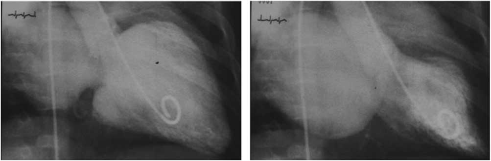
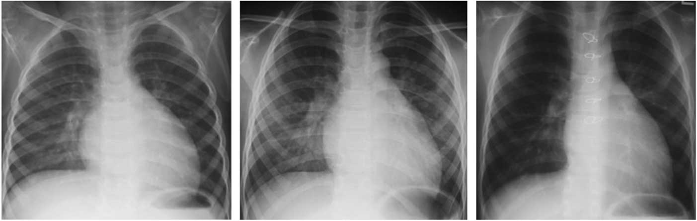
She underwent cardiac catheterisation on the 109th day of illness. The left ventricular end-diastolic pressure and mean pulmonary wedge pressure were 18 and 14 mmHg, respectively. The left ventricular end-diastolic volume and left ventricular ejection fraction were 29 ml (172% of normal) and 77%, respectively (Figs 1 and 2). Endomyocardial biopsy of the right ventricle showed no inflammatory cells. She was maintained on angiotensin-converting enzyme inhibitor, furosemide, and spironolactone in the outpatient clinic. The left ventricular end-diastolic dimension by two-dimensional echocardiography was 48.6 mm (136% of normal) at 5 years old. She underwent cardiac catheterisation again at the age of 6 years (Fig 2). The left ventricular end-diastolic pressure and mean pulmonary wedge pressure were 13 and 10 mmHg, respectively. The left ventricular end-diastolic volume and left ventricular ejection fraction were 111 ml (177% of normal) and 67%, respectively. She underwent mitral valve annuloplasty with resection of the posterolateral leaflet. The left ventricular end-diastolic dimension by two-dimensional echocardiography decreased to 41.2 mm (98% of normal) and her mitral regurgitation improved from severe to trivial. No medications were required post surgery. She has been doing well for 16 years.
Left ventriculogram at 9 months old in patient 2. Severe mitral regurgitation was detected. The left atrium and ventricle were dilated.

Chest X-ray two before and after operation in patient 2. (Left) The cardiothoracic ratio was 56% in 2-year-old, and the double shadow because of enlargement of the left atrium was shown. (Middle and right) Chest X-P at 6 years. The cardiothoracic ratio improved from 54 to 51% after surgery.

Patient 3
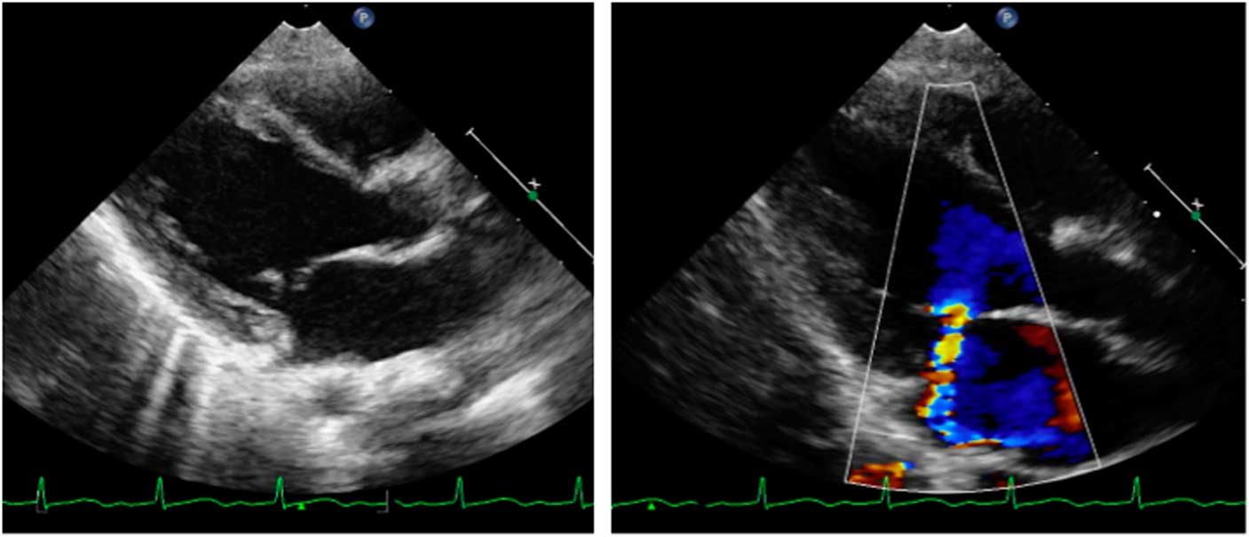
An 8-month-old girl infant, body weight 9 kg, was referred to our hospital on the 4th day of acute typical Kawasaki disease, and aspirin was started. Mild regurgitation of both mitral and tricuspid valves was present. The maximum values of C-reactive protein and white blood cell were 5.78 mg/dl and 19,900 u/ml, respectively. On the 5th day, immediately after intravenous immunoglobulin of 0.1 g/kg/day was administered, fever was alleviated; however, fever with rash reappeared on the 10th day of illness. On the 11th day, after receiving intravenous immunoglobulin of 1 g/kg/day, her fever subsided. There were no coronary aneurysms. The degree of mitral regurgitation was trivial by two-dimensional echocardiography on the 37th day. After 1 month, she had fever for 3 days followed by rash. She was diagnosed as exanthema subitum in the nearby clinic. After the episode, she had had no symptoms. On the 90th day, a systolic heart murmur was detected at her regular outpatient clinic visit. Slight to moderate mitral regurgitation owing to ruptured chordae tendineae of the antero-middle leaflet of the mitral valve was detected by two-dimensional echocardiography (Fig 3). The cardiothoracic ratio in chest X-ray was 48%, and a double shadow was found. The left ventricular end-diastolic dimension and left ventricular ejection fraction by two-dimensional echocardiography were 32.6 mm (115% of normal) and 79%, respectively. Spironolactone and angiotensin-converting enzyme inhibitor were started. When 2 years old, her left ventricular end-diastolic dimension by two-dimensional echocardiography was 33.0 mm (105% of normal). Spironolactone was discontinued.
Two-dimensional echocardiogram at 1-year-old in patient 3. (Left) Prolapse of chordae tendineae in the anterior leaflet was detected. (Right) Mitral regurgitation was found. The grade of mitral regurgitation was slight to moderate.

Discussion
A Japanese study of about 95 patients with ruptured mitral chordae tendineae was reported in 2014.Reference Shiraishi, Nishimura and Sakaguchi 6 The age of patients ranged from 21 days to 16 months. A total of 10 patients with ruptured mitral chordae tendineae related to acute Kawasaki disease were included in that study. Its incidence with acute Kawasaki disease was estimated to be about 1/10,000. Ruptured mitral chordae tendineae had occurred both in the acute phase and the convalescent phase. The number of patients in the acute phase and the convalescent stage were 3 and 7 patients, respectively. The number affecting the convalescent stage was more than that of the acute phase in that study. Hypercytokinaemia because of acute Kawasaki disease vasculitis is considered as one of the causes of infant ruptured mitral chordae tendineae. On the contrary, the occurrence of ruptured mitral chordae tendineae in the late period of more than 4 months after the onset of acute KD had not been reported. Furthermore, there was no report about ruptured mitral chordae tendineae in the acute Kawasaki patients more than 24 months old.
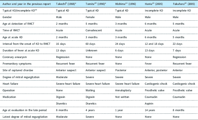
Acute Kawasaki disease is characterised by hypercytokinaemia and a systemic vasculitis of medium-sized vessels. The hypercytokinaemia leads to various systemic symptoms and signs in multiple organs including the heart. Acute Kawasaki disease vasculitis can develop pericarditis, myocarditis, endocarditis, and valvulitis, so features are not restricted to the coronary arteries.Reference Tsuda, Uchiyama and Abe 7 Initially systemic inflammation owing to acute Kawasaki disease involves the small vessels and peaks on the 10th day of illness, then the inflammation regresses until the 20th illness day leaving no residue.Reference Fujiwara and Hamashima 8 Heart murmur was detected in the third week of acute Kawasaki disease in most of the patients with ruptured mitral chordae tendineae in our experience and the previous reports (Tables 1 and 2). The most ruptured mitral chordae tendineae seems to occur at that time. Further, inflammation after the initial acute Kawasaki disease vasculitis is often recurrent. It is well known that the recurrent fever is one of the factors that makes coronary artery aneurysm. Our patients had had recurrent fever vasculitis after alleviation of the initial Kawasaki disease vasculitis. Although it is unclear whether the recurrent fever is related to acute Kawasaki disease or not, the repeat inflammatory attacks might predispose to ruptured mitral chordae tendineae (Tables 1 and 2).Reference Takechi, Tani and Ozaki 9 – Reference Fukuhara, Toyohara and Tazato 13 On the contrary, ruptured mitral chordae tendineae without recurrent fever in the convalescent stage has been reported.Reference Mishima, Asano and Saito 11 Further, the occurrence of ruptured mitral chordae tendineae in all phases of acute phase of Kawasaki disease has been described, not only in the convalescent stage but also in acute phase.Reference Haniu, Kitano, Kurosaki, Watanabe, Tsuda and Echigo 12 The initial severe hypercytokinaemia can also cause ruptured mitral chordae tendineae.
Characteristics and treatment in patients with raptured chordae tendineae of mitral valve.

ACEI=angiotensin converting enzyme inhibitor; KD=Kawasaki disease; LVDd=left ventricular end-diastolic dimension; RMCT=ruptured mitral chordae tendineae
* Discontinue
Characteristics and treatment in patients with raptured chordae tendineae of mitral valve in the previous reports.

KD=Kawasaki disease; RMCT=ruptured mitral chordae tendineae
* As the cause of mitral regurgitation in two patients, RMCT was strongly suspected
** The diagnosis of KD was based on diagnostic guidelines prepared by the Japanese Kawasaki disease society
In the acute phase of Kawasaki disease, mild regurgitation of the mitral and tricuspid valves occurs in 30–40% patients, particularly in small infants rather than older children.Reference Suzuki, Kamiya and Tsuchiya 1 Not only valvulitis but also a ruptured mitral chordae tendineae is one of the causes of mitral regurgitation, a possible outcome of valvulitis caused by Kawasaki disease. In the past when two-dimensional echocardiography had not developed, the diagnosis of a ruptured mitral chordae tendineae might be difficult. Severe mitral regurgitation caused by valvulitis resulting from acute Kawasaki disease was reported in infants, particularly under 6 months of age.Reference Imakita, Sasaki, Misugi, Miyazawa and Hyodo 4 Furthermore, the age at ruptured mitral chordae tendineae caused by Kawasaki disease in the previous reports was within 6 months old (Table 2). The risk of ruptured mitral chordae tendineae seems to depend on the age at the onset of acute Kawasaki disease. Hence it is believed that the weakness of the chordae tendineae owing to inflammation is related to the younger ages.
We have experienced repeat ruptured mitral chordae tendineae 12 and 18 days after the onset of disease, in patients whom incomplete Kawasaki disease had been suspected (Table 2).Reference Haniu, Kitano, Kurosaki, Watanabe, Tsuda and Echigo 12 He was a 6 months old boy who had persisting fever and bilateral conjunctivitis injection. He developed suddenly cardiogenic shock because of mitral regurgitation caused by ruptured mitral chordae tendineae of the anterior and posterior leaflets of mitral valve, and underwent mitral valve replacement with a prosthetic valve. Our experience is that idiopathic ruptured mitral chordae tendineae suddenly leads to severe heart failure or cardiogenic shock in infants around 6 months and the left ventricle is unable to rapidly compensate for the increased volume. As a result, the hemodynamic state becomes unstable, which may sometimes be fatal. Most of the infants had had fever suspected to be infection as the premonitory symptom. The causes of infantile ruptured mitral chordae tendineae remain unknown, and its occurrence is only reported in Japan. The causes of ruptured mitral chordae tendineae might be related to racial or genetic factor, in the view of immunologic response for infection.
In the Japanese survey of 2014 about ruptured mitral chordae tendineae, eight deaths (8.4%) among 95 patients had occurred.Reference Shiraishi, Nishimura and Sakaguchi 6 The severity of heart failure depends on the extent of ruptured mitral chordae tendineae. In the treatment of sudden and progressive heart failure because of ruptured mitral chordae tendineae, anti-acute heart failure must be considered with inotropic support and respiratory management. Strict management of fluid volume to prevent pulmonary oedema is also important. In the cases with intractable heart failure, surgery should be considered early. Emergency surgery is often needed to save the patients. However, mitral valvuloplasty in small infants is difficult in some cases, particularly because of the degeneration of the valve. Artificial chordae tendineae for reconstruction would be needed, particularly in the anterior leaflet. Mitral valve replacement with a prosthetic valve should be avoided in small children, if possible. The latter the time of surgery, the better the outcome. Therefore, the initial management for ruptured mitral chordae tendineae is very important. If acute heart failure can be stabilised, the left ventricular end-diastolic volume after ruptured mitral chordae tendineae gradually increases to compensate for volume load of the left ventricle with ageing. The indication of surgery for mitral valve depends on volume overload of the left ventricle. Angiotensin-converting enzyme inhibitor and diuretic should be recommended to prevent remodelling of the left ventricle.
Conclusion
We suspect that ruptured mitral chordae tendineae in infant within 6 months occurs because of weakness of the chordae tendineae, secondary to injury by inflammation because of acute vasculitis and the recurrent fever. The correct diagnosis of ruptured mitral chordae tendineae and its careful management are important to prevent occasional deaths, particularly at convalescent stage.
Financial Support
This research received no specific grant from any funding agency or commercial or not-for-profit sectors.
Conflicts of Interest
None.
Ethical Standards
All procedures performed in studies involving human participants were in accordance with the ethical standards of the institutional committee and with the 1964 Helsinki declaration and its later amendments or comparable ethical standards.
Acknowledgements
The authors thank Professor Peter Olley and Dr Setsuko Olley for their kind consultation on English language.