Introduction
Sarcocystis neurona, Neospora spp. and Toxoplasma gondii are related protozoans that can cause encephalitis in animals. These protozoans were considered T. gondii until the 1980s because of morphologic similarities in their replicating stages. Transmission electron microscopy (TEM) is needed to distinguish their replicating stages (tachyzoites and merozoites). These protozoans are also related molecularly (Table 1).
Genomes of 3 related apicomplexans

Although T. gondii has been known to cause clinical diseases in animals for more than a century, nothing was known of clinical toxoplasmosis-like illness in horses until 1970, except that horses can develop antibodies to T. gondii (Dubey and Beattie, Reference Dubey and Beattie1988). In the 1970s an equine clinical syndrome was referred to by three different names ‘segmental myelitis’ (Rooney et al., Reference Rooney, Prickett, Delaney and Crowe1970), ‘equine protozoan encephalomyelitis’ (Beech, Reference Beech1974) and ‘equine protozoal myeloencephalitis (EPM)’ (Mayhew et al., Reference Mayhew, de Lahunta, Whitlock and Pollock1976); the term EPM has been used by most authors. Until 1973, the aetiology of EPM was unknown. The author (referred as I) has been involved in the biology of EPM since 1973. In the present paper, the history and biology of EPM, neosporosis and toxoplasmosis in horses are reviewed with a personal perspective of discoveries. I would like to acknowledge the contributions of many with whom I have collaborated for decades that made these discoveries possible; the collaborators include D. D. Bowman, C. K. Fenger, M. E. Grigg, D. E. Granstrom, A. N. Hamir, D. K. Howe, D. S. Lindsay (now deceased), S. M. Reed, B. M. Rosenthal, C. A. Speer and W. J. A. Saville (to whom this review is dedicated).
Sarcocystis neurona and EPM
Aetiology
Protozoa identified
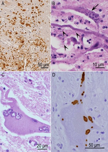
Protozoa were recognized in lesions of encephalitis in horses in 1973 by three groups of researchers and I was associated with all three groups. I got a lucky break in my career when I was appointed, in the summer of 1973, Assistant Professor of Veterinary Parasitology (Department of Pathology) at the Ohio State University (OSU), Columbus, Ohio, USA. The very first week in the department, I was shown slides from a medicolegal case of fatal myeloencephalitis in a horse considered to have died of toxoplasmosis. This 500-kg, 4-year-old paint stallion (S4858) was admitted to the OSU Veterinary Hospital for treatment of lameness of the right hindlimb. Two days prior to admission, the horse was hit by a car from behind while being transported in a trailer. Despite a 2-week treatment, the horse died and was immediately necropsied. The horse belonged to a wealthy owner, legal action was pursued as to the cause of death and public interest in this case was intense. Because there was no published record of clinical toxoplasmosis in horses, I vigorously followed up this case and three other cases that were in the collection of the Department of Pathology. I wanted to publish findings right way but waited because the editor of the Journal of American Veterinary Medical Association asked me to review a paper by Cusick et al. (see below). At the same time, Dr Dodd (Beech and Dodd – see below) asked for my help with the identification of protozoa they had found in horses, including the cases described by Rooney. The paper by Cusick et al. (Reference Cusick, Sells, Hamilton and Hardenbrook1974) was published in January 1974 and it is the first record of finding protozoa in lesions associated with EPM. They misidentified the parasite as T. gondii. Retrospectively, it is clearly due to S. neurona. The paper by Beech and Dodd (Reference Beech and Dodd1974) appeared next, and they called the parasite T. gondii-like; very few organisms were present in lesions. Unlike these two reports, numerous organisms were seen in the cases reported by Dubey et al. (Reference Dubey, Davis, Koestner and Kiryu1974), particularly in horse S4858 (Fig. 1); I have never seen such massive parasitization in EPM cases in the next 50 years; I named the parasite S. neurona from this horse (discussed later – Dubey et al., Reference Dubey, Davis, Speer, Bowman, de Lahunta, Granstrom, Topper, Hamir, Cummings and Suter1991). It remained unknown if stallion S4858 had asymptomatic S. neurona infection aggravated by the automobile trauma and symptomatic treatment with anti-inflammatory drugs.
Histological sections of spinal cord of a horse (S4858) with EPM. Sarcocystis neurona was first named from the parasite observed in this horse with massive infection of the spinal cord. (A) Severe parasitization of spinal cord (all red-stained structures are merozoites and schizonts). Immunostaining with S. neurona antibody. (B) Myelin degeneration and numerous parasites in an axon (arrow). Merozoites (arrowheads) are 2–3 μm long (arrowheads) and difficult to see. The empty spaces are due to myelin degeneration. Haematoxylin and eosin (HE) stain. (C) Neuron with several schizonts. HE stain. (D) Intraneuronal merozoites (arrowheads) and schizonts (arrow) in an intact neuron. Immunostaining with S. neurona antibody. (From Dubey et al., Reference Dubey, Calero-Bernal, Rosenthal, Speer and Fayer2016.)

Although a definitive identification could not be made in 1974, it was stressed early that the parasite was not T. gondii (Dubey, Reference Dubey1974; Dubey et al., Reference Dubey, Davis, Koestner and Kiryu1974). In the next 20 years, I had an opportunity to review slides from many EPM horses, but never succeeded in culturing the organism. In 1991, the EPM organism was grown for the first time in cell culture in my laboratory (Davis et al., Reference Davis, Speer and Dubey1991; Dubey et al., Reference Dubey, Davis, Speer, Bowman, de Lahunta, Granstrom, Topper, Hamir, Cummings and Suter1991). The credit for this idea in part belongs to Dr Dwight Bowman of Cornell University, USA. He obtained a horse suspected of EPM. The horse was given corticosteroids and necropsied when its condition deteriorated; fresh spinal cord was sent to me for in vitro cultivation. Fortunately, viable S. neurona was cultivated from this horse tissue in a first attempt. I asked Dr Stanley W. Davis, a postdoctoral fellow in my laboratory, to inoculate monolayer cultures of bovine monocytes and cardiopulmonary arterial endothelial (CPA) cells with the spinal cord homogenate from the horse. We were fortunate to have inoculated two cell lines because we later found that unlike other species of Sarcocystis, the EPM organism does not develop in CPA cells.
The EPM organism was named S. neurona (Dubey et al., Reference Dubey, Davis, Speer, Bowman, de Lahunta, Granstrom, Topper, Hamir, Cummings and Suter1991). I had decided to name the organism S. neurona based on its morphology in histological sections of horse tissues. I was criticized for naming the parasite without finding other life cycle stages (sarcocyst and sporocyst). My decision to name the EPM organism S. neurona was simply to provide a clear identifiable name on it, rather than calling it T. gondii-like. In vitro cultivation of S. neurona made it possible to better describe its morphology, develop a diagnostic test, characterize its genome and search for a definitive host.
Discovery of the definitive host
Dr David Granstrom and his Ph.D. student, Clara K. Fenger, an equine clinician, led the project for discovering the host. They proposed that the Virginia opossum (Didelphis virginiana) was the likely definitive host. It was a great discovery and how it came about is even more interesting. Fenger used polymerase chain reaction (PCR) targeting a small subunit ribosomal RNA to detect DNA from feces of carnivores. The assay had previously been developed by Granstrom et al. (Reference Granstrom, MacPherson, Gajadhar, Dubey, Tramontin and Stamper1994) to amplify S. neurona DNA for clinical diagnosis of EPM. Amplification was obtained with Sarcocystis sporocysts from opossum feces (Fenger et al., Reference Fenger, Granstrom, Langemeier, Stamper, Donahue, Patterson, Gajadhar, Marteniuk, Xiaomin and Dubey1995). Unfortunately, opossums are also a host to other Sarcocystis species, especially Sarcocystis falcatula, and some scientists even were quick to synonymize S. neurona with S. falcatula based on then available genetic data (Dame et al., Reference Dame, MacKay, Yowell, Cutler, Marsh and Greiner1995). In a subsequent experiment, horses fed sporocysts from naturally infected opossums developed clinical signs consistent with EPM (Fenger et al., Reference Fenger, Granstrom, Gajadhar, Williams, McCrillis, Stamper, Langemeier and Dubey1997). Molecularly, S. neurona is very closely related to S. falcatula-like Sarcocystis species in opossums (Gondim et al., Reference Gondim, Soares, Tavares, Borges-Silva, de Jesus, Llano and Gondim2019). However, biologically these are different parasites; S. neurona is not infective to avian species but is lethal to gamma interferon gene knockout (KO) mice and S. falcatula is not infective to KO mice but is infective to several avian species (Dubey, Reference Dubey2000).
Discovery of the S. neurona sarcocyst and the intermediate host
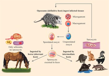
The complete life cycle of S. neurona was resolved in 2000 when the sarcocyst stage was identified in cats (Dubey et al., Reference Dubey, Saville, Lindsay, Stich, Stanek, Speer, Rosenthal, Njoku, Kwok, Shen and Reed2000). The discovery is intriguing because the prey host (cat) was the least likely to act as an intermediate host and develop sarcocysts. I was trying to induce clinical EPM in cats as a model for experimental therapy. In that vein, I fed S. neurona sporocysts from a naturally infected opossum to two cats on 6 December 1999; the opossum was infected with only S. neurona and S. falcatula or Sarcocystis speeri sporocysts were absent. The cats were laboratory-raised from a closed colony. The cats remained asymptomatic despite administration of corticosteroids and the fact that sarcocysts were found in muscles of both cats euthanized 45 and 57 days later. Because the cats had never eaten meat, and I had never seen any protozoan tissue cysts in these colony-raised cats, I was convinced that I had witnessed S. neurona sarcocyst formation. I could not sleep and telephoned Dr Bill Saville at the Ohio State University asking if he possessed any laboratory-raised opossums. Dr Saville advised that the opossums would not be available for at least 6 months and he asked, ‘why the sudden need for opossums’. At that moment I did not share my discovery of sarcocysts in the cat. The 6-month wait for opossums was difficult for me, but I had learned not to let the cat out of the bag. Meanwhile, I infected four additional cats using sporocysts from the same opossum and waited to feed sarcocysts from cats to opossums as described (Dubey et al., Reference Dubey, Saville, Lindsay, Stich, Stanek, Speer, Rosenthal, Njoku, Kwok, Shen and Reed2000). The opossums fed sarcocysts from experimentally infected cats excreted sporocysts that were infectious to ponies and induced fatal encephalitis in KO mice. Finding the sarcocyst stage in an experimental host allowed for production of bonafide sporocysts of S. neurona for infections in ponies and raccoons. I telephoned Dr G. E. Esch, editor of the Journal of Parasitology for his consideration to publish the results on a fast track. He accepted the paper within a day of submission and scheduled publication in the next issue of the journal. Thus, there were many contributors to this discovery, including all coauthors. Literally, it took the ‘whole village’, including the late Dr G. E. Esch, to complete the life cycle of S. neurona (Fig. 2).
Life cycle of S. neurona. Opossums are the definitive host and horses are aberrant intermediate hosts; raccoons, sea otters, armadillos, domestic cats and skunks are some of the natural intermediate hosts. Sarcocystis neurona parasitizes and causes lesions (dark areas) in the brain and spinal cord of horses and several other hosts. Only sexual cycle occurs in opossums. Only schizonts are found in horses. Sarcocysts are produced in intermediate hosts.

Overview of S. neurona biology (1974–2015)
This section is abstracted from three reviews (Dubey et al., Reference Dubey, Lindsay, Saville, Reed, Granstrom and Speer2001c, Reference Dubey, Howe, Furr, Saville, Marsh, Reed and Grigg2015, Reference Dubey, Calero-Bernal, Rosenthal, Speer and Fayer2016) and most citations are omitted for brevity.
Hosts
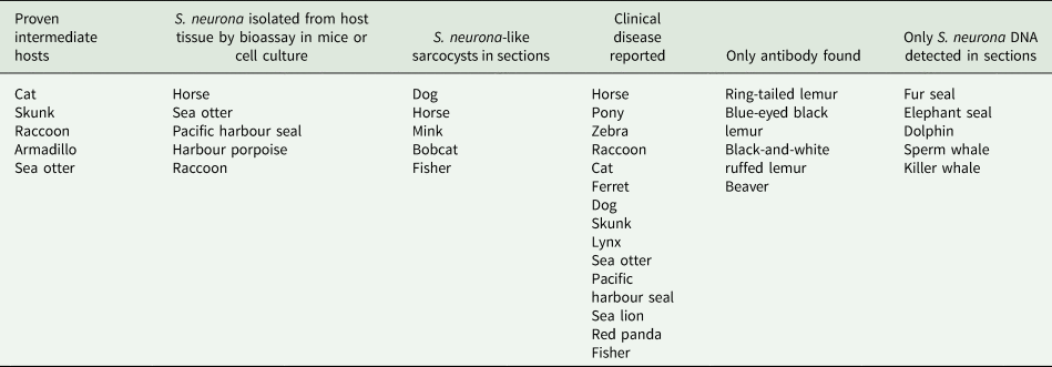
Sarcocystis neurona has a wide host range relative to other species in the genus Sarcocystis (Table 2). The North American opossum (Didelphis virginiana) and the South American opossum (Didelphis albiventris) are its known definitive hosts. Several other animal species are its intermediate or aberrant hosts (Fig. 2). In some hosts, only schizonts have been identified with certainty and those species where only schizonts are found are considered aberrant hosts. Intermediate hosts are those in which sarcocysts mature. Based on bioassay in laboratory-raised opossums, skunks, armadillos, raccoons, domestic cats and sea otters are all proven intermediate hosts. Additionally, S. neurona-like sarcocysts were reported in the muscles of 1 horse, 2 domestic dogs, 1 mink (Mustela vison) and 1 bobcat (Lynx rufus). Whether brown-headed cowbird (Molothrus ater) is an intermediate host for S. neurona is uncertain.
Intermediate or aberrant hosts of S. neurona a

a From Dubey et al. (Reference Dubey, Howe, Furr, Saville, Marsh, Reed and Grigg2015).
Structure and life cycle
Schizonts and sarcocysts are tissue stages in intermediate hosts. Viable S. neurona has been isolated from brain and spinal cord of horses. Numerous cell lines can support the growth of S. neurona. Its schizonts divide by endopolygeny where the nucleus becomes multilobed before merozoites are formed; schizonts of different stages of maturity can be found in a single cell. Sarcocysts have been experimentally induced in domestic cats and raccoons. Sarcocysts are microscopic. The sarcocyst wall is up to 2.5 μm thick and has slender villar protrusions (vps) that are up to 2.8 μm long. The vps have microtubules that extend from tip to the base but have no granules. Few of these microtubules extend deeper in the ground substance. Bradyzoites in sections are 4.8–6.5 × 1.0–1.3 μm in size and contain only 2 rhoptries.
On the basis of experimental studies on raccoons and KO mice, it is postulated that after ingestion of sporocysts, sporozoites excyst in the small intestine, invade intestinal tissue and following parasitaemia, invade other tissues. However, in naturally infected horses with S. neurona, schizont is the only stage identified and schizonts were found only in the brain and spinal cord. Both neural and inflammatory cells in the central nervous system (CNS) can be parasitized. Mature schizonts in the CNS are up to 30 μm long and they can be oval, round, elongated or irregular in shape. Ultrastructurally, merozoites are either stumpy (7.7 × 3.1 μm2) or slender (7.3 × 1.7 μm2) and they contain the same organelles as described in merozoites of other Sarcocystis species, including the absence of rhoptries. Sarcocystis neurona sporocysts are 11.3 × 8.2 μm2 in size. Sporozoites are slender and have 2–4 rhoptries but no crystalloid body. Sporocysts of S. neurona are 10.7 × 7.0 μm2 in size.
Currently, there is no documented case of transplacental S. neurona infection in horses.
Genome characterization and cell biology
One of the major advances has been the characterization of the whole S. neurona genome (Blazejewski et al., Reference Blazejewski, Nursimulu, Pszenny, Dangoudoubiyam, Namasivayam, Chiasson, Chessman, Tonkin, Seshadri Swapna, Hung, Bridgers, Ricklefs, Boulanger, Dubey, Porcella, Kissinger, Howe, Grigg and Parkinson2015) with several surprises. Its genome is more than twice in size than that of T. gondii and Neospora. Sarcocystis neurona lacks rhoptry kinase genes; its genome is 130 222 184 bp but the chromosome number is unknown (Table 2). It has surface antigens that are similar to T. gondii and surface proteins and are termed SnSAG/SRSs. The major SnSAGs are located on the surface of the extracellular merozoite stage of S. neurona and are present throughout the intracellular development of the schizont as well and these have been used in the development of enzyme-linked immunosorbent assays (ELISAs) for diagnosis.
Serologic prevalence
Serologic surveys have revealed widespread exposure in horses, depending on the distribution of opossums in the area, type and age of horses sampled, geographical location and the serological test used (Dubey et al., Reference Dubey, Howe, Furr, Saville, Marsh, Reed and Grigg2015). Infections are thought to be acquired postnatally because transplacental or lactogenic transmission of S. neurona in horses has not been documented. Opossums are the only known definitive hosts for S. neurona, and they are found only in the Americas. Theoretically, therefore, horses born and raised in other countries should have no exposure to S. neurona.
Clinical infections
The clinical signs can vary from acute to insidious onset of focal or multifocal signs of neurologic disease involving the brain, brainstem, spinal cord or any combination of the areas of the CNS. Horses can have difficulty in standing, walking or swallowing, and the disease can progress very rapidly. The onset can be gradual or sudden and horses have been known to fall while racing. Gait abnormalities are often a result of damage to the brainstem or spinal cord and can be quite variable depending on the location and severity of the lesion. The most frequent sign is gait abnormality, related to CNS involvement. The most frequent brain or cranial nerve deficits observed in horses appear to be head tilt, depression, facial nerve paralysis and difficulty swallowing, upper airway dysfunction such as dorsal displacement of the soft palate and laryngeal hemiplegia.
EPM is a disease of well-cared for horses, 3–6 years of age and is usually not associated with poor nutrition or known concurrent infections. The pathogenesis of S. neurona in horses is unclear because it has been difficult to reliably induce the disease experimentally in horses.
Diagnosis
Ante-mortem diagnosis of EPM is difficult. Clinical signs, detection of antibodies, parasites or parasitic antigens or proteins can help in diagnosis. Evaluation of serum antibodies is useful only in a negative way because it is widely prevalent in asymptomatic horses; a negative test may rule out EPM. However, detection of intrathecal antibodies in cerebrospinal fluid (CSF) is helpful in diagnosis. PCR testing of equine CSF may provide information regarding the presence of S. neurona DNA in the CNS. However, the sensitivity of the PCR test appears to be much lower than initially estimated, possibly because intact merozoites rarely enter CSF. Nonetheless, PCR test may be a useful adjunct for the post-mortem diagnosis of EPM in selected cases when it is used on tissues collected at necropsy. Definitive diagnosis is only possible by demonstration of parasites immunohistochemically in lesions of encephalomyelitis; S. neurona has not been identified in tissues of asymptomatic horses.
Treatment
Several anti-protozoal drugs, including sulphonamides and pyrimethamine, Ponazuril (Marquis®, Merial Inc., Duluth, Georgia, USA), diclazuril (Protazil®, Merck Animal Health, Madison, New Jersey, USA) and decoquinate have been used for the past four decades. Treatment often requires prolonged therapy and although anti-protozoal treatment is highly effective, but full resolution of neurologic signs is not 100%.
Prevention and control
Prevention of contamination of feed and water with opossum feces is essential to prevent EPM in animals. Opossums can excrete millions of sporocysts in their feces for months. Sporocysts are resistant to environmental influences, including most used disinfectants. Heating to 60°C for 1 min will kill sporocysts, but exposure to 55°C for 5 min will not. Currently there is no vaccine for EPM.
Update of S. neurona biology (2015–2021)
Seroprevalence and risk factors in asymptomatic horses
The first nationwide study revealed a high serologic prevalence of S. neurona (James et al., Reference James, Smith, Conrad, Packham, Guerrero, Ng and Pusterla2015). Archived sera from 5250 horses from 4 regions of the USA (northeast 650, south 1910, mid-west 1302 and west 1388), originally collected for leptospirosis survey, were tested for S. neurona antibodies by using an indirect fluorescent antibody test (IFAT) with a cut-off of 1:40. Among the risk factors, geography, age and breed were the main factors. Seroprevalence varied from 68% in the west, 81% in the mid-west, 82% in the northeast and 84% in the south. Prevalence increased with age; infections were higher in horses older than 5 years. Among the 6 breeds of horses, the highest prevalence was in warmbloods 86%, followed by Thoroughbreds (82%), draft (80%), Arabian (78%) and paint (76%). A recent study reported coinfection of arboviruses and S. neurona infection in horses from Amazonian Brazil; of the 213 horses, antibodies to S. neurona were detected in 85 (39.9%), T. gondii in 1 and Neospora caninum in 1 (Gomes et al., Reference Gomes, Jansen, Machado, Pena, Fumagalli, Silva, Alves, Roque and Figueiredo2019).
Clinical S. neurona infections
Sarcocystis neurona-associated EPM has been diagnosed histologically at post-mortem in hundreds of horses in the USA since 1973. A recent report confirmed that most cases of EPM in the USA are due to S. neurona; of the 49 cases of EPM, 48 were due to S. neurona and 1 was due to Neospora hughesi (Schale et al., Reference Schale, Howe, Yeargan, Morrow, Graves and Johnson2018). Compared with the USA, only a few cases have been diagnosed in horses from South America (Dubey et al., Reference Dubey, Lindsay, Saville, Reed, Granstrom and Speer2001c, Reference Dubey, Howe, Furr, Saville, Marsh, Reed and Grigg2015). Recently, S. neurona has been identified immunohistochemically in the CNS sections of 11 of 13 horses in Brazil (Henker et al., Reference Henker, Bandinelli, de Andrade, Bianchi, Sonne, Driemeier, Soares and Pavarini2020); in 6 horses, diagnosis was also confirmed by PCR targeting the ITS1 gene.
Diagnosis
A panel of clinicians and researchers provided an update on the diagnosis of EPM and treatment (Reed et al., Reference Reed, Furr, Howe, Johnson, MacKay, Morrow, Pusterla and Witonsky2016; Saville et al., Reference Saville, Reed, Dubey, Granstrom, Morley, Hinchcliff, Kohn, Wittum and Workman2017). They stressed the importance of a thorough neurological examination, exclusion of other causes of EPM-like diseases, serologic testing and use of molecular methods. They tabulated information on various tests, commercial laboratories performing the tests, interpretations of results, specificity and sensitivity of each test. The IFAT and ELISAs are the two tests most employed. They discussed usefulness and pitfalls of different ELISAs. In general, both IFAT and ELISAs efficiently detected S. neurona antibodies both in serum and CSF (Reed et al., Reference Reed, Furr, Howe, Johnson, MacKay, Morrow, Pusterla and Witonsky2016). Another study revealed that proteins not related to S. neurona (C-reactive protein and amyloid A) were not elevated in the cases of EPM and had no diagnostic value (Mittelman et al., Reference Mittelman, Stefanovski and Johnson2018). Although S. neurona is morphologically related to T. gondii-like parasites, these parasites are antigenically and molecularly distinct from T. gondii (Dubey et al., Reference Dubey, Harrison, Dangoudoubiyam, Bandini, Cheng, Kosber, Agop-Nersesian, Howe, Samuelson, Ferguson and Gubbels2017b) and a species of Sarcocystis, S. falcatula (Borges-Silva et al., Reference Borges-Silva, de Jesus, Ferreira and Gondim2020).
Even with all advances in immunodiagnostic and molecular methods, ante-mortem diagnosis of EPM remains a challenge. Overall, the detection of intrathecally produced S. neurona antibodies is the most useful criterion for ante-mortem diagnosis of EPM. The detection of S. neurona DNA in CSF is also useful but the results can vary with sensitivity and specificity of the method in clinical settings. In a retrospective study of CSF samples from 210 horses with neurological signs, by using a highly sensitive reverse transcription (RT)-PCR method, S. neurona DNA was detected in 25 horses that were negative for intrathecally derived S. neurona [isothermal titration calorimetry (ITC)] antibodies (Bernardino et al., Reference Bernardino, Smith, Conrad, Packham, Tamez-Trevino, Barnum and Pusterla2021). In 30 horses, ITC antibodies were detected but had no evidence for parasite DNA. Only 13 horses had both ITC antibodies and parasite DNA. Because of the retrospective nature of the study there was no post-mortem confirmation of EPM (Bernardino et al., Reference Bernardino, Smith, Conrad, Packham, Tamez-Trevino, Barnum and Pusterla2021). The authors speculate that detection of parasite DNA in horses without ITC antibodies might have been related to the clinical stage of EPM and whether horses were treated with antiprotozoal drugs (Bernardino et al., Reference Bernardino, Smith, Conrad, Packham, Tamez-Trevino, Barnum and Pusterla2021). Further studies are needed to explore the diagnosis by using RT-PCR.
Treatment search for new therapies
Finding new therapies for EPM and their evaluation is difficult and search for better medicines continues (Lindsay et al., Reference Lindsay, Nazir, Maqbool, Ellison and Strobl2013; Ojo et al., Reference Ojo, Dangoudoubiyam, Verma, Scheele, Derocher, Yeargan, Choi, Smith, Rivas, Hulverson, Barrett, Fan, Maly, Parsons, Dubey, Howe and Van Voorhis2016; Van Voorhis et al., Reference Van Voorhis, Doggett, Parsons, Hulverson, Choi, Arnold, Riggs, Hemphill, Howe, Mealey, Lau, Merritt, Maly, Fan and Ojo2017). Fortunately, S. neurona is biologically related to other apicomplexans such as T. gondii and Plasmodium spp. that cause serious clinical illness in humans; therefore, drugs found effective for these diseases can be screened for S. neurona therapy. For example, the antimalarial drug, pyrimethamine, is used for therapy of all 3 diseases, EPM, malaria and toxoplasmosis. Recently, Bowden et al. (Reference Bowden, Land, O'Connor and Fritz2018) used high-throughput screening to find the compound dantrolene possibly useful in treating S. neurona-associated EPM. A group of bumped kinase inhibitors are a potential for EPM therapy targeting calcium-dependent protein kinases (Ojo et al., Reference Ojo, Dangoudoubiyam, Verma, Scheele, Derocher, Yeargan, Choi, Smith, Rivas, Hulverson, Barrett, Fan, Maly, Parsons, Dubey, Howe and Van Voorhis2016; Van Voorhis et al., Reference Van Voorhis, Doggett, Parsons, Hulverson, Choi, Arnold, Riggs, Hemphill, Howe, Mealey, Lau, Merritt, Maly, Fan and Ojo2017). Ultimately, potential therapies identified by in vitro (cell culture) methods must be tested in vivo (Dubey et al., Reference Dubey, Fritz, Lindsay, Shen, Kwok and Thompson2001a; Lindsay et al., Reference Lindsay, Nazir, Maqbool, Ellison and Strobl2013).
In vivo testing for S. neurona is expensive because EPM is difficult to induce in horses. To my knowledge, the KO mouse model is the only reliable method for initial animal screening against EPM (Dubey et al., Reference Dubey, Fritz, Lindsay, Shen, Kwok and Thompson2001a). Diclazuril is one such drug that has efficacy in cell culture, in KO mice and in naturally infected horses (reviewed in Dubey et al., Reference Dubey, Howe, Furr, Saville, Marsh, Reed and Grigg2015). For treatment, diclazuril is administered at 1 mg kg−1 body weight in alfalfa-based pellets that are top dressed in the daily grain ration. At lower doses of 0.5 mg kg−1 body weight, diclazuril has the potential for prophylaxis because of attainment of sufficient levels in plasma and CSF (Hunyadi et al., Reference Hunyadi, Papich and Pusterla2015). In an extension of their earlier research, the authors assessed plasma levels of the drug in adult horses administered at a low dose of 0.5 mg kg−1 of 1.56% diclazuril pellets for 3–4 days (Hunyadi et al., Reference Hunyadi, Papich and Pusterla2018). They found that the half-life of the drug was 1.6 days and the levels of plasma concentration were sufficient to suppress the growth of diclazuril (Hunyadi et al., Reference Hunyadi, Papich and Pusterla2018). Coadministration of ponazuril in corn oil attained higher levels of ponazuril in serum and CSF (Furr and Kennedy, Reference Furr and Kennedy2020).
Genetic manipulation
Toxoplasma gondii has been used widely by molecular biologists as a model for genetic manipulation of intracellular pathogens. Some of the technologies developed for T. gondii manipulation have been adapted to S. neurona (Howe et al., Reference Howe, Yeargan, Simpson and Dangoudoubiyam2018). These manipulations can be applied to develop better chemotherapies and to study intracellular events. For example, transgenic clones of S. neurona expressing different coloured fluorescent proteins allowed us to study synthesis of different organelles during the division of S. neurona (Vainshnava et al., Reference Vaishnava, Morrison, Gaji, Murray, Entzeroth, Howe and Striepen2005; Gaji et al., Reference Gaji, Zhang, Breathnach, Vaishnava, Striepen and Howe2006).
Neospora spp. infections
Overview of Neospora spp. biology (1988–2016)
In 1988, a new parasite Neospora caninum was named (Dubey et al., Reference Dubey, Carpenter, Speer, Topper and Uggla1988). Until then, this parasite was misdiagnosed as T. gondii. A year later, N. caninum was recognized as a cause of abortion in cattle (Thilsted and Dubey, Reference Thilsted and Dubey1989). Two years later, Neospora sp. was identified in the lungs of an aborted horse fetus (Dubey and Porterfield, Reference Dubey and Porterfield1990). Biologically, N. caninum and T. gondii are distinct. Felids are the definitive hosts of T. gondii and canids (dogs, coyotes and wolves) are the definitive hosts for N. caninum. Additionally, N. caninum is a major cause of abortion in cattle, whereas T. gondii is not proven to be an abortifacient in cattle.
In 1998, a new species of Neospora, N. hughesi was described from a horse in California, USA (Marsh et al., Reference Marsh, Barr, Packham and Conrad1998). Currently, N. hughesi has been reported only from horses. It is uncertain at present whether there is 1 or 2 species of Neospora that infect horses. Therefore, the available information on N. hughesi and neosporosis in horses was summarized previously (Dubey et al., Reference Dubey, Hemphill, Calero-Bernal and Schares2017a) and is abstracted here.
Morphologically, tachyzoites and tissue cysts of N. caninum and N. hughesi are indistinguishable; oocyst stage of N. hughesi is unknown (Dubey et al., Reference Dubey, Hemphill, Calero-Bernal and Schares2017a). There is a serologic cross-reactivity between N. caninum and N. hughesi infections (Vardeleon et al., Reference Vardeleon, Marsh, Thorne, Loch, Young and Johnson2001; Packham et al., Reference Packham, Conrad, Wilson, Jeanes, Sverlow, Gardner, Daft, Marsh, Blagburn, Ferraro and Barr2002). Molecular differences support validity of these species (Khan et al., Reference Khan, Fujita, Randle, Regidor-Cerrillo, Shaik, Shen, Oler, Quinones, Latham, Akanmori, Cleaveland, Innes, Ryan, Šlapeta, Schares, Ortega-Mora, Dubey, Wastling and Grigg2019). Currently, there is no evidence for the presence of pNc5 gene-specific for N. caninum in N. hughesi (Dubey et al., Reference Dubey, Hemphill, Calero-Bernal and Schares2017a).
Serologic prevalence
Antibodies to Neospora spp. have been reported worldwide and data until 2016 have been summarized previously (Dubey et al., Reference Dubey, Hemphill, Calero-Bernal and Schares2017a). Most surveys were from Brazil and the USA. In Brazil, seroprevalence varied widely from 0 to 85% but a valid comparison is not possible because of different serological tests and cut-offs used. In most of these surveys, N. caninum tachyzoites were used as antigens. Two surveys from the USA are noteworthy because of the large numbers of horses tested and use of N. hughesi tachyzoites in an IFAT (Pusterla et al., Reference Pusterla, Mackie, Packham and Conrad2014a, Reference Pusterla, Tamez-Trevino, White, VanGeem, Packham, Conrad and Kass2014b; James et al., Reference James, Smith, Conrad, Packham, Guerrero, Ng and Pusterla2015). Of 3123 serum samples submitted to a diagnostic facility in California, 38 (1.2%) had an IFA titre of 1:320 (Pusterla et al., Reference Pusterla, Tamez-Trevino, White, VanGeem, Packham, Conrad and Kass2014b). Of 1785 horses from 18 states, 34% (1785 of 5250) apparently healthy horses were seropositive, but the IFA cut-off used was not stated (James et al., Reference James, Smith, Conrad, Packham, Guerrero, Ng and Pusterla2015). Antibodies to Neospora were detected in 2 of 616 (0.3%) horses in Turkey (Zhou et al., Reference Zhou, Cao, Sevinc, Sevinc, Ceylan, Liu, Wang, Moumouni, Jirapattharasate, Suzuki, Nishikawa and Xuan2017).
Isolation of viable Neospora spp. from horses
Viable Neospora was isolated from three clinical horses in the USA: from an 11-year-old gelding from California (Marsh et al., Reference Marsh, Barr, Madigan, Lakritz, Nordhausen and Conrad1996, Reference Marsh, Barr, Packham and Conrad1998), a 20-year-old gelding from Oregon (Dubey et al., Reference Dubey, Liddell, Mattson, Speer, Howe and Jenkins2001b) and a 13-year-old mare from Alabama (Cheadle et al., Reference Cheadle, Lindsay, Rowe, Dykstra, Williams, Spencer, Toivio-Kinnucan, Lenz, Newton, Rolsma and Blagburn1999). Recently, genomes of these isolates have been compared with genomes of N. caninum (Khan et al., Reference Khan, Fujita, Randle, Regidor-Cerrillo, Shaik, Shen, Oler, Quinones, Latham, Akanmori, Cleaveland, Innes, Ryan, Šlapeta, Schares, Ortega-Mora, Dubey, Wastling and Grigg2019).
Transplacental infection
Unlike cattle, transplacental Neospora infection in horses appears to be rare. Three types of evidence for congenital infections are summarized. To my knowledge, there is only 1 confirmed neosporosis abortion in horses. It was a retrospective study. The mare aborted 2 months before term. Toxoplasma gondii antibodies were not found in 1:10 dilution of the mare's serum tested by using a modified agglutination test. Two pieces of lung fixed in 10% neutral-buffered formalin were studied histologically. Individuals and groups of tachyzoites were found in macrophages and in unidentified cells in alveoli. The parasite did not stain with T. gondii antibodies in an immunohistochemical reaction (Dubey and Porterfield, Reference Dubey and Porterfield1986). After the discovery of Neospora in 1988, the parasite was identified as Neospora immunohistochemically (Dubey and Porterfield, Reference Dubey and Porterfield1990).
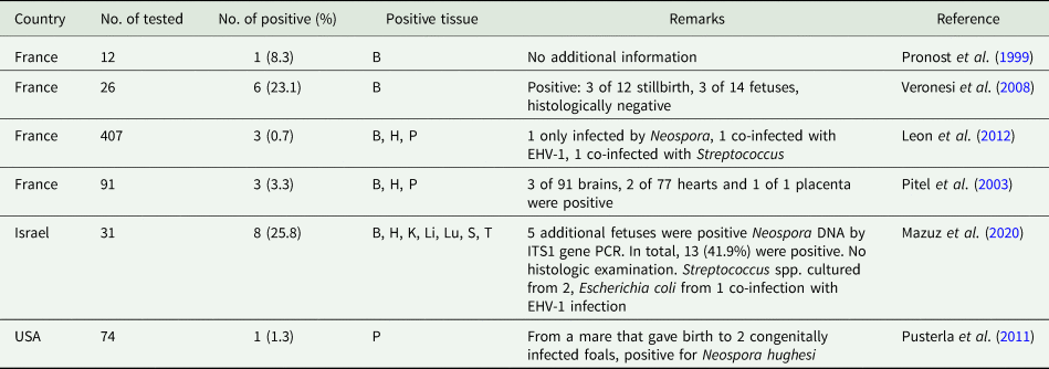
Neospora spp. DNA (based on pNc5 gene) has been found in tissue fetuses from horses in France, Israel and the USA (Table 3). As there was no histological confirmation of lesions and parasites, it is uncertain if the abortion was due to neosporosis.
Detection of Neospora DNA (PCR, pNc-5 gene) in aborted equine fetusesa

a Modified from Dubey et al. (Reference Dubey, Hemphill, Calero-Bernal and Schares2017a).
B, brain; H, heart; K, kidney; Li, liver; Lu, lung; P, placenta; S, spleen; T, thoracic fluid; EHV-1, equine herpes virus 1.
The presence of antibodies in pre-suckling foal sera is suggestive of transplacental infection. However, some maternal antibodies can cross the placental barrier in horses (Duarte et al., Reference Duarte, Conrad, Barr, Wilson, Ferraro, Packham, Carpenter and Gardner2004a, Reference Duarte, Conrad, Wilson, Ferraro, Packham, Bowers-Lepore, Carpenter and Gardner2004b). Antibodies to Neospora were detected in pre-suckling foal sera in Brazil (Locatelli-Dittrich et al., Reference Locatelli-Dittrich, Dittrich, Richartz, Gasino Joineau, Antunes, Pinckney, Deconto, Hoffmann and Thomaz-Soccol2006; Antonello et al., Reference Antonello, Pivoto, Camillo, Braunig, Sangioni, Pompermayer and Vogel2012; Pivoto et al., Reference Pivoto, de Macêdo Junior, da Silva, Ferreira, Silva, Pompermayer, Sangioni, Mineo and Vogel2014).
In a study of mares from 4 farms in California, USA for 3 years, antibodies to Neospora were not detected in pre-suckling sera of 366 foals from 261 mares, and 69 of 324 (21.2%) mares were seropositive to N. hughesi at the time of parturition (Duarte et al., Reference Duarte, Conrad, Barr, Wilson, Ferraro, Packham, Carpenter and Gardner2004a). Additionally, Neospora was not found in fetal tissues or placenta from 22 abortions by histology on these farms (Duarte et al., Reference Duarte, Conrad, Barr, Wilson, Ferraro, Packham, Carpenter and Gardner2004a).
In another study, antibodies to N. hughesi were detected in 17 of 74 (22.9%) mares (Pusterla et al., Reference Pusterla, Conrad, Packham, Mapes, Finno, Gardner, Barr, Ferraro and Wilson2011). Pre-suckling sera from 74 foals and their 58 dams were tested for N. hughesi antibodies. Only 3 foals were seropositive in pre-suckling sera (IFAT 1:1280, 1:2560, 1:20 480). These seropositive foals were from 2 mares (A and B) with IFAT titres of 1:640 and 1:10 240. Two congenitally infected foals were born to the same mare, and N. hughesi DNA was detected in its placenta. Neospora hughesi can be transmitted in subsequent pregnancies. Mares A and B from the above study (Pusterla et al., Reference Pusterla, Conrad, Packham, Mapes, Finno, Gardner, Barr, Ferraro and Wilson2011) gave birth to additional 4 congenitally infected foals in subsequent pregnancies (Pusterla et al., Reference Pusterla, Mackie, Packham and Conrad2014a). All 7 congenitally infected foals in these 2 studies were healthy. Colostrally acquired antibodies to N. hughesi disappeared or declined after 3 months post-suckling (Pusterla et al., Reference Pusterla, Conrad, Packham, Mapes, Finno, Gardner, Barr, Ferraro and Wilson2011).
Recently, in a Brazilan study, amniotic fluids (AF) from 31 mares in third gestational period were tested simultaneously for the three parasite DNA's; Neospora DNA (ITS1, Nc 5) was detected in amniotic fluids of five and placentas of four, S. neurona DNA was detected in one placenta and T. gondii DNA was not detected in any sample (Cabral et al., Reference Cabral, Sato, de Sousa, Rossa, Sanches, Bortoleto and Dittrich2022).
A recent study from Israel presented both serological and molecular evidences of Neospora abortion in horses. Paired samples were collected from aborting mares and their fetuses (Mazuz et al., Reference Mazuz, Mimoun, Schvartz, Tirosh-Levy, Savitzki, Edery, Blum, Baneth, Pusterla and Steinman2020). Most mares were Arabian and from 27 farms; abortions occurred during the second and third trimesters. Antibodies to N. caninum were assayed by using an IFAT (cut-off 1:50); antibodies were detected in 22 of 31 (70.9%) mare sera and in 1 fetal fluid. Neospora DNA was detected by PCR using Nc-5 gene and/or ITS gene (Table 3). Neospora DNA was found in 13 of 31 (41.9%) fetuses; in 10 of 22 from seropositive mares and in 3 of 9 seronegative mares. Neospora DNA was detected in several fetal tissues (Table 3). This study amply documented transplacental Neospora infection, but the aetiology of abortion remained uncertain due to the absence of lesions verified histologically (Mazuz et al., Reference Mazuz, Mimoun, Schvartz, Tirosh-Levy, Savitzki, Edery, Blum, Baneth, Pusterla and Steinman2020).
Clinical infections
Clinical post-natal neosporosis has been diagnosed in 7 horses, all from the USA or Canada; 6 cases were 10–20-year-old horses (reviewed in Dubey et al., Reference Dubey, Hemphill, Calero-Bernal and Schares2017a). One of these cases was a 1-month-old foal. The foal had circled its mare to the right from birth, indicative of neurological disorder. The foal was blind in one eye and had minimal vision in the second eye. Histologically, it had granulomatous encephalitis, marked by gliosis especially in the mid-brain and optic nerve. Thick-walled Neospora tissue cysts, but no tachyzoites, were detected in the brain (Lindsay et al., Reference Lindsay, Steinberg, Dubielzig, Semrad, Konkle, Miller and Blagburn1996). Six cases of neosporosis with neurological signs were diagnosed in aged horses, 3 of them had other non-protozoal complications (Cheadle et al., Reference Cheadle, Lindsay, Rowe, Dykstra, Williams, Spencer, Toivio-Kinnucan, Lenz, Newton, Rolsma and Blagburn1999; Daft et al., Reference Daft, Barr, Collins and Sverlow1996; Marsh et al., Reference Marsh, Barr, Madigan, Lakritz, Nordhausen and Conrad1996; Hamir et al., Reference Hamir, Tornquist, Gerros, Topper and Dubey1998; Wobeser et al., Reference Wobeser, Godson, Rejmanek and Dowling2009). Visceral neosporosis was reported in 1 horse (Gray et al., Reference Gray, Harmon, Sales and Dubey1996). The horse had anaemia, was icteric, had abdominal lymphadenopathy and foci of thrombosis in lymph nodes and lungs. Unlike other cases, numerous Neospora tachyzoites were present in the small intestines and in perinodal connective tissue (Gray et al., Reference Gray, Harmon, Sales and Dubey1996).
In addition to histologically confirmed cases of neosporosis, clinical neosporosis was diagnosed in 4 equids, all from California (Finno et al., Reference Finno, Aleman and Pusterla2007, Reference Finno, Eaton, Aleman and Hollingsworth2010). All of these were initially presented with signs reminiscent of S. neurona-induced encephalomyelitis, including neurological symptoms, muscle atrophy and facial nerve involvement. Detailed laboratory investigations excluded S. neurona infection. The diagnosis of neosporosis bas based on serological data (Finno et al., Reference Finno, Aleman and Pusterla2007).
Toxoplasma gondii infections
Toxoplasma gondii infection is not clinically important in horses but is of public health significance because horse meat is consumed in several countries.
Serological prevalence
It has been known for more than 7 decades that horses can develop T. gondii antibodies (Feldman and Miller, Reference Feldman and Miller1956). Experimentally, horses fed T. gondii oocysts developed very high levels of antibodies that were persistent for several months (Dubey and Desmonts, Reference Dubey and Desmonts1987). Antibodies to T. gondii were found in horses worldwide and data until 2021 were summarized previously (Dubey and Beattie, Reference Dubey and Beattie1988; Dubey, Reference Dubey2010, Reference Dubey2022; Dubey et al., Reference Dubey, Murata, Cerqueira-Cézar and Kwok2020). Recently, T. gondii antibodies have been reported in 20 of 72 (27.8%) horses from Turkey (Aciöz et al., Reference Aciöz, Bozkaya and Babür2021) and in 36 of 192 (18.7%) horses from Brazil (Arruda et al., Reference Arruda, Freitas, Carrijo, Paz, Silva, Sudré, Marques-Santos, Fonseca, Amendoeira and Millar2020).
Isolation of viable T. gondii
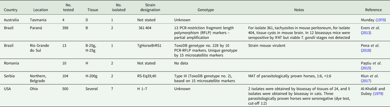
Viable T. gondii was isolated from tissues of horses from Australia, Brazil, Romania, Serbia and the USA (Table 4). It is noteworthy that some of the parasitologically proven horses had very low T. gondii antibodies or were seronegative (Table 4).
Isolation of viable T. gondii from horses by mouse bioassay

B, brain; D, diaphragm; H, heart.
Clinical infections
As stated earlier, clinical toxoplasmosis has rarely been documented in horses. In a survey of 101 horses with neurologic disease simulating EPM, there was no evidence for T. gondii association with EPM (Schale et al., Reference Schale, Howe, Yeargan, Morrow, Graves and Johnson2018). However, a study in California, USA reported that in EPM-affected horses, higher IFAT T. gondii titres (1:320–1:640) were more prevalent than in case-controlled horses without EPM (James et al., Reference James, Smith, Packham, Conrad and Pusterla2017).
I am aware of only 1 histologically verified report of clinical toxoplasmosis in a horse (Kimble et al., Reference Kimble, Gomez, Szule, Dubey, Buchanan and Porter2021). An adult American Quarter Horse gelding with a history of weight loss had an acute onset of colic, fever, soft feces and elevated liver enzymes. At necropsy, there were gastric mucosal masses and evidence of caecal necrosis. Histologically, the masses were lymph nodes with granulomatous inflammation and areas of liquefactive necrosis. Within and surrounding necrotic areas were free and intra-histiocytic clusters of tachyzoites. Similar but milder inflammation was evident in the spleen, lung and liver. Diagnosis was confirmed by immunohistochemistry and TEM examination (Kimble et al., Reference Kimble, Gomez, Szule, Dubey, Buchanan and Porter2021). Histologically, lesions in this case resembled visceral neosporosis (Gray et al., Reference Gray, Harmon, Sales and Dubey1996).
Public health significance
Horse meat is consumed in several countries and has been epidemiologically linked to clinical toxoplasmosis in 3 human subjects in France who ate raw imported horse meat (Pomares et al., Reference Pomares, Ajzenberg, Bornard, Bernardin, Hasseine, Dardé and Marty2011). These illnesses were noteworthy because of the severity. A 74-year-old man died of toxoplasmosis despite anti-toxoplasma therapy. The second case was in a 24-year-old who gave birth to an infant with toxoplasmosis. The third case was in a 21-year-old whose pregnancy was terminated because the infant had severe toxoplasmosis (Pomares et al., Reference Pomares, Ajzenberg, Bornard, Bernardin, Hasseine, Dardé and Marty2011).
Conclusions
Although much has been learnt regarding EPM in the last five decades, there are many gaps in knowledge of this illness. Sarcocystis neurona is the main cause of EPM. Schizont is the only stage found in horses and it is uncertain if this stage persists for a long time and whether clinical relapse is associated with reactivation or reinfection. Treatment for EPM is expensive and newer medicines are needed to treat EPM. Neospora spp. is another protozoan associated with EPM but it is rarely compared with S. neurona infections. Of the two known species of Neospora, only N. hughesi has been isolated in vitro from affected equine tissues. Although N. caninum DNA has been found in aborted equine tissues, it is uncertain if both species of Neospora occur in horses. Currently, T. gondii is not considered as a cause for EPM in horses. The main importance of T. gondii infection in horses is its public health significance because humans have become ill after eating undercooked horse meat infected with T. gondii.
Acknowledgements
The author thanks Oliver Kwok and Fernando Murata for technical assistance with this review. Generous advice of Drs D. K. Howe, J. Poulos and N. Pusterla is greatly appreciated.
Financial support
This study was funded by USDA Project ‘Detection and Control of Foodborne Parasites for Food Safety: 8042-32000-113-00D’.
Conflict of interest
No conflict of interest.
Ethical standards
No experiments were performed.