Introduction
The development of the sea urchin archenteron and its attachment to the blastocoel roof have been of interest to developmental biologists for over a century (Herbst, Reference Herbst1900; Ernst, Reference Ernst1997), and are still not fully understood. Sea urchin embryos are transparent, and the migrations and changing interactions of cells with other cells and extracellular matrix have been studied in great detail. These embryos are an easily accessible system for studying broad principles of development, and one designated by the National Institutes of Health (NIH) as a model system that helps to explain processes related to human health and disease (Davidson & Cameron, Reference Davidson and Cameron2002; Davidson, Reference Davidson2006).
During gastrulation of the sea urchin, the vegetal plate invaginates and forms a cavity or tube called the archenteron, which extends across the blastocoel toward the animal pole (Ettensohn, Reference Ettensohn1984). Filopodia from the secondary mesenchyme cells extend and adhere to cells in the roof of blastocoel, and the archenteron is pulled and elongated across the blastocoel and finally attaches to the blastula roof (Ettensohn & McClay, Reference Ettensohn and McClay1988; Harden, Reference Harden1989; Ettensohn, Reference Ettensohn1990). Within the extracellular matrix associated with the secondary mesenchyme cells, glycans have been shown to mediate sea urchin gastrulation (Ingersoll & Ettensohn, Reference Ingersoll and Ettensohn1994).
Our laboratory has provided evidence for a role of specific carbohydrates in archenteron development and attachment in the sea urchin embryo (Khurrum et al., Reference Khurrum, Hernandez, Esklaei, Badali, Coyle-Thompson and Oppenheimer2004; Idoni et al., Reference Idoni, Ghazarian, Metzenberg, Carroll, Oppenheimer and Carroll2010; Singh et al., Reference Singh, Karabidian, Kandel, Metzenberg, Carroll and Oppenheimer2014), an easily accessible model for studying cell-cell and cell matrix interactions. These studies demonstrated that free sugars and specific glycosidases have highly specific effects on archenteron development, and highlighted the roles of complex glycans and their putative receptors and lectin-like targets in the embryo. In the most recent study, Smith & Oppenheimer (Reference Smith and Oppenheimer2013) showed that l-rhamnose had a significant effect on early development, interfering with complete attachment of the archenteron to the blastocoel roof at low concentrations. This was puzzling because l-rhamnose-containing glycans are expressed in bacterial cell walls and capsules, in fungi, and plants, but there are no known biosynthetic or salvage pathways for l-rhamnose in animals.
While l-rhamnose-containing glycans may originate in other kingdoms, animals have well characterized receptors for l-rhamnose that are implicated in cell development and other functions. Faury et al. (Reference Faury, Ruszova, Molinari, Mariko, Raveaud, Velelny and Robert2008) reported an l-rhamnose-binding lectin in human fibroblasts that stimulates intracellular Ca2+ signaling, affecting cellular proliferation and extracellular matrix biosynthesis. A recent report (Ohta et al., Reference Ohta, Ido, Kusano, Miura and Miura2014) suggests that l-rhamnose might be biochemically active in pupae of the melon fly in the synthesis of a polysaccharide termed ‘dipterose’, and so there remains the possibility that this sugar is incorporated into sea urchin glycans by an unrecognized mechanism. Vertebrate and invertebrate animals express rhamnose-binding lectins (RBL) at many developmental stages, some of which have agglutinating activity and may be involved in innate immunity against bacteria (Ozeki et al., Reference Ozeki, Matsui, Suzuki and Titani1991; Tateno et al., Reference Tateno, Saneyoshi, Ogawa, Muramota, Kamiya and Saneyoshi1998; Watanabe et al., Reference Watanabe, Tateno, Nakamura-Tsuruta, Kominami, Hirabayashi, Nakamura, Watanabe, Kamiya, Naganuma, Ogawa, Naudé and Muramoto2009; Ogawa et al., Reference Ogawa, Watanabe, Naganuma and Muramoto2011). In nature, and even in controlled laboratory experiments, embryos develop in the presence of commensal and pathogenic bacteria and have cellular defenses to maintain their developmental programme (Hamdoun & Epel, Reference Hamdoun and Epel2007).
In this study, we broadened our examination of the role of l-rhamnose by treating embryos with the enzyme α-l-rhamnosidase, which removes terminal l-rhamnoses from glycans. We and others have shown previously that the interior of sea urchin embryos is accessible to enzyme-sized molecules without microinjection (Latham et al., Reference Latham, Martinez, Cazares, Hamburger, Tully and Oppenheimer1998; Itza & Mozingo, Reference Itza and Mozingo2005), and so treatment of whole embryos with α-l-rhamnosidase would expose large parts of the interior to this specific glycosidase. We assessed the morphology of over 11,000 living, swimming embryos treated with α-l-rhamnosidase, and found significant inhibition of archenteron development, in much the same way as did low levels of the free sugar l-rhamnose (Smith & Oppenheimer, Reference Smith and Oppenheimer2013). The inhibitory effect of the enzyme was eliminated by enzyme denaturation, and by inhibition of the enzyme with high levels of l-rhamnose. These findings suggest a functional role for l-rhamnose-containing glycans during sea urchin development. Such l-rhamnosidic glycans could be involved in a specific set of cellular adhesions, representing a novel use of the sugar in the animal kingdom, or may represent one of the earliest known targets of innate immune defenses during embryogenesis.
Materials and methods
α-l-Rhamnosidase
α-l-Rhamnosidase (EC 3.2.1.40) was purchased as an ammonium sulfate suspension from Megazyme International (Wicklow, Ireland) (catalog E-RHAMS, Lot 110501a). Megazyme reports that, relative to its α-l-rhamnosidase activity, the product has <0.001% activity of α-l-arabinofuranosidase, α-l-arabinopyranosidase, α-d-galactosidase, α-d-galactosidase, α-d-glucosidase, β-d-glucosidase, α-d-mannosidase, β-d-mannosidase, α-d-xylosidase, and β-d-xylosidase. This indicates that the enzyme is essentially free of other glycosidase activities. A 1/100 dilution of the enzyme was dialyzed (3500 MWCO Mini Dialysis Unit, Pierce) against ice-cold sterile distilled H2O to remove traces of ammonium sulfate, and low-molecular-weight contaminants, and then assayed for enzyme activity and protease contamination.
Enzyme activity was determined as recommended by the manufacturer, in an assay buffer of 100 mM sodium phosphate (Sigma Aldrich #71500, 30412), pH 6.5 at 50°C, with 1 mg/ml bovine serum albumin (Sigma Aldrich #A7906) and 5 mM 4-nitrophenyl-α-l-rhamnopyranoside (Sigma Aldrich #N7763). Diluted enzyme (0.2 ml) and 2× assay buffer (0.2 ml) were pre-equilibrated at 50°C for 5 min, before being combined and incubated for an additional 10 min at 50°C. The reaction was stopped with 3 ml 2% (w/v) tribasic sodium phosphate dodecahydrate, and the absorbance measured at 410 nm, with the extinction coefficient of p-nitrophenol assumed to be 18.1 mM−1 cm−1. One unit of α-l-rhamnosidase activity is defined as the amount of enzyme required to release 1 μmol of p-nitrophenol per minute, under these reaction conditions.
Possible protease contamination of the enzyme was assessed by its activity against Azocoll (Sigma Aldrich #A4341), which releases an azo dye upon proteolysis (recorded by spectrophotometry at 520 nm). Next, 25 mg of Azocoll was incubated with 1.0–1.2 units of α-l-rhamnosidase, in 5 ml of 100 mM potassium phosphate (Sigma Aldrich #P5379), pH 7.8, for periods of 0–12 h at 37°C, and the results were compared with a standard curve prepared using a Bacillus licheniformis protease (Sigma Aldrich #P5380). The protease contamination in 1 unit of α-l-rhamnosidase was below the limit of detection of the Azocoll assay (1 × 10⁻⁴ unit-equivalents of the B. licheniformis standard).
A denatured enzyme control was prepared by incubation of a sample of enzyme at 100C for 24 h. l-Rhamnose-inhibited enzyme was prepared by pre-mixing 1/1000, 1/500, 1/200, and 1/100 dilutions of dialyzed enzyme (2–23 units/μl) with 13 nmol of l-rhamnose per unit enzyme.
Gel electrophoresis
α-l-rhamnosidase enzyme purity was assessed by sodium dodecyl sulphate (SDS) polyacrylamide gel electrophoresis (Laemmli, Reference Laemmli1970), and an 8% stacking gel, and 10% separation gel (Sigma Aldrich #08091, with 50 mM dithiothreitol added to the sample before denaturation). Molecular weight markers (Dual Color Precision Plus Protein™ Pre-stained Standards) were purchased from Bio-Rad (#161–0374). The samples were subjected to electrophoresis at 180 V for 60 m, and stained with Coomassie Blue dye (Bio-Rad #161–0406), and photographed.
Embryo treatments
Sea urchins were obtained from South Coast Bio-Marine (San Pedro, CA, USA) and maintained in refrigerated aquaria at 12°C in artificial seawater (ASW: 98.88 g NaCl, 2.68 g KCl, 5.44 g CaCl2·H2O, 18.64 g MgCl2·6H2O, 25.16 g MgSO4·7H2O, and 0.72 g NaHCO3, pH 8.0, per 4 l). Gametes were collected after injection of adult Lytechinus pictus with 0.55 M KCl, and mixed for fertilization. Embryos were cultured at 15°C in ASW until they reached the swimming blastula stage (typically 24 h). At this time 75 μl of embryos were transferred as a suspension to each well of a 96-well microplate with approximately 20–40 embryos in each well (Razinia et al., Reference Razinia, Carroll and Oppenheimer2007; Smith & Oppenheimer, Reference Smith and Oppenheimer2013).
A volume of 25 μl of experimental enzyme or control (ASW) solutions were added to the embryos in each well, and the microplates were incubated for 22–24 h at 15°C until the controls developed completely attached archenterons, following which 10 μl of 10% formaldehyde was added to each well. The specific activity of the enzyme at 15°C was estimated by the manufacturer to be approximately 52 U/mg, or 25% of its activity under standard assay conditions at 50°C. Before fixation most control and experimental embryos were living and swimming. Fixed embryos were observed and photographed with a light compound microscope and a Canon camera (SD14001S) (Melville, NY, USA). The morphology of each archenteron was classified as: completely attached to the blastocoel roof (CA), incompletely attached (IA), non-invaginated (NA), or exogastrulated (EXO). Contingency tables and Pearson's chi-squared tests were used to determine the significance of associations between embryo treatments and morphological types. In sum, 11,128 embryos were assessed in this study.
Results
α-l-Rhamnosidase purity
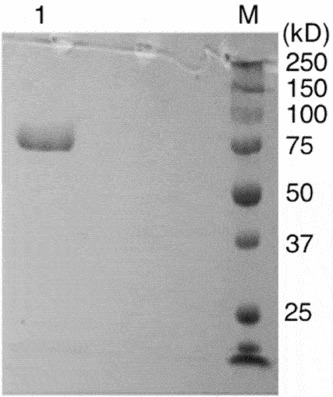
We independently assessed the purity and activity of the α-l-rhamnosidase (EC 3.2.1.40), by gel electrophoresis and biochemical analyses. The enzyme produced a single band of the expected size (75.4 kDa) by SDS gel electrophoresis under reducing conditions (Fig. 1). The gel had a single prominently staining band and no minor bands, consistent with the reported enzyme purity. All assays were performed using dialyzed samples of enzyme, calibrated for enzymatic activity using the colorimetric substrate 4-nitrophenyl-α-l-rhamnopyranoside. The α-l-rhamnosidase was obtained as an ammonium sulfate suspension, and we determined that trace amounts of ammonium sulfate and other low molecular weight contaminants (predicted to remain after thorough dialysis) have no detectable effects on embryogenesis (data not shown). The enzyme had no detectable proteolytic activity, as determined by analysis on Azocoll (described in the Materials and methods section, data not shown). Additional information about enzyme specificity, provided by the manufacturer, is included in the Materials and methods section.
SDS polyacrylamide gel electrophoretic analysis of α-l-rhamnosidase. Lane 1: 2 μg of α-rhamnosidase enzyme (10 μl of 0.02% w/v suspension). Lane M: Pre-stained molecular weight markers. The gel was not cropped and shows a single prominent protein band.

Effect of α-l-rhamnosidase on archenteron development
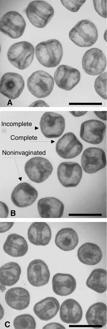
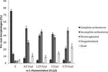
The sea urchin embryo is sufficiently permeable that enzyme-sized molecules can enter freely (Latham et al., Reference Latham, Martinez, Cazares, Hamburger, Tully and Oppenheimer1998; Itza & Mozingo, Reference Itza and Mozingo2005). We treated blastula-stage embryos with samples of α-l-rhamnosidase varying between 0 and 23 U/μl (0 to 5.75 U/μl after mixture with embryos), and allowed development to continue for 22–24 h before the embryos were fixed with formaldehyde and analyzed morphologically. Control embryos incubated in ASW had mostly (75 ± 2.2%) developed archenterons completely attached to the blastocoel roof by this stage (Fig. 2A ), whereas embryos incubated with 1.25 U/μl α-l-rhamnosidase had predominantly incomplete and non-invaginated archenterons (Fig. 2B).
Light micrographs of formaldehyde-fixed L. pictus embryos, at 48 h development. Scale bar: 100 μm. (A) Untreated embryos, showing the typical morphology of complete archenteron attachment. (B) Embryos treated with 1.25 units/μl α-rhamnosidase, showing a mixture of ‘Incomplete’ and ‘Complete’ archenteron attachment, and ‘Non-invaginated’ embryos (arrows). (C) Embryos treated with 1.25 units/μl α-l-rhamnosidase pre-mixed with 15 mM l-rhamnose, showing a restoration of complete archenteron attachment.

The effects of α-l-rhamnosidase on archenteron development are broadly seen across the range 0.5 U/μl to 5.75 U/μl, with a gradual decrease in embryo viability to 75% (±3.3) as enzyme dose increases to 5.75 U/μl (Fig. 3). In an experiment with 5992 embryos, the incomplete attachment and non-invagination morphologies were typically in the range of 21% (±2.3) to 50% (±3.7) each, with complete attachment reduced to only 4.3% (±1.0) to 7.2% (±1.7). Heat denatured α-l-rhamnosidase enzyme generated results indistinguishable from ASW controls (data not shown).
Morphologies of embryos treated with α-rhamnosidase. At each enzyme treatment level, from 0 to 5.75 U/μl, embryos were scored as having (from dark to light bar shading) a complete or incomplete archenteron, a non-invaginated or exogastrulated morphology, or scored as dead. Error bars indicate ± standard error (SE), based on n = 1916 (0 U/μl), 1051 (0.5 U/μl), 1208 (1.25 U/μl), 1121 (3.0 U/μl) and 696 (5.75 U/μl) embryos. The percentage showing complete archenteron development was significantly different between the control sample (0 U/μl) and all enzyme treated samples (P < 0.001), by two-sided independent t-test.

Mitigation of α-l-rhamnosidase effects by cocultivation with free l-rhamnose
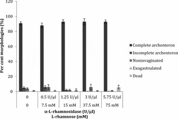
Archenteron development is normal when 30 h embryos are incubated in the presence of high levels (30 mM) l-rhamnose (Smith and Oppenheimer, Reference Smith and Oppenheimer2013), and we tested whether the free sugar (the reaction product) could reverse the effects of α-l-rhamnosidase on embryonic development. By pre-mixing the α-l-rhamnosidase with 13 nmol of l-rhamnose per unit enzyme, we found that enzymatic activity against the colorimetric substrate 4-nitrophenyl-α-l-rhamnopyranoside was reduced to approximately 1.1 to 1.4% of its original level in each dilution. In trials on live blastula-stage embryos, the l-rhamnose inhibited the effects of α-l-rhamnosidase allowing normal development and nearly uniform complete attachment of the archenteron (Fig. 2C ) in 88% (±3.2) to 93% (±4.1) of embryos. In an experiment with 2066 embryos (Fig. 4), there were small numbers of embryos with IA archenterons, 2% (±1.1) to 6% (±1.8), and non-invaginated archenterons, 1% (±0.5) to 6% (±3.7). The viability of the embryos was uniformly high, 94% (±2.4) to 99% (±0.7). The concentration of l-rhamnose in these trials varied from 0 to 75 mM, spanning the high range of levels used in the prior experiments with l-rhamnose alone (Smith & Oppenheimer, Reference Smith and Oppenheimer2013), and the normal embryonic development was consistent with those results. The results in Figures 3 and 4, where differences in archenteron morphology were observed between experimental and control embryos, were found to be significant (P < 0.001). Figures 2 and 3 show that the enzyme primarily caused archenteron disattachment, not archenteron exogastrulation noted in previous studies (Smith & Oppenheimer, Reference Smith and Oppenheimer2013).
Morphologies of embryos treated with α-rhamnosidase and l-rhamnose. The enzyme was pre-mixed with l-rhamnose before dilution and application to the embryos, and the final enzyme (U/μl) and l-rhamnose (mM) concentrations are indicated along the axis. Embryos were scored as having (from dark to light bar shading) a complete or incomplete archenteron, a non-invaginated or exogastrulated morphology, or scored as dead. Error bars indicate ± standard error (SE), based on n = 890 (0 U/μl, 0 mM), 395 (0.5 U/μl, 7.5 mM), 324 (1.25 U/μl, 15 mM), 288 (3.0 U/μl, 37.5 mM) and 169 (5.75 U/μl, 75 mM) embryos. The percentage showing complete archenteron development was not significantly different between the control sample (0 U/μl, 0 mM) and all enzyme + l-rhamnose treated samples (P > 0.05), by two-sided independent t-test.

Discussion
We have shown previously that sea urchin gastrulation is strongly affected by l-rhamnose (Smith & Oppenheimer, Reference Smith and Oppenheimer2013) but not the related 6-deoxyhexose l-fucose (Rashidi et al., Reference Rashidi, Yaghoobian and Oppenheimer2011). The presence of millimolar levels of l-rhamnose during the developmental period of 24–48 h post-fertilization leads to exogastrulation in 20–30% of the embryos. At higher levels of 10–20 mM l-rhamnose, incomplete attachment of the archenteron was a notable result in 30–60% of the embryos. When embryos were treated with l-rhamnose at a later stage and for a shorter time (30–48 h post-fertilization), they were refractory to the higher levels of l-rhamnose, with only 4–16% showing exogastrulation and 1–2% incomplete attachment, but the embryos still had 20–30% exogastrulation with the lower millimolar levels of l-rhamnose.
There is no known pathway for de novo synthesis of l-rhamnose in animals, nor is there a characterized salvage pathway for incorporating l-rhamnose into glycans. However, it has recently been suggested that dipterose, a novel l-rhamnose-rich polysaccharide in melon fly pupae, is the product of a previously unrecognized biosynthetic pathway in insects and not residual plant polysaccharides consumed and stored during the larval stage (Ohta et al., Reference Ohta, Ido, Kusano, Miura and Miura2014). We cannot exclude the possibility that sea urchins also possess novel pathways for l-rhamnose utilization, but such pathways would have escaped notice during genome sequencing and comprehensive biochemical analyses to date.
Despite its uncertain origin, many animal cells specifically recognize l- or d-rhamnose through surface receptors or RBL that are thought to be involved in cell signaling or innate immunity. The genome of the sea urchin Strongylocentrotus purpuratus, for example, encodes a number of putative RBL (UniGene Spu.40441, Spu.31763, Spu.31453) (Sea Urchin Genome Sequencing Consortium, 2006), and Sea Urchin Egg Lectin (SUEL) derived from the urchin Anthocidaris crassispina (Ozeki et al., Reference Ozeki, Matsui, Suzuki and Titani1991) was the first characterized member of the RBL family. Rhamnose-binding lectins may play a role in sea urchin defense (Sakai et al., Reference Sakai, Edo, Nakagawa, Shinohara, Nishiitsutsuji and Ohura2013), as they add an erythrocyte agglutinating activity to venoms, and also more broadly in innate immunity by the ability of RBL to agglutinate bacteria that express rhamnosidic glycans in their cell walls (Ogawa et al., Reference Ogawa, Watanabe, Naganuma and Muramoto2011).
If sea urchins express glycans with α-l-rhamnosides through a novel biosynthetic or salvage pathway in animals, then our treatment of embryos with α-l-rhamnosidase enzyme may have interfered with a novel cellular interaction. Conversely, if the target rhamnosidic glycan was bacterial in origin then the α-l-rhamnosidase enzyme might have inhibited gastrulation in several ways. Bacteria are known to play symbiotic or commensal roles in animal development (McFall-Ngai et al., Reference McFall-Ngai, Hadfield and Bosch2013), and it is possible that rhamnosidic glycan-expressing bacteria are involved in sea urchin development. The α-l-rhamnosidase enzyme could have disturbed a natural symbiosis, causing derangement during gastrulation. Bacteria may instead interfere with sea urchin development (Hamdoun & Epel, Reference Hamdoun and Epel2007), with the RBL of the urchin protecting the embryo from infection. Enzyme treatment could then have made the pathogenic bacteria functionally invisible to the RBL of the urchin's innate immune system, leading to overgrowth and possible disruption of the developmental program.
We have shown that α-l-rhamnosidase enzyme strongly interferes with sea urchin development, an effect that is inhibited by simultaneous addition of l-rhamnose. This finding, along with our previous work (Smith & Oppenheimer, Reference Smith and Oppenheimer2013), suggests that rhamnosidic glycans are present in sea urchin embryos and that their cleavage directly or indirectly affects development. More studies will be needed to determine whether these glycans help to guide archenteron attachment, which would be an unprecedented use of l-rhamnose in animal development, or activate an innate immunity that is manifested at the earliest stages of development. We believe that this study offers intriguing insights into sea urchin embryo biology that have not been previously explored.
Financial support
This work was directly or indirectly supported by an NSF Presidential Award (0731633), Sydney Stern Memorial Trust, NIH Score (S06 48680), RISE, MARC, the Joseph Drown Foundation, California State University Northridge, and Julie Gorchynski, M.D. Scholarship Awards.