INTRODUCTION
Adrenergic signalling in the myocardium contributes to the control of heart rate (chronotropy), strength of contraction (inotropy), and rate of relaxation (lusitropy) by changing the intracellular Ca2+ levels or by altering the sensitivity of critical regulatory proteins to Ca2+ (Dzimiri, Reference Dzimiri1999; Barki-Harrington et al. Reference Barki-Harrington, Perrino and Rockman2004; George and Pitt, Reference George and Pitt2006). Signalling is mediated predominantly by 2 distinct β-adrenergic receptors (β-AR), β1 and β2, which differ in their abundance, distribution, and downstream signal transducers (Steinberg, Reference Steinberg2004; Brodde, Reference Brodde2008). Approximately 75% of the cardiac β-AR are β1, which couple to the stimulatory (Gs) heterotrimeric G protein. The less-abundant β2 receptors couple to both Gs and Gi (inhibitory) proteins. Nevertheless, stimulation of either β1 or β2 activates adenylyl cyclase to increase intracellular cAMP levels. In turn, cAMP activates protein kinase A (PKA), resulting in the phosphorylation of key elements of the contractile apparatus and of proteins that control internal Ca2+ levels (Chakraborti et al. Reference Chakraborti, Chakraborti and Shaw2000; George and Pitt, Reference George and Pitt2006). Several alterations of this system have been described for many cardiac diseases such as dilated cardiomyopathy (Movsesian and Bristow, Reference Movsesian and Bristow2005; Jahns et al. Reference Jahns, Boivin and Lohse2006), cardiac hypertrophy (Esposito et al. Reference Esposito, Rapacciuolo, Naga Prasad and Rockman2002), heart failure (Keys and Koch, Reference Keys and Koch2004), hypertension and a number of other cardiac diseases (reviewed by Dzimiri, Reference Dzimiri1999), most of them with a low β-AR density among several additional alterations.
Chagas disease is a common cause of cardiomyopathy resulting in premature or sudden death and disability across much of Latin America (Barrett et al. Reference Barrett, Burchmore, Stich, Lazzari, Frasch, Cazzulo and Krishna2003), where Trypanosoma cruzi, the protozoan parasite responsible for this disease, continues to represent a health threat for an estimated 28 million people (Barrett et al. Reference Barrett, Burchmore, Stich, Lazzari, Frasch, Cazzulo and Krishna2003; WHO, 2002, 2007). The course of the infection includes an initial or acute phase, with evident parasitaemia and symptoms that persist for 1 or 2 months; followed by a chronic phase, that in most cases presents as an indeterminate form (asymptomatic), but may evolve into the cardiac or into the digestive forms, or both cardiac and digestive forms together. About 10–30% of the infected patients develop a symptomatic disease, usually after decades of chronic asymptomatic infection (Rassi et al. Reference Rassi, Rassi and Little2000). The cardiac chronic form is the most expressive manifestation of Chagas disease, both because of its frequency and because of its severity. It generally appears between the second and fourth decades of life, 5–15 years after the initial infection. The most common signs and symptoms of chronic Chagas cardiopathy are arrhythmia, cardiac failure, atrioventricular and branch blocks, and thromboembolism (Coura, Reference Coura2007). The chronic chagasic cardiomyopathy is an often fatal outcome of this infection, with a worse prognosis than other cardiomyopathies (Bestetti and Muccillo, Reference Bestetti and Muccillo1997; Freitas et al. Reference Freitas, Chizzola, Paes, Lima and Mansur2005). A diffuse myocarditis, intense myocardial hypertrophy, damage and fibrosis, in the presence of very few circulating T. cruzi forms, are the histopathological hallmarks (Higuchi et al. Reference Higuchi, De Morais, Pereira Barreto, Lopes, Stolf, Bellotti and Pileggi1987). It is also common to find dilatation of the cavities, with the presence of thrombi, fibrosis, and thinning of the ventricular apices, particularly in the left ventricle (Coura, Reference Coura2007). Serious alterations in the sympathetic and parasympathetic nervous systems have also been described (Sterin-Borda and Borda, Reference Sterin-Borda and Borda1994).
Previous studies from our laboratory demonstrated important changes in some components of the cardiac β-adrenergic signal transduction system in the acute phase and the chronic indeterminate form of T. cruzi infection in mice (Lo Presti et al. Reference Lo Presti, Bustamante, Rivarola, Fernández, Enders, Fretes, Levin and Paglini-Oliva2006, Reference Lo Presti, Rivarola, Bustamante, Fernández, Enders, Levin, Juaneda, Fretes, Triquell and Paglini Oliva2008). This system acquires additional relevance since it has been demonstrated that antibodies developed during the infection not only bind, but also induce activation of β1-AR by mechanisms involving the molecular signalling pathway elicited by the agonists (Labovsky et al. Reference Labovsky, Smulski, Gómez, Levy and Levin2007). Since the cardiac β-adrenergic signal transduction system is the most powerful regulator of cardiac function, in the present work, we studied some of its components in an early and a late stage of the cardiac chronic form of the infection, in order to determine whether those previously found changes have intensified and are responsible for inducing the progressive cardiopathy.
MATERIALS AND METHODS
Experimental infection
Blood trypomastigote forms obtained from mice infected with Trypanosoma cruzi (Tulahuen strain or SGO-Z12 isolate) were used to inoculate 328 Albino Swiss mice (each weighting 30±1 g) (Bustamante et al. Reference Bustamante, Rivarola, Fernández, Enders, Fretes, De Luca D'Oro, Palma and Paglini-Oliva2003a). The Tulahuen strain has been extensively used and studied under experimental conditions in our laboratory, while the SGO-Z12 isolate has been obtained from an endemic area and represents an example of a strain that is actually in circulation in the sylvatic and domestic cycles of the infection. Both parasite strains used had been found to be cardiotropic (Montamat et al. Reference Montamat, Doro, Gallerano, Sosa and Blanco1996), developing therefore, the cardiac form of the infection. Each mouse was inoculated intraperitoneally with 50 bloodstream trypomastigotes of each strain; this parasite load has been previously found to be sufficient to reproduce the acute phase and the different forms (indeterminate and cardiac) of the chronic phase of the experimental T. cruzi infection (Bustamante et al. Reference Bustamante, Rivarola, Fernández, Enders, Fretes, De Luca D'Oro, Palma and Paglini-Oliva2003a). The number of parasites/ml of blood was estimated in a Neubauer haemocytometer.
The mice were divided in 3 groups: infected with trypomastigote forms of T. cruzi Tulahuen strain (Tul, n=164), and infected with trypomastigote forms of T. cruzi SGO-Z12 isolate (Z12, n=114). A non-infected group was also studied (NI, n=50).
The experimental procedures were carried out in accordance with the Guide for the Care and Use of Laboratory Animals published by the U S National Institute of Health, publication N° 85-23 (revised 1996).
The evolution of the experimental infection was followed by electrocardiography, cardiac histopathological studies and survival until the cardiac chronic phase of the infection, which was studied at 2 time-points: an early stage (135 days p.i.) and a late stage (365 days p.i.). We have previously described (Bustamante et al. Reference Bustamante, Rivarola, Fernandez, Enders, Fretes, Palma and Paglini-Oliva2003b) that the cardiac chronic phase of the infection is characterized by the presence of inflammatory infiltrates, fibrosis and fibre fragmentation in the cardiac tissue, alterations that are present by day 135 p.i. in mice infected with either parasite strain. In the present models, the studies were carried out at 2 different time-points of this phase in order to follow the changes produced by the different parasite strains and to determine whether they intensify with the evolution of the chronic infection. An aged-matched uninfected group was analysed at the same time-points described for the infected groups.
Survival
The survival of each group was monitored every day. The animals used for the different experiments were not included in the survival curve.
Electrocardiograms
Electrocardiogram (ECG) tracings were obtained with a digital electrocardiographic unit (CardioCom – Model CC12DER MCP) under Ketamine ClH (Ketalar®, Parke Davis, Warner Lambert Co, USA) anaesthesia (10 mg/kg), before infection and in the early and late chronic stages of the experimental disease. The electrocardiographic tracings were obtained with 6 standard leads (contact electrode) (dipolar leads DI, DII, DIII and unipolar leads aVR, aVL, aVF), recording at 50 mm/s with amplitude set to give 1 mV/10 mm. The electrocardiographic parameters analysed were: cardiac frequency, length of the PR segment and duration of the Q-T interval. Data were then transferred to a computer for further analysis. Wave durations (in sec) were calculated automatically by the software (CardioCom) after cursor placement.
Histopathological studies
Mice were sacrificed by decapitation, after Ketamine ClH (Ketalar®, Parke Davis, Warner Lambert Co, USA) anaesthesia (10 mg/kg). The hearts were removed, fixed in buffered 10% formaldehyde (pH 7·0), and embedded in paraffin. Each heart was cut horizontally into 5 μm sections from the apex to the auricles and stained with Haematoxylin-Eosin. A total of 50 slices from each group was analysed and at least 30 areas from each slice were examined with a 40 X objective. The same procedures were followed for skeletal muscle samples obtained from the posterior legs of the mice from the different groups.
In order to study the cardiac β-adrenergic system and the cardiac function in the chronic stage of the infection (135 and 365 days p.i.), we determined: the primary messenger (epinephrine and norepinephrine) levels in plasma; the β receptor density, affinity and function; the cardiac concentration of the second messenger (cAMP) given its importance for the activation of the PKA and the subsequent phosphorylation of the proteins involved in cardiac contraction; and the cardiac contractility as a response to the ligand binding to the receptor. Additionally, we determined the presence of autoantibodies in the infected animals that interact with the cardicac β-AR, and the functional consequence of this interaction upon cardiac contractility.
Assays of catecholamines
In order to exclude the influence of stress in the obtention of the samples, 15 days before the animals were sacrificed, the mice were handled daily by the same investigator to minimize stress reactions to manipulation, reproducing the extraction procedure, without sacrificing the animals. In early and late chronic stages their blood was poured into heparinized plastic tubes kept on ice and centrifuged. Individual plasma samples were frozen and stored for subsequent determination of epinephrine and norepinephrine concentration.
The catecholamines in 500 μl aliquots of plasma were partially purified by batch alumina extraction, separated by reverse-phase high-pressure liquid chromatography (RF-HPLC) using a 4·6×250 mm Zorbax R x C18 column (New England Nuclear, Du Pont, Boston, MA, USA) as previously described (Lo Presti et al. Reference Lo Presti, Bustamante, Rivarola, Fernández, Enders, Fretes, Levin and Paglini-Oliva2006).
Cardiac β-AR binding
The density and affinity of β-receptors was measured in microsomal fractions of right and left ventricles from the NI, Tul and Z12 groups in early and late chronic stages of the experimental infection. 3H/dihydroalprenolol (3H/DHA, specific activity 3·515×1015 Bq/mol from NEN Life Science Products) was used as radioligand in β-adrenergic receptors' binding assays as described previously (Lo Presti et al. Reference Lo Presti, Bustamante, Rivarola, Fernández, Enders, Fretes, Levin and Paglini-Oliva2006). Specific binding was defined as the difference in radioactivity bound in the absence or presence of 1 μm propranolol. The dissociation constant (Kd) and maximum 3H/DHA binding (Bmax) were determined by saturation curve and Scatchard analysis using GraFit (Erithacus Software).
Cyclic AMP quantification
The ventricles obtained from the different experimental groups were homogenized at 4°C in 1 ml of cold trichloroacetic acid (0·06 g/ml). The homogenate was centrifuged at 1500 g for 10 min and the supernatant fraction was separated and extracted 3 times with 5 ml of water-saturated-ethyl ether. The ether phase was discarded and the aqueous phase was heated at 80°C to remove all traces of ether. The amount of cAMP was determined using a commercial ELISA kit (Cayman Chemicals). In order to study the participation of the phosphodiesterases (PDEs), caffeine (a PDEs inhibitor – 100 μm) was added to 2 samples from each group before the addition of trichloroacetic acid. If these enzymes were working properly, caffeine would inhibit them and therefore incresase the cAMP concentration.
Cardiac contractility and functional studies of the β-ARs using epinephrine and norepinephrine
The ventricles from the different experimental groups were placed in a glass chamber containing Krebs Ringer bicarbonate medium (pH 7·4) gassed with 95% O2 and 5% CO2 and maintained at a temperature of 32°C. The ventricles were then attached to a force transducer (Statam Universal Cell) and a basal tension of 5 mN was applied leaving the tissues to equilibrate in these conditions for 30 min. Then a 15 V stimulus was applied at a frequency of 30 pulses/min and a duration of 12 msec each for another 30 min. The stimulation pattern was maintained throughout the experimental period. The contractility was registered at this moment, transfered to a PC and measured in mN. The effect of different concentrations of epinephrine (between 2·5×10−6m and 1·5×10−5m) and norepinephrine (between 10−8m and 10−5m) (Sigma Chemical Co.) upon the β-ARs was studied through dose-response curves immediately after. The minimal and maximal epinephrine and norepinephrine concentrations used correspond to the threshold and the maximal effect obtained. The effect of the drugs was considered as the difference between the basal contractility and the response reached in the presence of the epinephrine or norepinephrine.
Determination of the presence of β-AR autoantibodies
IgG purification
The IgG fraction from the NI, Tul and Z12 groups in the early and late stages of the cardiac chronic phase of the infection was purified by affinity chromatography. The technique was performed by Protein G- Sepharose 4 Fast Flow (GE Healthcare) according to the manufacturer's instructions. Briefly, sera from the different groups were dialysed overnight in binding buffer (NaH2PO4, pH 7) at 4°C under agitation. After that, sera were centrifuged at 10000 g for 30 min at 4°C. Protein G-Sepharose™ 4 Fast Flow was loaded into the column and it was washed with 60 ml of binding buffer. The supernatant was passed down a 1 ml column of protein G-Sepharose. The column was washed with 60 ml of binding buffer and eluted with 0·1 m glycine-HCl buffer (pH 2·5). Eluted fractions were re-collected in 1 m Tris Buffer (pH 9) and analysed by spectrophotometry at 280 nm. Finally, the fractions were dialysed overnight in phosphate buffer saline.
Effect of IgG fraction on cardiac contractility
Ventricles from NI mice, placed in a glass chamber containing Krebs Ringer bicarbonate medium as described for the contractility studies, were exposed to the IgG fraction (600 μl) from Tul and Z12 groups (from both stages of the cardiac chronic phase) and their effect on the cardiac contractility registered in order to determine the presence of β-AR autoantibodies. We used a constant amount of IgG fraction for all the groups in order to determine if the antibody concentration was different between them. To confirm the interaction with the β-AR, competitive experiments were performed by adding norepinephrine (5 μm) to tissues previously incubated with the antibodies for 15 min.
Statistical analysis
Data were compared by ANOVA and multiple comparison by Fisher Test (catecholamine levels, cAMP levels, contractility studies and electrocardiographic parameters) and Bonferroni test (β receptors' affinity and density), multivariate analysis of variance by Hotelling test (receptors' functional studies), Kaplan Meier Survival test (survival) and Pearson Square Chi test (ECG percentages); the significance level was set at 0·05.
RESULTS
Chronic experimental Trypanosoma cruzi infection evolution
Survival
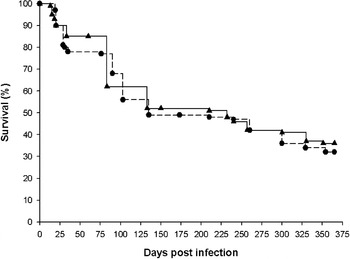
Both infected groups presented a similar survival along the chronic phase of the infection, with 32% of the Tulahuen (n=164) and 36% of the SGO-Z12 (n=114) infected mice surviving until 365 days after the inoculation with the parasites. The survival of the infected groups during the experimental infection can be observed in Fig. 1.
Survival of Tulahuen-infected (n=164) (•) and SGO-Z12-infected (n=114) (▴) mice until the late chronic stage of the experimental Trypanosoma cruzi infection (365 days p.i.). This figure was obtained using the Kaplan Meier Survival test. Data are representative of 7 independent experiments each with 30–45 mice.

ECG analysis
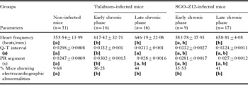
The groups infected with either parasite strain exhibited a higher percentage (P<0·05) of mice with some kind of electrocardiographic alteration when compared to the uninfected controls and to the acute phase of the infection (Lo Presti et al. Reference Lo Presti, Bustamante, Rivarola, Fernández, Enders, Fretes, Levin and Paglini-Oliva2006), but similar between them, in both the early and the late chronic stages of the experimental infection. In either stage of the cardiac chronic phase and with either parasite strain, the most frequent alterations were the atrioventricular blockades (AVB), as shown by a prolonged PR segment and the intra-ventricular blockades (IVB), being a cause of a prolonged Q-T interval, despite the higher frequency presented by either group. In the early chronic stage, 44% of the Tulahuen-infected mice and 11% of the SGO-Z12-infected mice presented AVB (P<0·05), while in the late stage, the percentages were similar in both groups (19 and 18% respectively). The IVB, on the other hand, were more frequent in the SGO-Z12 group (45% of the mice) than in the Tulahuen group (25%) in the early stage of the chronic phase (P<0·05). In the late stage, the percentages were similar in both groups (Tul: 19%; Z12: 12%). The SGO-Z12 group also presented other types of arrhythmias (disorders in heart beat and absence of P waves) in 33% of the mice in the early chronic stage and in 24% of the mice in the late chronic stage of the infection.
The parameters analysed and the electrocardiographic results are summarized in Table 1.
Electrocardiographic analysis of non-infected and Trypanosoma cruzi (Tulahuen strain and SGO-Z12 isolate)-infected groups in an early (135 days p.i.) and a late (365 days p.i.) chronic stage of the experimental infection
(Values show mean±standard error. Data are representative of 3–5 independent experiments each with 5–10 mice. The statistical comparisson was made within the values from each line. Different letters in brackets [a vs b] show a significant difference between the different groups in the values of each parameter (percentages by Pearson Chi square test, P<0·05; the rest of the parameters by Fisher test, P<0·05).)

Histopathological studies
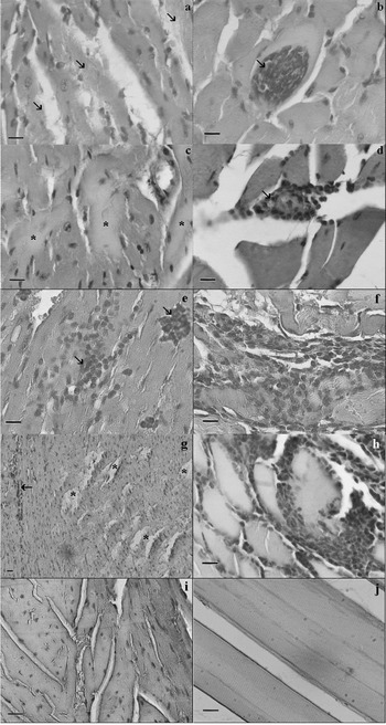
In the early chronic stage of the infection, all the hearts from the Tulahuen group presented disperse lympho-monocitary infiltrates, fibre dissolution (Fig. 2A) and isolated necrosis. The lympho-monocitary infiltrates from the skeletal muscle samples, on the other hand, were more intense, with structural disorganization, multiple necrosis and other alterations produced by the presence of the parasites (amatigote nests; Fig. 2B). In the late chronic stage, this group presented similar cardiac alterations, with few and small inflammatory infiltrates, especially in the right ventricle. Thickening of the epicardium, fibrosis and necrosis (Fig. 2C) in the interventricular septum were also observed. The skeletal muscle presented structural disorganization, necrosis, intense inflammatory infiltrates (Fig. 2D) and perivasculitis at this late stage.
Histological sections (a, b, c, d) from Tulahuen-infected mice. (a) Cardiac section, 135 days p.i. Fibre dissolution can be observed (arrows). (b) Skeletal muscle section, 135 days p.i. The arrow shows an amastigote nest. (c) Cardiac section, 365 days p.i. Areas with necrosis can be observed (stars). (d) Skeletal muscle section, 365 days p.i. The arrow shows an inflammatory infiltrate. Histological sections (e, f, g, h) from SGO-Z12-infected mice. (e) Cardiac section, 135 days p.i. The arrows show inflammatory infiltrates. (f) Skeletal muscle section, 135 days p.i. Area with intense myositis. (g) Cardiac section, 365 days p.i. Fibre fragmentation (stars) and an inflammatory infiltrate (arrow) can be observed. (h) Skeletal muscle section, 365 days p.i. Area with an intense inflammatory infiltrate. Histological sections (i, j) from uninfected mice. (i) Cardiac section. No alterations can be observed. (j) Skeletal muscle section. No alterations can be observed. The bars correspond to 50 μm.

The hearts from the SGO-Z12-infected group, in the early chronic stage of the infection, also showed few small lympho-monocitary infiltrates (Fig. 2E) and fibre disorganization, whereas the skeletal muscle presented remains of old lesions produced by the presence of the parasite, scarce and isolated necrosis and myositis (Fig. 2F). In the late chronic stage, this group presented fibre fragmentation and inflammatory infiltrates both in the myocardium (Fig. 2G) and the skeletal muscle samples (Fig. 2H).
Cardiac and skeletal muscle sections from uninfected mice are shown for comparison (Fig. 2I and J).
The cardiac β-adrenergic system
Plasma catecholamines
In both infected groups, the plasma catecholamine levels diminished with the evolution of the infection when compared to the uninfected controls (NI (n=8), epinephrine: 8·89±1·41 ηg/ml; norepinephrine: 17·35±1·50 ηg/ml). Epinephrine levels were significantly lower in both infected groups in the late stage of the chronic phase of the infection (Tul (n=6): 0·29±0·12 ηg/ml; Z12 (n=10): 0·44±0·14 ηg/ml; P<0·05), while norepinephrine levels were significantly different in the early chronic stage (Tul (n=6): 4·55±0·53 ηg/ml; Z12 (n=10): 8·65±0·99 ηg/ml; P<0·05) and in the late chronic stage (Tul (n=7): 0·12±0·05 ηg/ml; Z12 (n=9): 0·58±0·14 ηg/ml; P<0·05) of the experimental infection. No significant differences were observed between the parasite strains except for norepinephrine levels that were higher in SGO-Z12 mice in the late chronic stage.
Receptor quantitative study: cardiac β-AR binding
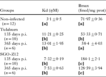
Table 2 shows the β-AR density and affinity from the uninfected and Tulahuen strain or SGO-Z12 isolate-infected groups in an early and a late stage of the chronic experimental infection. As can be observed, the groups infected with either parasite strain presented a decrease in β-AR affinity, represented in the table as the dissociation constant (Kd), when compared to the affinity presented by the uninfected controls and by mice in the acute phase of the infection (Lo Presti et al. Reference Lo Presti, Bustamante, Rivarola, Fernández, Enders, Fretes, Levin and Paglini-Oliva2006) (P<0·05).
β-Adrenergic receptor affinity and density of non-infected and Trypanosoma cruzi (Tulahuen strain and SGO-Z12 isolate)-infected groups at an early (135 days p.i.) and a late (365 days p.i.) chronic stage of the experimental infection
(Values show mean±standard error. The statistical comparison was made within the values from each column. Different letters in brackets [a vs b vs c] show a significant difference between the different groups in the values of each parameter (Bonferroni test, P<0·05).)
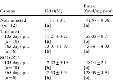
The β-AR density, however, presented a different behaviour according to the parasite strain. While the SGO-Z12 group showed an increase in receptor density (Bmax) in both the early and late stages when compared to the uninfected controls (P<0·01), the Tulahuen group presented a decrease in this parameter in both stages (P<0·05). Noteworthy is the fact that the SGO-Z12 group presented the highest β-AR density in the indeterminate form of the chronic stage of the infection (Lo Presti et al. Reference Lo Presti, Rivarola, Bustamante, Fernández, Enders, Levin, Juaneda, Fretes, Triquell and Paglini Oliva2008), decreasing thereafter but still being higher than in the uninfected group.
Functional studies of the β-ARs using epinephrine (EPI) and norepinephrine (NE)
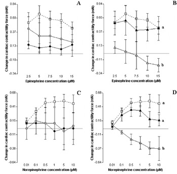
Increasing concentrations of EPI and NE were used in order to study the response of the β-ARs to the first messenger. The uninfected group (Fig. 3, white squares) presented a clear positive inotropism until an EPI concentration of 7·5×10−6m and a NE concentration of 5×10⁻⁷m. Higher concentrations of the agonists did not produce any further significant increment in the cardiac contractility.
Comparative study of the effects produced by epinephrine and norepinephrine on the contractile force. (A) Effect of epinephrine upon uninfected ventricles (□; n=13), and infected with the Tulahuen strain in an early (•; n=4) and a late stage ( ; n=4) of the chronic Trypanosoma cruzi infection. (B) Effect of epinephrine upon uninfected ventricles (□; n=13), and infected with the SGO-Z12 isolate in an early (▴; n=9) and a late stage (
 ; n=3) of the chronic T. cruzi infection. (C) Effect of norepinephrine upon uninfected ventricles (□; n=11), and infected with the Tulahuen strain in an early (•; n=2) and a late stage (; n=2) of the chronic T. cruzi infection. (D) Effect of norepinephrine upon uninfected ventricles (□; n=11), and infected with the SGO-Z12 isolate in an early (▴; n=5) and a late stage (; n=3) of the chronic T. cruzi infection. Bars show standard error. Different letters (a vs b) show significant difference between the studied groups (Hotelling test, P<0·05).

The ventricles from the Tulahuen strain infected group (Fig. 3A) in the early chronic phase of the infection (n=4) presented a response to EPI similar to that found for the uininfected hearts, although the positive inotropic effect was not as evident as in this later group. No significant changes in the response to EPI were found in the late stage of the chronic phase of the infection.
The ventricles from the SGO-Z12 isolate infected group (Fig. 3B) on the other hand, in the early chronic phase of the infection (n=9) presented a response to EPI similar to that found for the uninfected hearts but a significantly dimished response in the late stage (P<0·05).
When NE was added to the hearts from Tulahuen-infected mice, the positive inotropism was not as clear as in the uninfected group in neither the early nor the late stages of the chronic phase of the infection (probably due to the low number of animals that survived until these periods) (Fig. 3C). The SGO-Z12 group, on the other hand, presented a similar response to the uninfected group in the early stage of the experimental infection and a significantly diminished response in the late stage (Fig. 3D; P<0·05), as found with the addition of EPI.
Experiments in the presence of propranolol (1 μm for 30 min previous the addition of the agonist) were also performed in order to block the β1-ARs and confirm their participation. As expected, the β1 blocker inhibited the effect of the addition of either EPI or NE (data not shown) to the hearts from both infected groups. Noteworthy is the fact that the SGO-Z12 ventricles from the late stage of the infection, showed a similar response in the presence or absence of the β1 blocker.
Cyclic AMP quantification
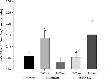
The group infected with the SGO-Z12 isolate presented cardiac cAMP levels that augmented with the evolution of the cardiac chronic form of the infection, but remained similar to those detected in the uninfected group; these levels however, were significantly lower than those previously found in the acute (P<0·05) (Lo Presti et al. Reference Lo Presti, Bustamante, Rivarola, Fernández, Enders, Fretes, Levin and Paglini-Oliva2006) and the indeterminate form (P<0·01) (Lo Presti et al. Reference Lo Presti, Rivarola, Bustamante, Fernández, Enders, Levin, Juaneda, Fretes, Triquell and Paglini Oliva2008) of the experimental infection.
The cAMP levels from the group infected with the Tulahuen strain were also similar to those detected in the uninfected group, but they were significantly higher (P<0·05) in the early than in the late stage of the chronic phase of the experimental infection. These levels were also significantly lower than those previously found in the indeterminate form (P<0·01) (Lo Presti et al. Reference Lo Presti, Rivarola, Bustamante, Fernández, Enders, Levin, Juaneda, Fretes, Triquell and Paglini Oliva2008) of the experimental infection.
The second messenger levels in an early and a late stage of the chronic phase of the experimental infection can be observed in Fig. 4.
Cardiac cAMP levels from the uninfected (n=12), and Trypanosoma cruzi Tulahuen strain-infected and SGO-Z12 isolate-infected groups in the early (Tulahuen n=5; SGO-Z12 n=6) and late (Tulahuen n=6; SGO-Z12 n=6) stages of the cardiac chronic phase of the experimental infection (bars show standard error). Different letters (a vs b) show significant difference between the studied groups (Fisher test, P<0·05).

In order to study the participation of the PDEs, we added caffeine (a PDEs inhibitor – 100 μm) prior to the cAMP quantification. As expected, and since the PDE were inhibited, the addition of caffeine to the uninfected hearts produced an increase in the cAMP concentration (0·12±0·03 nmoles/L mg protein) (P<0·05) when compared with the same group in the absence of the inhibitor (0·059±0·008). However, the addition of caffeine to either of the infected hearts had no effect upon the second messenger levels (Tul 135 days p.i. (n=2): 0·22±0; Tul 365 days p.i. (n=2): 0·018±0·015; Z12 135 days p.i. (n=2): 0·079±0; Z12 365 days p.i. (n=2): 0·061±0), which lead us to think that the PDEs are diminished or their fuction altered. Nevertheless, these results are not conclusive due to the low numbers of animals used as a result of the low numbers of animals reaching the cardiac chronic stage of the infection.
Cardiac contractility studies
In the early chronic stage of the infection, the hearts from the mice infected with the Tulahuen strain showed a cardiac contractility similar to that found in the uinfected controls (Tul: 0·48±0·04 mN; NI: 0·64±0·1 mN). This group presented an augmented contractility in the late stage of the chronic phase (Tul: 1·05±0·21 mN; P<0·05) similar to that found in the acute phase of the experimental infection (Lo Presti et al. Reference Lo Presti, Bustamante, Rivarola, Fernández, Enders, Fretes, Levin and Paglini-Oliva2006).
The SGO-Z12-infected hearts showed a contractile force similar to the uninfected controls in both stages analysed (Z12 early stage: 0·9±0·14 mN; Z12 late stage: 0·8±0·15 mN) but higher than in previous stages (Lo Presti et al. Reference Lo Presti, Bustamante, Rivarola, Fernández, Enders, Fretes, Levin and Paglini-Oliva2006, Reference Lo Presti, Rivarola, Bustamante, Fernández, Enders, Levin, Juaneda, Fretes, Triquell and Paglini Oliva2008).
Effect of β-AR autoantibodies upon cardiac contracttility
To determine if β-AR autoantibodies present in the IgG fraction from chronically infected animals (with either parasite strain) exert biological actions upon cardiac contractility through β-AR activation, we tested changes in the cardiac contractile behaviour of uninfected mice hearts exposed to such antibodies.
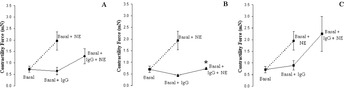
As shown in Fig. 5, the addition of the IgG fraction from either group (NI, Tul or Z12) did not produce any significant change in the basal cardiac contractility (basal+IgG). However, it resulted in a decrease in the contractile response when NE was added to the medium, particularly with the IgG fraction from the SGO-Z12 group (P<0·05), which would confirm the interaction of the antibodies with the β-AR leading to a blockage in the adrenergic response. Even though an apparent blockage can be observed with the antibodies generated by the Tulahuen strain, we could not demonstrate a significant effect like that induced by the SGO-Z12 antibodies.
Comparative effect of the addition of the different IgG fractions to uninfected hearts upon cardiac contractility. (A) IgG from Tulahuen-infected mice in the cardiac chronic phase of the infection. (B) IgG from SGO-Z12-infected mice in the cardiac chronic phase of the infection. (C) IgG from uninfected mice. (*) Shows significant difference between the response to NE in the presence and the absence of the IgG (Fisher test, P<0·05).

DISCUSSION
Chronic Chagas heart disease is a slowly developing inflammatory cardiomyopathy that affects approximately 30% of the infected individuals and is the most lethal endemic infectious disease in the Western hemisphere (Rassi et al. Reference Rassi, Rassi and Little2000; Cubillos-Garzón et al. Reference Cubillos-Garzón, Casas, Morillo and Bautista2004; Coura, Reference Coura2007). The main life-threatening manifestations of this chronic disease are heart failure, arrhythmias and thromboembolism, among other alterations in cardiac conduction that become more frequent with the evolution of the infection (Teixeira et al. Reference Teixeira, Nascimento and Sturm2006). In our murine models infected with 2 different parasite strains, the most frequent electrocardiographic alterations in the cardiac chronic phase were the atrioventricular and the intra-ventricular blockades, as well as a higher cardiac frequency. These blockades are the typical electrocardiographic alterations of this stage of the infection, and the higher cardiac frequency is in accordance with the results found by Rocha and collaborators (Reference Rocha, García, Jiménez, Hernández, Senra, Lina, Cyrino, Bouskela, Soares, Ribeiro dos Santos and Campos de Carvalho2006) in another murine model, corresponding to a difference between the model and the human disease, where bradychardia is the most common finding. This higher frequency could result from local mechanisms related to the higher degree of inflammation observed in the hearts from the infected animals (Rocha et al. Reference Rocha, García, Jiménez, Hernández, Senra, Lina, Cyrino, Bouskela, Soares, Ribeiro dos Santos and Campos de Carvalho2006), which presented inflammatory infiltrates and amastigote nests like our infected mice. The changes observed in the cardiac frequency in both infected groups, however, would not be related to plasma catecholamine levels since they were lower than in the uninfected group.
It has been demonstrated that in the human body, T. cruzi can parasitize any tissue; however, the intensity of infection varies from case to case, depending on the hosts and parasites genetic backgrounds (Nunes et al. Reference Nunes, Sobral and Coutinho1992; Teixeira et al. Reference Teixeira, Nascimento and Sturm2006; Manoel-Caetano and Silva, Reference Manoel-Caetano and Silva2007). Some parasite isolates concentrate infection in the mononuclear phagocytic system, while others distribute themselves randomly in non-phagocytic tissues where they evade the immune system. Since smooth and striated muscles are usually heavily invaded (Teixeira et al. Reference Teixeira, Nascimento and Sturm2006), we studied cardiac and skeletal muscle samples in order to see the alterations produced by the infection and whether the parasites were located in these organs. In both infected groups, the main microscopic findings in the heart were the inflammatory infiltrates characteristic of this stage, that are usually present in every case (Teixeira et al. Reference Teixeira, Nascimento and Sturm2006). Fibre dissolution, fibrosis and necrosis were also present in both infected groups along the chronic stage of the experimental infection. The alterations found in the skeletal muscle were similar to those found in the heart, but more intense and with the presence of parasite nests. The small variations found between both infected groups can be attributed to the genetic variation and heterogeneity between the parasite strains and their differential tissue distribution.
Questions related to the mechanisms by which tissue lesions are formed in the course of the infection have long been, and still are, a matter of intense debate. The two primary hypotheses raised to explain how cardiac pathology develops are: that the persistence of the parasite at specific sites in the host results in chronic inflammatory reactivity (Tarleton and Zhang, Reference Tarleton and Zhang1999), and that T. cruzi infection induces an immune response also targetted to self tissues (Kierszenbaum and Hudson, Reference Kierszenbaum and Hudson1985; Kalil and Cuhna-Neto, Reference Kalil and Cunha-Neto1996). In both cases, the immune-based pathology results in the cumulative, focal destruction of tissues, and the signs and symptoms of the clinical disease. The finding of the parasite in our skeletal muscle samples is particularly important in this scenario, since it demonstrates the presence of the parasite in the host in the chronic phase of the infection, even when no circulating parasites were detected, which is in accordance with the hypothesis of the persistence of the parasite throughout the infection (Sterin-Borda and Borda, Reference Sterin-Borda and Borda1994; Zhang and Tarleton, Reference Zhang and Tarleton1999), and would encourage treatment at this stage.
Autoimmunity may occur either by molecular mimicry, in which an immune response to parasite proteins cross-reacts with host tissues, eventually leading to the development of pathology (Wood et al. Reference Wood, Hudson, Jessell and Yamamoto1982), or simply from bystander activation resulting from significant release of self antigens by parasite-mediated myocytolysis (Cossio et al. Reference Cossio, Bustouoabad, Paterno, Lotti, Casanova, Podesta, Bolomo, Arana and de Pasqualini1984). Both mechanisms could be involved in our models and would generate the autoantibodies detected by the contractility studies, since both infected groups presented parasites in the skeletal muscle samples, and showed cardiac fibre dissolution and necrosis from the early stage of the chronic phase, as shown by the histopathological studies, lesions that would release surface and cytosol molecules which would therefore become accessible and trigger the immune response leading to the production of such autoantibodies.
Evidence accumulated over the years has confirmed the existence of these circulating antibodies in T. cruzi infected patients and murine models, which bind to cardiac β-adrenergic and muscarinic cholinergic receptors, due to molecular mimicry between the carboxy-terminal region of T. cruzi ribosomal P proteins and the second extracellular loop of cardiac β1 and muscarinic receptors (Sterin-Borda and Borda, Reference Sterin-Borda and Borda1999; Wallukat et al. Reference Wallukat, Nissen, Morvinski and Müller2000; Sterin-Borda et al. Reference Sterin-Borda, Giordanengo, Joensen and Gea2003; Lopez Bergami et al. Reference Lopez Bergami, Gómez, Levy, Grippo, Baldi and Levin2005; Levin and Hoebeke, Reference Levin and Hoebeke2008). The interaction of these antibodies with cardiac receptors, would trigger intracellular signalling mechanisms that alter the physiological behaviour of the myocytes (Sterin-Borda and Borda, Reference Sterin-Borda and Borda1994). Autoantibodies that interact with the β-AR and block their activity are present in our models since the addition of the IgG fractions from infected animals (particularly with the SGO-Z12 isolate) inhibited the interaction of the receptors with the agonists (NE). The autoantibodies generated by the Tulahuen strain, however, are different from those generated by the SGO-Z12 isolate, since the latter ones induced a stronger blockage of the agonist-receptor interaction. This stronger interaction is also supported by the fact that the response to exogenous EPI or NE of SGO-Z12-infected ventricles from the late stage of the chronic infection, was similar to that registered in the presence of propranolol (β1 blocker), which reinforces the idea that these antibodies are blocking the receptors. In our models however, these autoantibodies do not seem to act as agonists since their addition did not modify the basal contractility; therefore their action would be different from that described by other authors (Sterin-Borda and Borda, Reference Sterin-Borda and Borda1994; Jahns et al. Reference Jahns, Boivin and Lohse2006), or they are probably being generated together with anti-muscarinic receptors that mask the response.
T. cruzi-infected mice showed important changes in some components of the cardiac β-adrenergic system in the acute (Lo Presti et al. Reference Lo Presti, Bustamante, Rivarola, Fernández, Enders, Fretes, Levin and Paglini-Oliva2006) and chronic indeterminate (Lo Presti et al. Reference Lo Presti, Rivarola, Bustamante, Fernández, Enders, Levin, Juaneda, Fretes, Triquell and Paglini Oliva2008) stages of the infection. Here, we described a cardiac functional disorder in the chronic phase of the experimental infection, which was similar in both infected groups, if one takes into account the survival and the electrocardiographic and histopatological alterations; but different if one considers the changes in the β-adrenergic system. Again, the differences found between the groups infected with either parasite strain can be ascribed to their genetic heterogeneity and the intraspecific variation detected in the T. cruzi population (Manoel-Caetano and Silva, Reference Manoel-Caetano and Silva2007).
The Tulahuen-infected mice presented lower EPI and NE plasma levels in both the early and late stages of the chronic infection; lower β-AR affinity and density (with a partial recovery of the latter in the late chronic stage); diminished response to exogenous NE; similar cAMP levels than the uninfected group but higher in the early than in the late chronic stage; impaired PDEs activity; and a basal cardiac contractility similar to non-infected controls in the early stage and augmented in the late chronic stage. This parasite strain induced the production of β-AR autoantibodies with weak interaction with the β-receptors. The SGO-Z12 mice, also presented lower NE plasma levels and EPI levels that diminished with the evolution of the chronic stage; lower β-AR affinity and an increased density; unchanged response to exogenous EPI and NE in the early stage and a diminished response in the late stage; higher cAMP levels in the late stage; impaired PDEs activity; and unchanged basal cardiac contractility in both stages of the chronic phase. This parasite strain induced the production of β-AR auto-antibodies with strong interaction with the β-receptors.
The lower catecholamine levels found in both infected groups could be a consequence of the presence of the parasite in the adrenal glands, which would impair their function (Lenzi et al. Reference Lenzi, Oliveira, Lima and Gattass1996). This diminished adrenergic input, together with the authonomic denervation postulated by several authors to destroy up to 80% of the cardiac nervous structures (Iosa et al. Reference Iosa, DeQuattro, Lee, Elkayam and Palmero1989; Correa-Araujo et al. Reference Correa-Araujo, Oliveira and Ricciardi Cruzm1991; Laucella et al. Reference Laucella, Rottemberg and De Titti1996; Lohse et al. Reference Lohse, Engelhart, Danner and Bohm1996; Machado et al. Reference Machado, Camargos, Guerra and Moreira2000), is considered to be the main cause of heart failure in this infection. Additional to this diminished agonist levels, both infected groups presented a decrease in the affinity of the cardiac β-AR. Despite this, the receptor density in the Tulahuen group was also diminished, which would worsen the alterations of the system and allow us to propose a more severe cardiac functional damage for this group, which could explain the higher mortality presented by the Tulahuen mice at the beginning of the infection (Lo Presti et al. Reference Lo Presti, Bustamante, Rivarola, Fernández, Enders, Fretes, Levin and Paglini-Oliva2006, Reference Lo Presti, Rivarola, Bustamante, Fernández, Enders, Levin, Juaneda, Fretes, Triquell and Paglini Oliva2008). Similar modifications, as well as the presence of autoantibodies, have been described for other dilated cardiomyopathies (Bristow, Reference Bristow1993; Levin and Hoebeke, Reference Levin and Hoebeke2008) in which a reduction in receptor number has been directly related to the severity of the disease (Brodde, Reference Brodde1991; Harding et al. Reference Harding, Brown, Wynne, Davies and Poole-Wilson1994). This receptor down-regulation could be partially induced by the presence of the autoantibodies, which were probably generated by molecular mimicry between the parasite antigens and the β1-AR (Sterin-Borda and Borda, Reference Sterin-Borda and Borda1999; Sterin-Borda et al. Reference Sterin-Borda, Giordanengo, Joensen and Gea2003; Levin and Hoebeke, Reference Levin and Hoebeke2008).
The SGO-Z12 group, on the other hand, presented a higher β-AR density in both stages; this implies that the homeostatic system in this group is at least partially maintained. However, this higher receptor density (1·78-fold) was not enough to totally compensate for the loss in their affinity (2·08-fold), since the receptor function was seriously compromised in the late stage of the chronic infection (presenting a diminished response to either EPI or NE).
Both infected groups presented higher cAMP levels (the Tul group in the early and the Z12 group in the late stages of the chronic phase). The lower levels of the late chronic stage of the Tulahuen group could be explained by the diminished primary messenger levels and the lower β-AR density and affinity observed in this group; the higher levels of the early stage, however, suggest the activation of additional signalling cascades leading to the production of the second messenger, as has been proposed by several authors (Barki-Harrington et al. Reference Barki-Harrington, Perrino and Rockman2004; Aoki et al. Reference Aoki, Cano, Pallegrini, Tanos, Guiñazú, Coso and Gea2006). Additionally, the higher cAMP levels presented by the SGO-Z12 group could be due to the spontaneous or constitutive activity of the overexpressed β-receptors (higher density), which could activate the signal cascade even in the absence of the agonist (presumably due to this spontaneous activity) (Lefkowitz et al. Reference Lefkowitz, Cotecchia, Samama and Costa1993; Left, Reference Leff1995; Milligan et al. Reference Milligan, Bond and Lee1995).
The higher cAMP levels (probably produced by a different signalling cascade activated as a result of the infection) could explain the receptor desensitization found (lower affinity) in both infected groups, since this second messenger would trigger a mechanism of heterologous desensitization mediated by the activation of PKA (Chakraborti et al. Reference Chakraborti, Chakraborti and Shaw2000).
Despite these high cAMP levels, both infected groups presented a cardiac basal contractility similar to that found in the uninfected group and a diminished response to exogenous NE. This diminished response was significant only for the SGO-Z12 group but not for the Tulahuen group, a result probably related to the high mortality of the later group in earlier stages. This decreased response is related to the binding of the antibodies, found in the sera of the infected groups, to the β-AR which inhibited, totally or partially, the interaction with the exogenous agonists and therefore altered their function, generating a response similar to that found in the presence of a β blocker such as propranolol. The different antibodies generated due to the distinctive genetic backgrounds of the strains would explain the differences in the response to exogenous EPI and NE between both infected groups. The unchanged contractility could be due to the fact that only the mice with less changes in the system survived to the late chronic stage (365 days p.i.) of the infection (Tul: 32%; Z12: 36%), therefore showing a conserved contractile force.
Only the Tulahuen mice presented a partial increment in cardiac contractility in the late stage of the chronic infection (when they have the lower cAMP levels), since the response to exogenous NE was also diminished. Both infected groups would, therefore, present additional alterations in the signalling system, between the second messenger and the contractile apparatus, probably in the phosphorylation of L-type calcium channels or the phosphorylation by PKA, both events necessary for cardiac contractility (Elizari, Reference Elizari1999). T. cruzi has been demonstrated to disrupt the myofibrillar organization and the intracellular calcium levels in mouse neonatal cardiomyocytes (Taniwaki et al. Reference Taniwaki, Machado, Massensini and Mortara2006), a process that is also involved in adult cardiomyocytes invaded by parasites (Rodriguez et al. Reference Rodríguez, Rioult, Ora and Andrews1995) and impairs the cardiac function (particularly the response of the system to EPI and NE). This is reflected in the fibre disorganization and fragmentation as well as in the structural alterations and necrosis found in both infected groups.
The present results demonstrate that the cardiac β-adrenergic system is seriously compromised in the chronic stage of the T. cruzi infection. The biochemical and molecular heterogeneity between the parasite strains induce different alterations in the signalling pathways of this system which would determine the severity of the cardiopathy. Some of these alterations intensify with the evolution of the chronic phase of the experimental infection (from early to late stages) and, since this system is the most powerful regulator of cardiac function, could be responsible for the progressive cardiac damage and the poor prognosis of the disease once the cardiac alterations have started. The present results enhance the existing knowledge of the mechanisms involved in the physiopathology of the chagasic myocardiopathy and could be useful for the prevention of Chagas disease. These alterations in the cardiac response to adrenergic stimulation reinforce the idea of a dysfunctional sympathetic activity during infection. The intensity of these cardiac autonomic disturbances may be also correlated with the clinical form of the disease.
This work was supported by grants from the Secretaría de Ciencia Técnica (SECyT) from Universidad Nacional de Córdoba and from Universidad Nacional de La Rioja and ANPCYT PICT N° 25428.