Introduction
The primary objectives of tympanoplasty are to repair tympanic membrane perforations, prevent recurrent infections and restore hearing function.Reference Gökçe Kütük and Özdaş1 Various surgical techniques and graft materials, including temporalis fascia, dura mater, perichondrium, fat and cartilage, have been used for this purpose.Reference Jalali, Motasaddi, Kouhi, Dabiri and Soleimani2, Reference Kaya, Benzer, Uslu, Bilgen and Kirazli3 Several modified approaches, such as three-point fixation and the over-underlay technique, have been proposed to enhance graft success.Reference Shim, Kim, Kim and Moon4, Reference Kartush, Michaelides, Becvarovski and LaRouere5
In 1998, Eavey introduced the butterfly cartilage inlay tympanoplasty, which employs a butterfly-shaped tragal cartilage–perichondrium graft via a transcanal approach.Reference Eavey6 While tympanoplasty was traditionally performed microscopically, the introduction of the endoscope in the 1990s enabled a wider field of view and minimally invasive access, leading to its increasing popularity.Reference Thomassin, Duchon-Doris, Emram, Rud, Conciatori and Vilcoq7
Although both endoscopic and microscopic techniques are widely used, comparative studies assessing long-term outcomes according to perforation size and location remain limited. This study aims to compare hearing outcomes and graft success between these two methods to guide surgical technique selection in different clinical contexts.
Methods
Participants and study design
A retrospective analysis was performed on patients who underwent tympanoplasty at the ENT Clinic between January 2022 and December 2024. Patient data were collected from hospital records, surgical logbooks and medical files. Cases involving additional otologic procedures (mastoidectomy, ossiculoplasty), cholesteatoma, revision surgery or inadequate follow-up were excluded. Eligible patients were retrospectively classified into two groups based on the surgical technique employed: Group 1, endoscopic butterfly inlay cartilage myringoplasty; and Group 2, microscopic over-underlay cartilage tympanoplasty. The study was approved by the University Non-Interventional Research Ethics Committee (Date: 27.11.2024, Decision No: 2024.11.18). Written informed consent was obtained from all participants.
Demographic data, operated ear, perforation characteristics and size, surgery duration and pre- and post-operative audiometry results were recorded. Perforations were classified as less than 50 per cent or greater than 50 per cent of the tympanic membrane area and as anterior or posterior in location. The perforation type was categorised as central, marginal or attic. Only patients with at least six months of post-operative follow-up were included. Graft success was defined as an intact tympanic membrane at six months.
Audiological evaluation
Pure-tone audiometry was performed pre- and post-operatively using an Otometrics Madsen Astera 2 audiometer in a soundproof room. Air conduction thresholds were measured at 250–8000 Hz and bone conduction at 500–4000 Hz. Four-frequency pure-tone averages (500, 1000, 2000 and 4000 Hz) were calculated. Hearing outcomes were assessed using mean air–bone gap (ABG) and ABG closure (difference between pre- and post-operative ABG values).
Surgical technique
All surgeries were performed under general anesthesia by a single surgeon using a tragal chondroperichondrial graft. In the endoscopic group, a transcanal approach was used with a 0° endoscope (4 mm, 18 cm). The graft was shaped in a butterfly configuration, placed between the medial and lateral surfaces of the tympanic membrane and supported with Gelfoam (Figure 1a,b,c).
In the microscopic group, a postauricular approach with a Wilde incision was used under an operating microscope. After elevating the tympanomeatal flap and preparing the graft, it was positioned using the over-underlay technique. Both incisions were closed with absorbable sutures, and standard dressings were applied.
Images showing the steps of endoscopic butterfly-inlay cartilage myringoplasty: (a) intra-operative view of an anterior tympanic membrane perforation, (b) view of the chondroperichondrium composite graft prepared for butterfly-inlay grafting and (c) intra-operative view of the perforation after endoscopic butterfly-inlay grafting.

Post-operative care
All patients were discharged the following day and prescribed oral antibiotics for one week. Tamponades were removed after one week, and follow-ups were scheduled at two weeks, 1, 3, 6, and 12 months post-operatively.
Statistical analysis
Data were analysed using IBM SPSS Statistics version 21.0 (SPSS Inc., Chicago, IL, USA). The normality of variables was assessed with the Kolmogorov–Smirnov test and visual inspection of histograms. Categorical variables (gender, perforation side, area, size and graft success) were compared using the chi-squared test or Fisher’s exact test, as appropriate. Continuous variables with normal distribution were analysed using the independent samples t-test, while the Mann–Whitney U test was applied for nonparametric comparisons. Results are presented as counts and percentages for categorical variables and as mean ± standard deviation (SD) for continuous variables. A two-tailed p value less than 0.05 was considered statistically significant.
Results
A total of 64 patients (34 females, 30 males; age range: 12–60 years) were included. Thirty-three patients underwent endoscopic butterfly inlay cartilage myringoplasty, and 31 underwent microscopic over-underlay cartilage tympanoplasty. The mean age was 41.09 ± 13.68 years in the endoscopic group and 39.74 ± 11.18 years in the microscopic group, with no significant difference between them (p > 0.05).
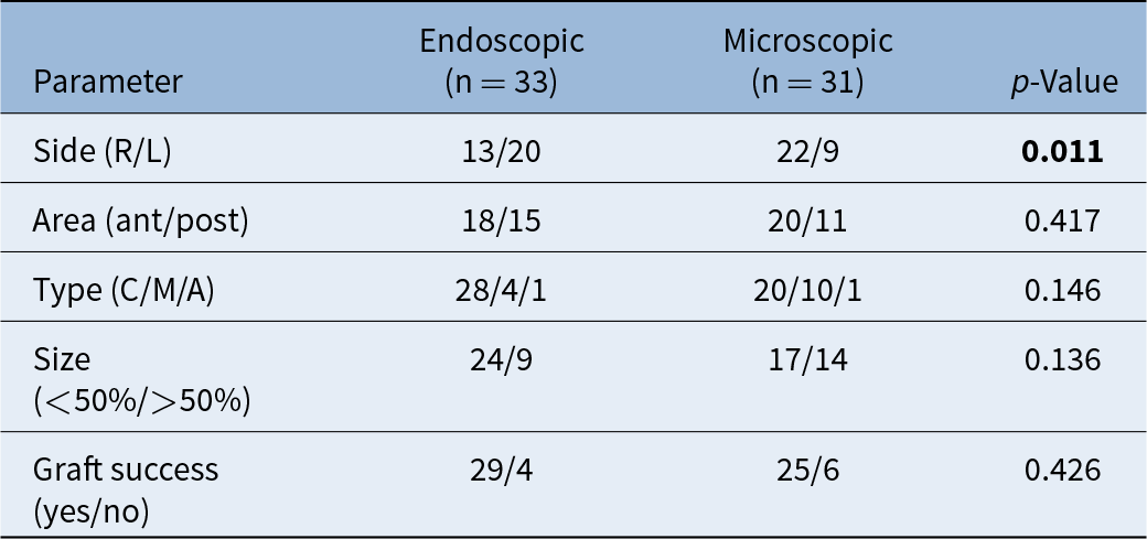
The mean follow-up period was 20.15 ± 6.86 months for the endoscopic group and 23.71 ± 10.45 months for the microscopic group (p > 0.05). Operative time was significantly shorter in the endoscopic group (65.15 ± 19.22 minutes) compared with the microscopic group (106.13 ± 39.80 minutes; p < 0.001) (Table 1).
Patient demographics and clinical characteristics

Note: Bold value indicates statistical significance.
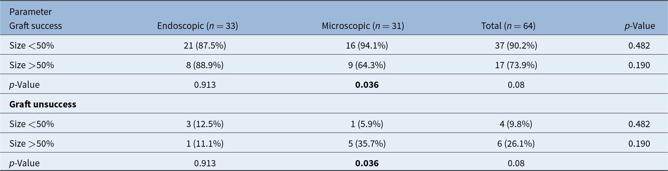
A statistically significant difference was observed between the groups regarding the operated ear side (right/left) (p = 0.011). No significant differences were observed between the groups regarding perforation location or perforation type (p > 0.05). Perforation size distribution (<50 per cent vs >50 per cent) was comparable between the endoscopic and microscopic groups (p = 0.136). Graft success was 87.9 per cent in the endoscopic group and 80.6 per cent in the microscopic group, with no statistically significant difference between the groups (p = 0.426) (Table 2). In the endoscopic group, 9 of 33 patients (27.3 per cent) had large perforations (>50 per cent), whereas 24 patients (72.7 per cent) had smaller perforations (<50 per cent). In the microscopic group, large perforations were observed in 14 of 31 patients (45.2 per cent), while 17 patients (54.8 per cent) had perforations involving less than 50 per cent of the tympanic membrane. The distribution of perforation size was comparable between the two groups (p = 0.136). Overall analysis showed higher graft success rates in smaller perforations (<50 per cent) compared with larger perforations (>50 per cent) (90.2 per cent vs 73.9 per cent); however, this difference did not reach statistical significance (p = 0.08). Subgroup analysis revealed that graft success significantly decreased with increasing perforation size in the microscopic group (p = 0.036), whereas no such association was observed in the endoscopic group (p = 0.913) (Table 3).
Definitions of tympanic membrane perforation types and graft success

R/L = right/left ear, ant/post = anterior/posterior, C/M/A = central/marginal/attic.
Note: Bold value indicates statistical significance.
Graft success according to perforation size and surgical technique

Note: Bold values indicate statistical significance.
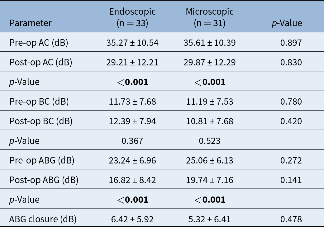
Both groups showed significant post-operative hearing improvement in air conduction and ABG values (p < 0.001), with comparable ABG closure (6.42 ± 5.92 dB vs 5.32 ± 6.41 dB; p > 0.05) (Table 4).
Comparison of audiological outcomes between and within endoscopic and microscopic techniques

ABG = air–bone gap; ABG closure = difference between pre-operative and post-operative ABG; AC = air conduction; BC = bone conduction.
Note: All scores are given as mean ± SD. Bold values indicate statistical significance.
Discussion
Although microscopic tympanoplasty remains widely used due to its bimanual capability and three-dimensional view, it may provide limited access to hidden areas of the middle ear, especially in narrow or curved ear canals.Reference Farahani, Shariatpanahi, Jahanshahi and Poorolajal8, Reference Kuo and Wu9 Endoscopic tympanoplasty offers a panoramic view and improved access without canaloplasty, though it requires one-handed operation and two-dimensional visualisation.Reference Mohindra and Panda10, Reference Lade, Choudhary and Vashishth11
Several studies and meta-analyses have demonstrated that endoscopic tympanoplasty is associated with shorter operative time compared to the microscopic approach.Reference Kawale, Landge, Garg and Kanani12–Reference Lee, Lee, Seo and Kim15 In the present study, the mean operative time was significantly shorter in the endoscopic group (65.15 ± 19.22 min) than in the microscopic group (106.13 ± 39.80 min), consistent with prior reports. This difference is likely due to smaller incisions and less extensive soft tissue dissection in the endoscopic technique.
The graft success rate of endoscopic butterfly inlay cartilage tympanoplasty has been reported in several studies. Akyigit et al. reported a 93.7 per cent success rate in children after one year, while Özgür et al. found early and late success rates of 97.8 per cent and 95.6 per cent, respectively, in adults.Reference Akyigit, Karlidag, Keles, Kaygusuz, Yalcın and Polat16, Reference Özgür, Dursun, Terzi, Erdivanlı, Coşkun and Oğurlu17 Eren et al. reported a rate of 95.5 per cent in a series of 56 patients.Reference Eren, Tugrul, Ozucer, Veyseller, Aksoy and Ozturan18 In this study, the graft success rate was 87.9 per cent, slightly lower than in previous reports, possibly due to the longer mean follow-up period (20.15 ± 6.86 months) compared to 6–12 months in other studies. Success rates for microscopic butterfly cartilage myringoplasty have been reported to range from 43 per cent to 100 per cent.Reference Özgür, Dursun, Terzi, Erdivanlı, Coşkun and Oğurlu17, Reference Lee, Kang, Lee, Kim and Lee19
In the present study, a statistically significant difference was observed between the groups regarding the operated ear side; however, no clinically relevant impact of this variable on graft success has been reported in the literature. No significant differences were found between groups in terms of perforation location (anterior/posterior) or perforation type (p > 0.05). Importantly, perforation size distribution (<50 per cent vs >50 per cent) was comparable between the endoscopic and microscopic groups, indicating that perforation size did not represent a confounding factor in the comparison of surgical outcomes. The overall graft success rate was 84.37 per cent.
According to a systematic review and meta-analysis by Tseng et al., the graft success rates for endoscopic and microscopic tympanoplasty were 85.1 per cent and 86.4 per cent, respectively, with no statistically significant difference between the two techniques.Reference Tseng, Lai, Wu, Yuan and Ding20 In line with the existing literature, our study demonstrated comparable graft success rates between the endoscopic (87.9 per cent) and microscopic (80.6 per cent) groups, with no statistically significant difference between the two techniques. While some studies have suggested that larger perforation size negatively impacts graft success,Reference Lee, Kelly and Mills21, Reference Onal, Uguz, Kazikdas, Gursoy and Gokce22 others have found no such correlation.Reference Wasson, Papadimitriou and Pau23, Reference Abdelhameed, Rezk and Awad24 In our cohort, graft success was higher in perforations involving less than 50 per cent of the tympanic membrane area (90.2 per cent) compared to those involving more than 50 per cent (73.9 per cent), although this difference did not reach statistical significance (p = 0.08). Notably, as perforation size increased, graft success significantly decreased in the microscopic group (p = 0.036), whereas no such trend was observed in the endoscopic group (p = 0.913). Given the comparable distribution of perforation sizes between groups, these findings suggest a potential technique-related difference in resilience to increasing perforation size rather than a selection bias.
Hearing improvement is a key indicator of surgical success in tympanoplasty. In the present study, comparison of pre-operative and post-operative pure-tone audiometry measurements—including air conduction (AC) thresholds and ABG values—showed statistically significant audiological improvement in both groups (p < 0.001). Kawale et al. found no significant difference in hearing restoration between the two techniques, reporting mean ABG closures of 10.56 ± 3.35 dB for the endoscopic group and 10.62 ± 3.66 dB for the microscopic group (p = 0.0001).Reference Kawale, Landge, Garg and Kanani12 Other studies have reported post-operative ABG improvements 9.4 dB by Özgür et al.Reference Özgür, Dursun, Terzi, Erdivanlı, Coşkun and Oğurlu17 Akyigit et al. observed a late post-operative ABG of 7.9 dB, while Eren et al. reported 10.8 dB.Reference Akyigit, Karlidag, Keles, Kaygusuz, Yalcın and Polat16, Reference Eren, Tugrul, Ozucer, Veyseller, Aksoy and Ozturan18 Nassif et al. also demonstrated hearing improvement in both groups, with ABG closure values of 6.2 dB for the microscopic technique and 6.6 dB for the endoscopic approach.Reference Nassif, Berlucchi and Radaelli de Zinis13 The literature consistently shows that post-operative ABG values improve significantly following both endoscopic and microscopic tympanoplasty. However, most studies, including the present one, report no statistically significant difference in auditory gain between the two approaches. In our study, mean ABG closure was 6.42 ± 5.92 dB in the endoscopic group and 5.32 ± 6.41 dB in the microscopic group, with no significant difference observed between them.
No correlation was found between the location of the perforation and the degree of hearing loss. Similarly, there was no correlation between the perforation site and post-operative hearing gain. This finding is consistent with previous reports in the literature.Reference Kaya, Benzer, Uslu, Bilgen and Kirazli3, Reference Karabulut, Mutlu, Sahin and Cirik25
This study had several limitations. First, the microscopic and endoscopic procedures were not performed in a strictly alternating or simultaneous manner, which may have introduced variability in surgical conditions. Second, the retrospective nature of the study inherently carries a risk of selection bias, including surgeon preference in selecting the surgical approach. However, given the comparable distribution of perforation sizes between groups, the potential impact of this bias on graft success comparisons appears limited. One of the strengths of this study is the relatively long follow-up period compared to many previous reports, allowing for a more reliable assessment of long-term graft success and hearing outcomes. Additionally, the study included a substantial number of patients in both the endoscopic and microscopic groups, enhancing the statistical power and validity of the comparisons. The use of objective audiometric data pre- and post-operatively further supports the robustness of our findings.
• Operative time was significantly shorter in the endoscopic group than in the microscopic group
• Graft success rates were comparable between endoscopic and microscopic techniques
• Both approaches provided comparable post-operative hearing improvement
• Graft success decreased with increasing perforation size in the microscopic group but remained unaffected in the endoscopic group
Conclusion
Both endoscopic butterfly inlay and microscopic over-underlay tympanoplasty effectively repair tympanic membrane perforations with comparable hearing outcomes. The endoscopic approach provides graft success rates comparable to the microscopic technique while offering a significantly shorter operative time, supporting its role as an effective minimally invasive alternative. Within the limitations of this retrospective study, the endoscopic technique may be considered a viable option in appropriately selected cases.
Declaration of competing interests
The authors declared no potential conflicts of interest with respect to the research, authorship, and/or publication of this article.
Ethical approval
The study was approved by the Kırıkkale University Non-Interventional Research Ethics Committee (Date: 27.11.2024, Decision No: 2024.11.18). Written informed consent was obtained from all participants.
Informed consent
Written informed consent was obtained from all individual participants included in the study.
Funding
The authors received no financial support for the research, authorship, and/or publication of this article.
Data availability statement
The data used to support the findings of this study are available from the corresponding author upon reasonable request.