Introduction
The presence of human mesenchymal stem cells (MSCs) in multiple tissue types as well as their multi-lineage differentiation and regenerative capacity has led to an increase in their clinical application with over 724 trials registered on ClinicalTrials.gov as of May 2017. The therapeutic benefit of MSC-administration in different pathologies (reviewed in [Reference Sharma, Pollock, Hubel and McKenna1]) can be at least partly explained by paracrine effects on endogenous regeneration that are mainly mediated by extracellular vesicles (EVs) released by MSCs.[Reference Gnecchi, He, Noiseux, Liang, Zhang, Morello, Mu, Melo, Pratt, Ingwall and Dzau2–Reference Kordelas, Rebmann, Ludwig, Radtke, Ruesing, Doeppner, Epple, Horn, Beelen and Giebel4] Thus, EVs are considered to represent a cost-efficient alternative to transplantation of MSCs with a potentially equally beneficial therapeutic outcome.
Notably, both transplantation of MSCs and manufacturing of MSC-derived EVs require large cell numbers, long cultivation time, and consequently large volumes of cell culture medium. Thus, culture expansion in vitro is a necessary step in order to obtain sufficient quantities of cells for the intended therapeutic application. However, it is well known that during initial and extended in vitro culture expansion, MSCs accumulate chromosomal aberrations, which may be caused by the extraction of the cells from their three-dimensional (3D) endogenous niche and two-dimensional (2D) in vitro culture conditions.[Reference Ben-David, Mayshar and Benvenisty5, Reference Bara, Richards, Alini and Stoddart6] Moreover, a large body of evidence suggests that prolonged 2D cultivation leads to a loss of multipotency and induces cellular senescence in MSCs.[Reference Turinetto, Vitale and Giachino7] Therefore, conventional 2D culture represents a bottleneck in broader use of MSC-based therapeutics and the upscaling of MSC-culture is a current technological and economic challenge.
To overcome these limitations, different 3D cultivation methods, more closely resembling the endogenous niche, have been applied to MSCs. Among others, MSCs can be cultivated in 3D within alginate hydrogels,[Reference Ho, Murphy, Binder, Vissers and Leach8] collagen-based matrices,[Reference Lund, Stegemann and Plopper9] fibrin-poly(ester-urethane) scaffolds,[Reference Gardner, Musumeci, Neumann, Eglin, Archer, Alini and Stoddart10] bacteria-derived cellulose,[Reference Favi, Benson, Neilsen, Hammonds, Bates, Stephens and Dhar11] methylcellulose,[Reference Cochis, Grad, Stoddart, Fare, Altomare, Azzimonti, Alini and Rimondini12] and the chondrosarcoma-derived Matrigel™.[Reference Yamaguchi, Ohno, Sato, Kido and Fukushima13] However, despite the obvious advantages over the 2D cultivation systems, most MSC 3D cultivation methods also have drawbacks. Specifically, alginate hydrogels require cross-linking, while retrieval of the cells from fibrin and collagen-based matrices can only be achieved by using enzymes also affecting mammalian cells. Matrigel™ is known to be affected batch-to-batch variability, cross-species immunogenicity and consequently difficulties with their use in a clinical setting.[Reference Serban, Liu and Prestwich14] In contrast, similar to other bacteria-derived biologics, bacteria-derived cellulose requires cost-intensive purification to avoid contaminations with endotoxins.
In this study, we examined the biocompatibility of multipotent human adipose tissue-derived and bone marrow-derived mesenchymal stem cells (adMSCs and bmMSCs) with the commercially available nanofibrillar cellulose (NFC) hydrogel GrowDex®, which has been demonstrated to be biocompatible with other cell types, including liver cells, liver progenitor cells, and human-induced pluripotent stem cells.[Reference Lou, Kanninen, Kuisma, Niklander, Noon, Burks, Urtti and Yliperttula15–Reference Malinen, Kanninen, Corlu, Isoniemi, Lou, Yliperttula and Urtti17] We demonstrate that both adMSCs and bmMSCs interact with the NFC, are evenly distributed within all three dimensions of the hydrogel, and are viable in 0.2% and 0.5% NFC hydrogel. Moreover, MSCs were successfully differentiated into osteogenic cells within the 3D hydrogel. Finally, we show that MSCs can be easily retrieved from the 3D hydrogel via an enzymatic digestion of the NFC and that the post-NFC MSCs show similar viability to 2D pre-cultivated MSCs.
Material and methods
NFC and calcofluor white staining
NFC hydrogel (Growdex®) and cellulase solution (GrowDase™) were kindly provided by UPM Biochemicals, Helsinki, Finland). The 0.2% NFC solution was prepared by diluting the 1.5% stock solution in Dulbecco's modified Eagle's medium (DMEM) (Sigma-Aldrich, Irvine, UK) and 100 µL of the hydrogel was transferred into a well of a low-adhesion 96-well plate using low-retention pipette tips (Sarstedt, Leicester, UK) followed by incubation with 25 µL of the Calcofluor white (0.01% aqueous solution of Calcofluor White M2R, 1 g/L, Evans blue, 0.5 g/L, Sigma-Aldrich) for 20 s at room temperature (RT). The staining was visualized using an inverted fluorescence microscope (VertA1, Carl Zeiss, Cambridge, UK) equipped with a DAPI filter.
Scanning electron microscopy (SEM)
Cell within 0.2% hydrogel or 0.5% NFC hydrogel alone were transferred into a 24 mm cell culture insert membrane (Corning, St. David's Park, UK; pore size 0.4 µm), incubated for 24 h at 37 °C and fixed with 2% paraformaldehyde and 2.5% glutaraldehyde for 1 h, washed three times with phosphate-buffered saline (PBS) for 30 min followed by dehydration in ethanol series then transferred on top of a stub. The samples were plunged into liquid nitrogen slash, and then freeze dried for 1.5 h followed by gold coating (25 nm). Analysis was performed using a scanning electron microscope (FEI Quanta FEG 600 SEM, Thermo Fisher, Paisley, UK). Fiber size was extracted from seven SEM images suing the ImageJ Fiji software. The histogram was created with GraphPad Prism software (GraphPad, La Jolla, California).
Human MSCs
Fully characterized human adipose tissue MSCs from non-diabetic adult lipoaspirates were obtained from Lonza (Slough, UK). Palatal adipose tissue MSCs and bone marrow MSCs were obtained from adult donors with written informed consent with an approval from the Ethics Committee of Dental Faculty of Selcuk University (approval number 2012-08). All MSCs have been characterized immunocytochemically and by tri-lineage differentiation assay as recommended by The International Society for Cellular Therapy.[Reference Dominici, Le Blanc, Mueller, Slaper-Cortenbach, Marini, Krause, Deans, Keating, Prockop and Horwitz18]
Cultivation of MSCs as 2D monolayer
MSCs were cultivated in DMEM high glucose, 1% L-glutamine, 1% penicillin/streptomycin (all from Sigma-Aldrich), and 20% heat-inactivated FBS (fetal bovine serum) (Sigma Aldrich, lot: 8204188981) [standard cultivation medium] at 37 °C and 10% CO2. The medium was changed every 2–3 days. For expansion, cells were detached by treatment with 0.05% trypsin/ethylenediaminetetraacetic acid (EDTA) (Sigma-Aldrich) and seeded in 175 cm2 tissue culture flasks (Sarstedt). All cells were used at passages 4–13 (except palatal adMSCs for live dead staining for the osteogenic differentiation assays at passage 18).
Cultivation of MSCs in NFC 3D matrix
After detachment by treatment with 0.05% trypsin/EDTA (Sigma-Aldrich), MSC suspensions were mixed with 1.5% NFC hydrogel to achieve 0.2% or 0.5% (w/v) hydrogels with desired cell density. After 30 min of incubation at 37 °C, standard culture medium was added to the top of the hydrogel.
MSCs were cultured in 0.2% and 0.5% NFC hydrogel at the seeding density of 105 cells/well in standard cultivation medium for up to 7 days at 37 °C and 10% CO2 unless otherwise stated. Medium was changed daily. Retrieval of cells from the NFC was performed by enzymatic digestion of the matrix with cellulase (GrowDase, UPM Biochemicals) for 24 h at 37 °C. Bright-field microscopy was done using an inverted microscope (VertA1, Carl Zeiss).
Liberation of MSCs from the NFC hydrogel
MSCs were seeded at 25 × 103 cells per 100 µL and cultured for 96 h in 0.2% and 0.5% NFC. The cellulase solution (GrowDase®, UPM, Helsinki, Finland) was prepared according to the manufacturer's instructions. Briefly, 300 µg of cellulase per 1 mg of NFC was incubated for 4 h at 37 °C and 10% CO2. Following NFC digestion, the suspension was removed and the well washed with PBS. Following centrifugation at 300 g for 10 min, cell counting was performed using a haemocytometer.
Cell viability assay (XTT)
Cell viability assays in 2D and 3D were performed using the Cell Proliferation Kit II (Sigma-Aldrich) according to the manufacturer's instructions. BM-MSCs and palatal adMSCs were used at passage 9, and adMSCs at passage 13. The fluorescence of the XTT metabolite was measured at an excitation wavelength of 450 nm and a reference wavelength of 650 nm on a Spectra Max 340PC plate reader (Molecular Devices, Wokingham, UK). Measurements were taken after 7 h of incubation.
PKH67 staining and fScan 3D fluorescence imaging
To visualize living cells within the NFC matrix, PKH67 Green Fluorescent Cell Linker Kit for General Cell Membrane Labelling (Sigma-Aldrich) was used. Briefly, 2 × 10⁵ adMSCs at passage 11 were labeled according to the manufacturer's instructions and embedded within 0.5% NFC hydrogel. For detection of living, labeled cells, the fScan 488 (Lein Applied Diagnostics, Reading, UK) 3D scanning confocal microscope was used. The 3D reconstruction was done using ImageJ Fiji software[Reference Schindelin, Arganda-Carreras, Frise, Kaynig, Longair, Pietzsch, Preibisch, Rueden, Saalfeld, Schmid, Tinevez, White, Hartenstein, Eliceiri, Tomancak and Cardona19] and the Volume Viewer plugin.
Live and dead staining and confocal laser scanning microscopy
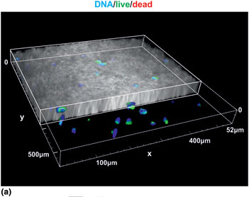
AdMSCs (passage 18) cultured in 0.2% NFC on translucent TC 24 well inserts (Sarstedt), stained with Live/Dead viability/cytotoxicity kit according to the manufacturer's instructions (Thermo Fisher), were fixed in 4% PFA for 20 min and counter-stained with DAPI (Sigma-Aldrich). The TC membrane was removed from the TC insert and mounted onto a slide in Mowiol 4-88 mounting solution (Sigma-Aldrich). Confocal images were gathered using the Nikon A1-R inverted confocal microscope with the Nikon Plan Apo VC 20× DIC N2 optic lens, running NIS Elements AR. NFC was visualized by phase contrast (TD), DAPI at an excitation/emission of 405/450 nm, Calcein 494/517 nm, and ethidium homodimer-1 at 528/617 nm with the Chroma 405/488/561/647 quad mirror. A z-stack depth of 50 µm (z-plane) was created for an area of 554 × 550 µm2 and a 3D reconstruction was generated with all the channels using the NIS Elements AR software (v4.0). The phase contrast channel showing the NFC was cut through the Y-plane.
Osteogenic differentiation of MSCs in 3D NFC hydrogels
For induction of osteogenesis, 2 × 10⁵ palatal-tissue (passage 18) and liposuction-derived adMSCs (passage 14) in normal cultivation medium were embedded in 0.2% NFC and transferred to 12-well cell culture inserts (Sarstedt). The medium was changed after 3 days to Stem Pro Osteocyte/Chondrocyte basal medium (Life Technologies) supplemented with StemPro Osteogenesis supplement (Life Technologies) according to the suppliers instructions. In control MSCs, the medium was changed to fresh standard medium. Cultures were maintained at 37 °C and were stained with Alizarin Red S after 21 days of differentiation. Medium was removed and cells were washed with PBS. Fixation in 4% PFA for 30 min was followed by three washing steps in distilled water. Alizarin Red staining solution (2 g Alizarin Red S in 100 mL distilled water, pH 4.1 with 0.1% NH4OH, filtered) was added and incubated for 45 min at RT in the dark. The staining solution was removed and unbound dye was removed by three washing steps with water.
Statistical analysis
All statistical analyses were performed using GraphPad Prism software (GraphPad, La Jolla, CA, USA). Data were compared using either a Student's t-test (two-tailed, confidence interval 95%), or one-way analysis of variance (ANOVA) with Bonferroni correction (CI 95%), where appropriate. At least three independent measurements were performed. P < 0.05 was considered statistically significant.
Results
NFC forms dense mesh-like structures with large pores
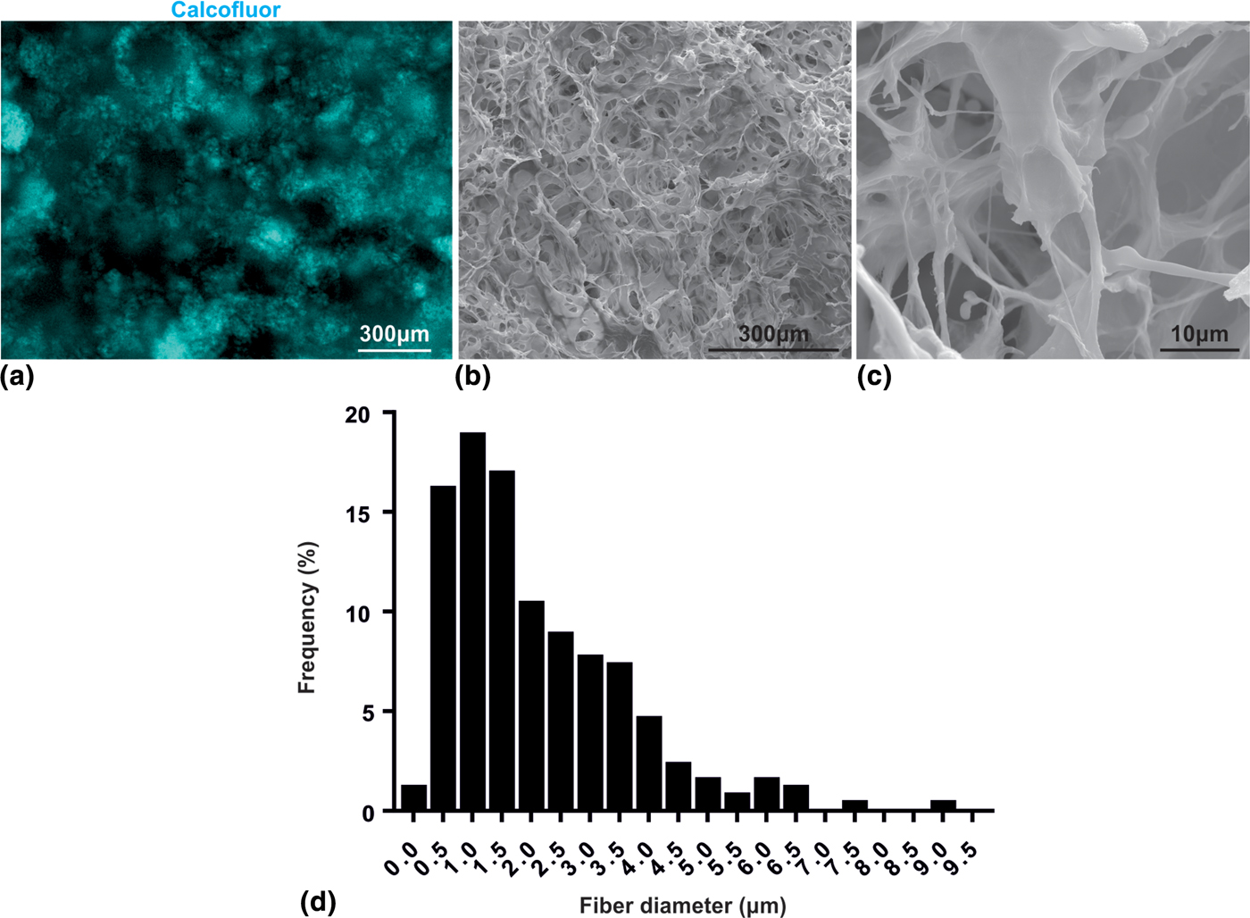
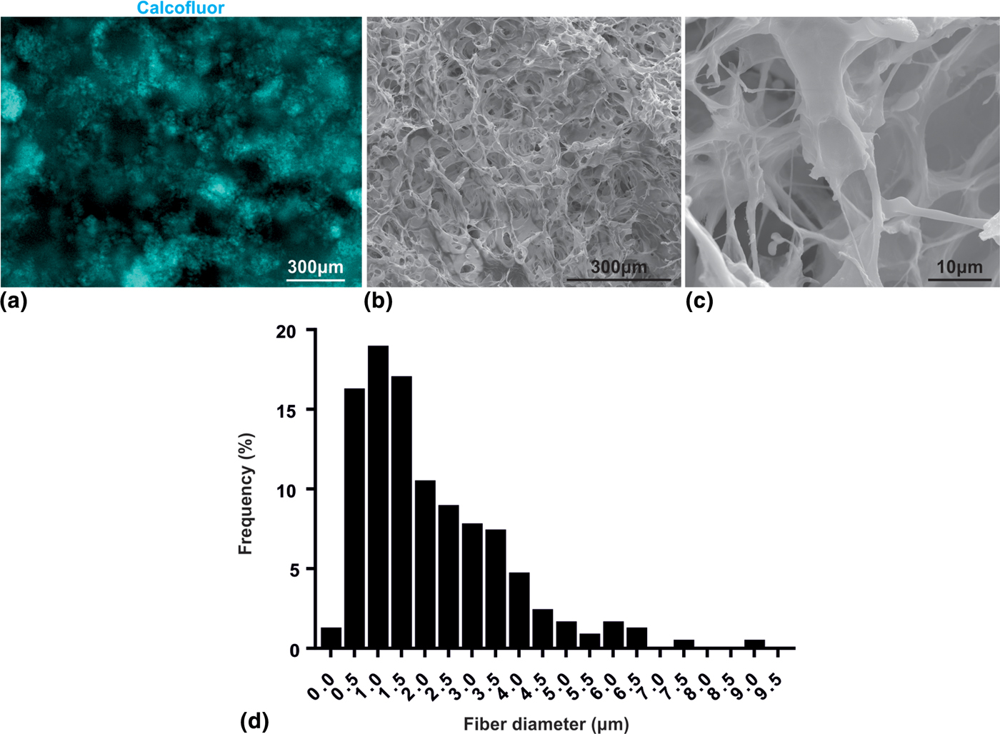
To visualize the cellulose fibers within the 3D NFC hydrogel, an aqueous solution of Calcofluor staining was used. Subsequent fluorescence microscopy analysis clearly showed that cellulose fibers are evenly distributed within the hydrogel [Fig. 1(a)]. To gather sub-structural data on the 3D NFC, the hydrogel was processed for SEM revealing a dense meshwork-like structure [Fig. 1(b)]. Higher magnification image showed a dense network of cellulose fibers of different size [Fig. 1(c)]. Analysis of the fiber diameter revealed an average diameter of 2.07 µm and heterogeneous distribution ranging from 0.08 to 9.1 µm. Notably, the majority of the fibers had a diameter smaller than 3.0 µm [Fig. 1(d)].
3D NFC hydrogel consists of a dense meshwork with heterogeneous pore sizes. (a) Calcofluor white staining and fluorescence microscopy was used to visualize the 3D NFC hydrogel under native conditions. A dense and evenly distributed cellulose fiber network was observed. Bar: 300 µm. (b, c) The ultrastructural appearance of the 3D NFC hydrogel was investigated by SEM revealing a dense meshwork-like surface structure. Higher magnification image (c) revealed presence of fibers of heterogeneous size. Bars: 300 and 10 µm, respectively. (d) NFC hydrogels contain nano- and microfibers with a heterogeneous diameter ranging from 0.08 to 9.1 µm.
3D NFC hydrogels are biocompatible with MSCs
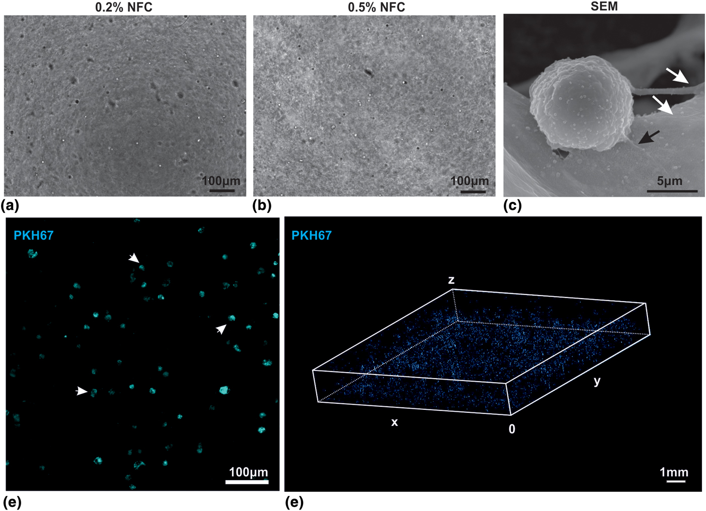
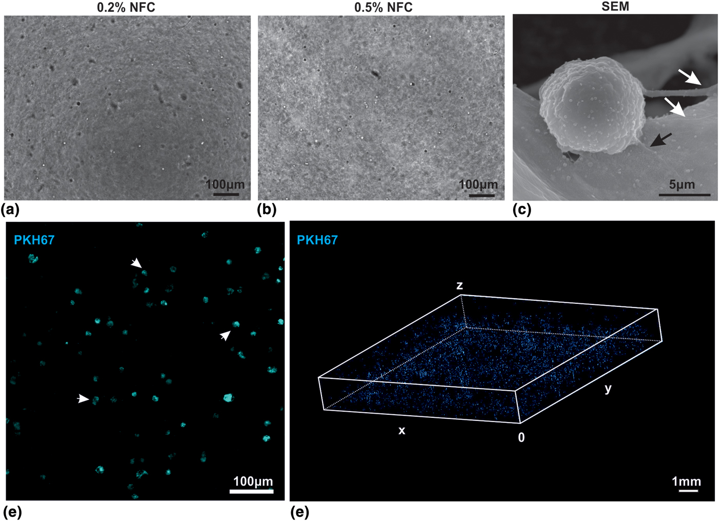
We next explored the feasibility of cultivation of human adMSCs in three dimensional NFC hydrogels at 0.2% and 0.5% NFC hydrogels. Most MSCs in the 0.2% NFC displayed a typical round morphology [Fig. 2(a)]. Cultivation of MSCs in 0.5% NFC resulted in a homogenous cell population with round cells similar to MSCs cultivated in 0.2% NFC. Scanning electron microscopy revealed that MSCs interact and bind to the NFC as evidenced by membrane protrusions at the matrix [Fig. 2(c)]. In order to visualize larger numbers of living MSCs within the NFC, MSCs were labeled with PKH67 [Fig. 2(d)] and imaged by high content laser scanning microscopy [Fig. 2(e)]. Here we demonstrate that living MSCs are isotropically distributed within the matrix over large areas of the well.
3D NFC hydrogels support in vitro growth of MSCs. (a) AdMSCs were cultivated in 0.2% NFC hydrogel in standard MSC cultivation medium revealing a typical fibroblast-like morphology and neurosphere-like clusters (arrow). Bar: 100 µm. (b) Cultivation of adMSCs in 0.5% NFC leads to a homogenous cell population with bipolar morphology (arrowheads). Bar: 50 µm. C. AdMSCs in 0.5% NCF hydrogel were fixed using paraformaldehyde and glutaraldehyde and processed for SEM. Data analysis revealed that MSCs interact with the NFC as evidenced by formation of membrane protrusions in direct contact with the cellulose fibers (arrows) Bar: 10 µm. (d, e) Living MSCs are homogenously distributed within the NFC matrix. (d) AdMSCs were labeled with PKH67 followed by embedding in 0.5% NFC hydrogel in inserts and fluorescence microscopy revealing high labeling efficiency and individual labeled cells within the hydrogel (arrows). Maximum intensity projection was generated in ImageJ Fiji (LUT: cyan hot). (e) High content 3D laser scanning microscopy using the fScan 488 device was applied to visualize the distribution of MSCs in all three dimensions of the NFC hydrogel. Subsequent image analysis in ImageJ Fiji (Volume Viewer Plugin, LUT: cyan hot) revealed isotropic and homogenous cell distribution. Scan area: 10 mm × 10 mm × 1.6 mm (x/y/z), bar: 1 mm.
3D NFC supports high viability and proliferation of MSCs
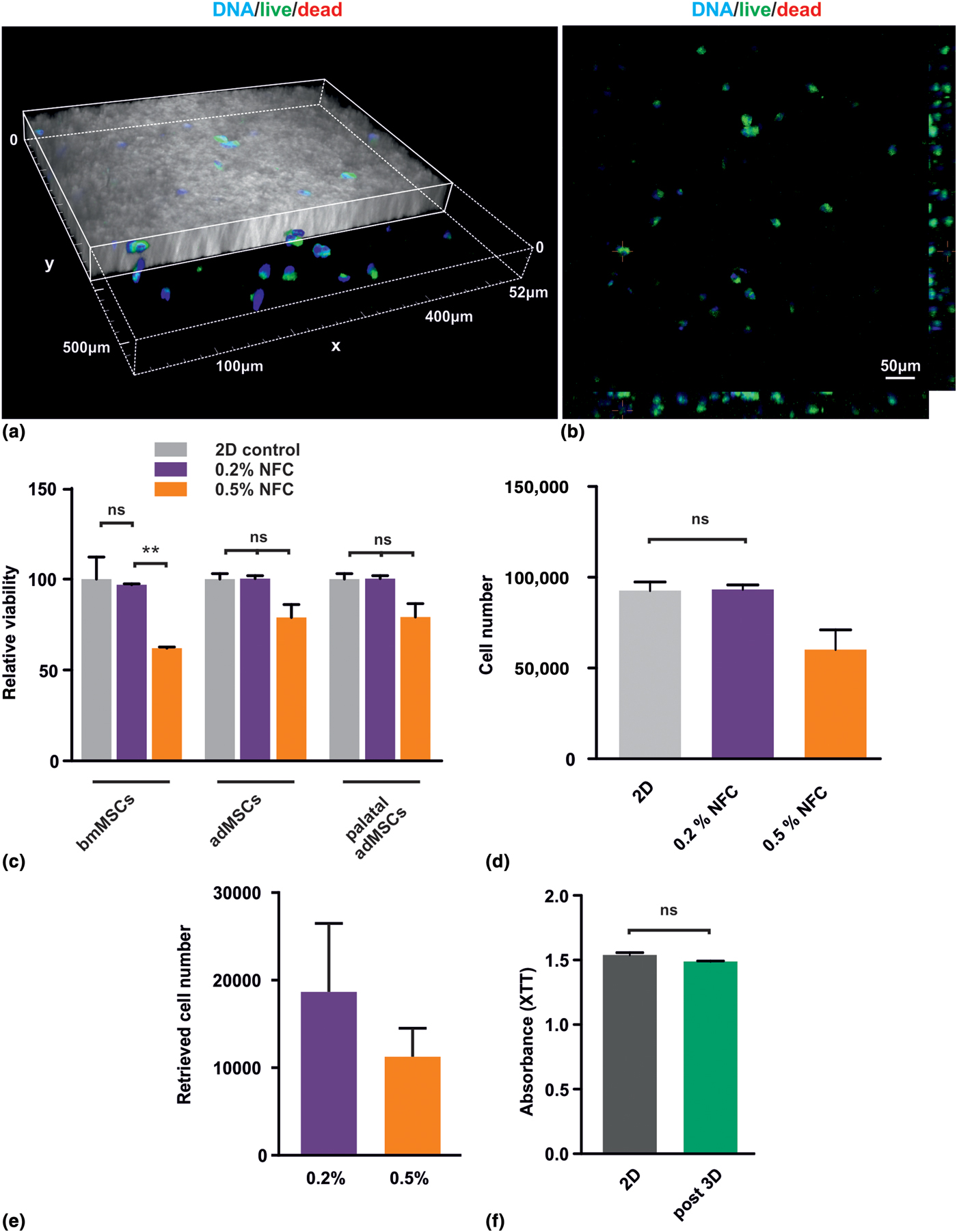
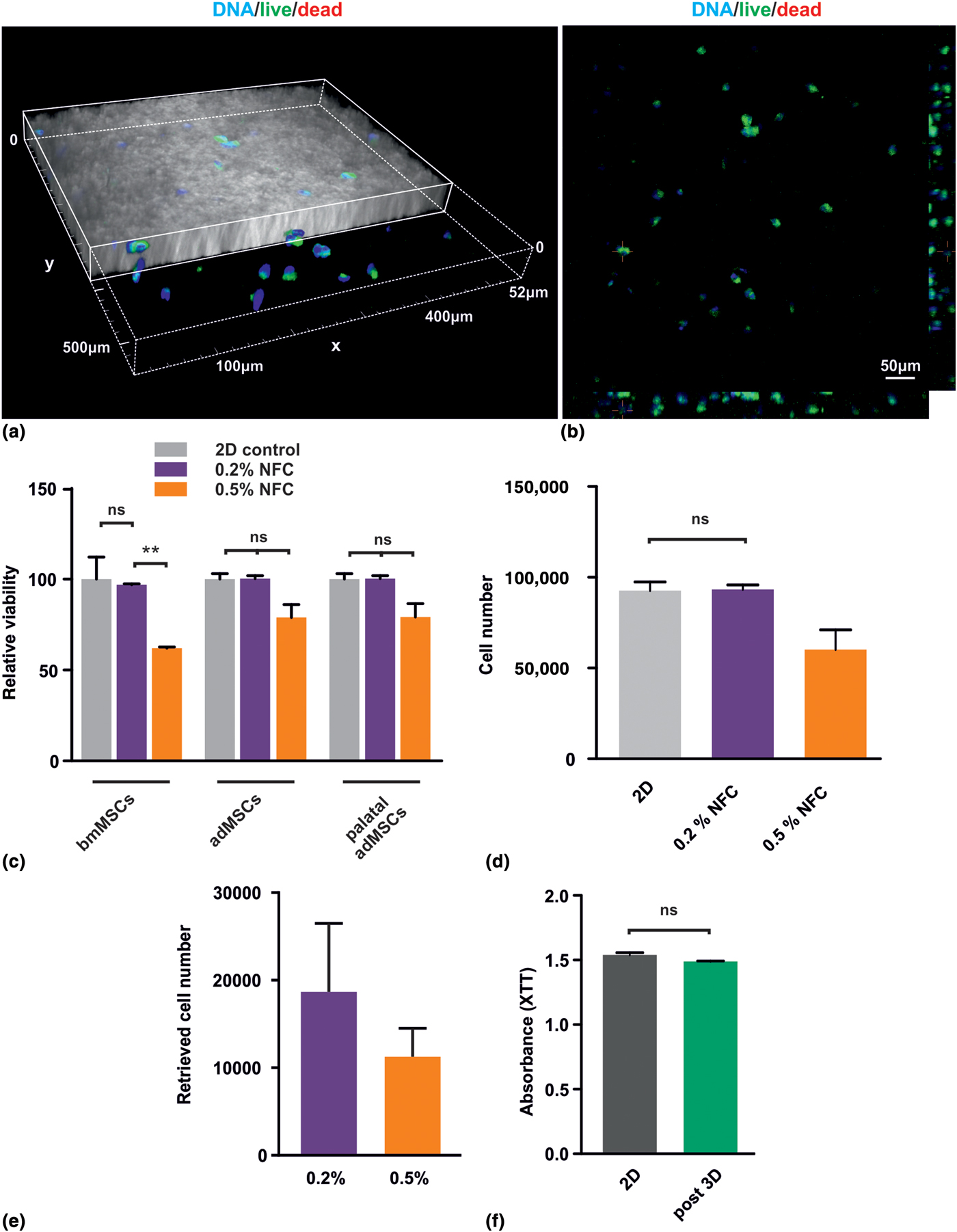
To assess the influence of 3D cultivation within the NFC hydrogel on cellular viability of MSCs, cells were cultured in 3D, stained with Calcein (living cells), ethidium homodimer-1 (dead cells), fixed and counter-stained for DAPI. Subsequent laser scanning microscopy and post hoc image analysis revealed that most cells within the hydrogel stained positively for Calcein [Figs. 3(d) and 3(e)]. Moreover, the cells were distributed evenly in all three dimensions of the hydrogel [Fig. 3(d)]. No pyknotic or fragmented nuclei were observed. To quantitatively determine the viability of 3D-cultivated MSCs, an XTT assay was performed [Fig. 3(e)]. BmMSCs, liposuction adMSCs, and palatal adMSCs cultivated in 0.2% NFC showed no significant changes in cellular viability compared with the 2D control. In contrast, all MSCs showed reduced viability in 0.5% NFC. However, the difference to the 2D control was only significant in the bmMSCs group. To investigate the ability of MSCs to proliferate within the NFC hydrogel, 36 × 103 adMSCs were embedded in 0.2% or 0.5% NFC followed by cultivation for 36 h and an XTT assay. A standard curve with defined cell numbers (10, 1000, 10,000, and 100,000 cells) was used to normalize the XTT absorption to cell numbers. Analysis of the normalized XTT absorption values revealed that there is no significant difference between proliferation of MSCs as 2D culture and within 0.2% NFC. Note that MSCs in 0.5% proliferate although at a lower rate than in 2D or within 0.2% NFC.
Different concentrations of 3D NFC hydrogels are biocompatible and support growth of viable human MSCs of different origin. (a, b) Confocal and phase contrast imaging of adMSCs within a 0.2% NFC, stained with Calcein (living cells), ethidium homodimer-1 (dead cells) and counter-stained with DAPI shows evenly distributed viable cells embedded within the NFC. 3D reconstruction (a) with part of the matrix cut through the y-plane reveals supported cells, while the maximum projection image (b) shows neither ethidium homodimer-1 signal nor pyknotic nuclei. (c) To assess the influence of 3D cultivation within the NFC hydrogel on cellular viability of human MSCs of different cellular origin, bone marrow MSCs, palatal adipose tissue-derived MSCs, and liposuction-derived adipose tissue MSCs were embedded in 0.2% or 0.5% 3D NFC hydrogel and assessed for viability (XTT assay). No significant difference in cellular viability was observed in all groups cultivated in 0.2% NFC compared with 2D controls. A slight but significant reduction of viability was observed in bmMSCs cultivated in the 0.5% NFC hydrogel. (d) MSCs proliferate within the NFC hydrogel. 36 × 103 adMSCs were embedded in 0.2% or 0.5% NFC followed by cultivation for 36 h and XTT. A standard curve with defined cell numbers (10, 1000, 10,000, and 100,000 cells) was used to normalize the XTT absorption to cell numbers. (e) 25,000 MSCs were seeded in 0.2% or 0.5% NFC followed by retrieval using cellulase. Approximately 100% were retrieved from 0.2% NFC, whereas 50% were retrieved from 0.5% NFC. (f) Viable MSCs can be retrieved from the 3D NFC hydrogel. AdMSCs were cultivated in 0.2% NFC hydrogel followed by enzymatic retrieval using cellulase. After replating under standard 2D conditions, an XTT assay was performed to assess the viability of the cells which had previously been cultivated in 3D NFC hydrogel (post 3D) compared with control cells (2D). ** P < 0.01.
Viable MSCs can be retrieved from the 3D NFC hydrogel
In order to determine if viable adMSCs can be retrieved from the 3D NFC hydrogel, cellulose was enzymatically digested using cellulase followed by harvesting of the suspension cells by centrifugation, reseeding under standard 2D conditions and a subsequent XTT assay [Fig. 3(d)]. Here we were able to show that adMSCs pre-cultivated in 3D show no significant differences in cellular viability compared with control cells (continuously cultivated as 2D monolayer).
3D NFC hydrogels are suitable for osteogenic differentiation of MSCs in 3D
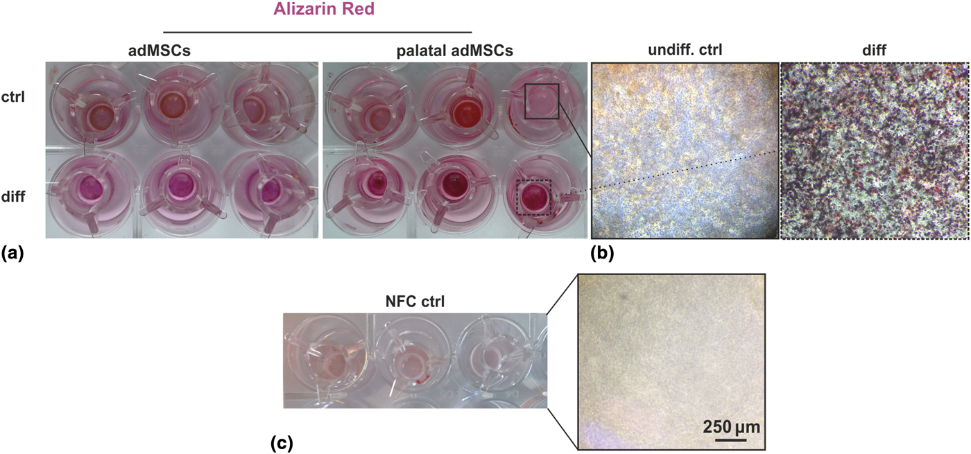
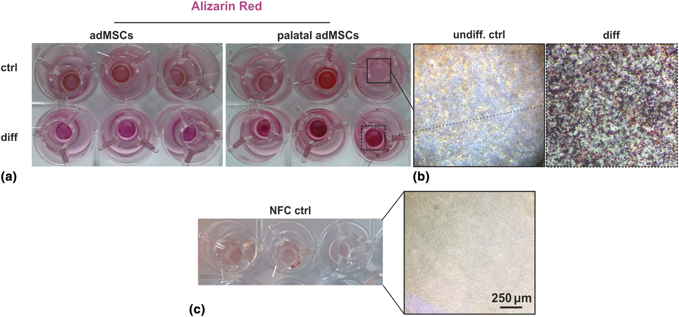
To assess the feasibility of osteogenic differentiation in 3D NFC, liposuction-derived adMSCs, and palatal tissue-derived MSCs were subjected to differentiation in 0.2% NFC for 21 days and stained for calcium deposition using Alizarin Red S. Here we demonstrate that human MSCs of both cellular origins deposit high amounts of calcium after 21 days of differentiation compared with undifferentiated cells in 0.2% NFC [Figs. 4(a) and 4(b)]. Note that differentiated samples show a typical deep red/magenta staining whereas undifferentiated samples and control scaffolds appear light/dim red due to unbound dye remaining in the NFC hydrogel [Fig. 4(c)].
3D NFC is suitable for osteogenic differentiation of MSCs in 3D. (a) Liposuction-derived adMSCs and as MSCs and adMSCs isolated from palatal adipose tissue were subjected to osteogenic differentiation in 0.2% NFC for 21 days and subsequently stained for calcium deposition with Alizarin Red S. Images of differentiated MSCs clearly show calcium deposition (magenta), whereas no differentiation was observed in control cells, which appeared red. (b) High-power images showing Alizarin Red S-labeled calcium deposits at the cellular level in palatal tissue-derived MSCs. (c) NFC control without cells showed only low level of Alizarin staining.
Discussion
This proof-of-concept study describes for the first-time 3D cultivation and differentiation of human MSCs within plant-derived 3D NFC hydrogels. We showed that the 3D NFC features fibers and pores of different sizes and is biocompatible with human MSCs of different cellular origin. In particular, we demonstrated that living MSCs are homogenously distributed in the hydrogel and that cultivation in 0.2% NFC does not negatively influence their cellular viability. Finally, we demonstrated that NFC is compatible with 3D differentiation of MSCs into the osteogenic fate.
Although MSCs represent one of the most promising adult stem cell types in regenerative medicine, their conventional expansion to obtain clinically significant cell numbers represents a significant roadblock toward routine use in a clinical setup. In particular, prolonged 2D cultivation of various adult stem cells harbors the intrinsic risk of chromosomal aberrations and tumorigenic transformation.[Reference Ben-David, Mayshar and Benvenisty5, Reference Bara, Richards, Alini and Stoddart6, Reference Kaus, Widera, Kassmer, Peter, Zaenker, Kaltschmidt and Kaltschmidt20] This could be at least partly explained by the removal of the stem cells from their endogenous niches, which is indeed 3D. In general accordance with this assumption, human breast epithelial cells were shown to develop into tumor-like cells when cultured in 2D, while reverting to normal behavior when cultured in a more native 3D microenvironment.[Reference Petersen, Rønnov-Jessen, Howlett and Bissell21] Thus, 3D cultivation of MSCs could reduce the risk of tumorigenic transformation during expansion in vitro. In addition to the increased risk of cellular transformation, cultivation of MSCs as a 2D monolayer is associated with significant reduction of multipotency and acquisition of early cellular senescence. In this context, 3D cultivation of adMSCs has been shown to induce autophagy and reduce general signs of senescence.[Reference Yang, Huang and Hsu22] Thus, 3D cultivation could provide a stable micro-environment for isolated MSCs ex vivo, maintaining their multipotency, preventing cellular senescence, and enabling high proliferation capacity and yield, while minimizing the culture time in vitro. In addition, cultivation of MSCs in 3D allows significantly higher cell numbers per volume of cell culture medium compared with conventional cell culture and stacks of flask-units commonly applied for large-scale production of MSCs. This would allow significant cost reduction in production of both clinically relevant numbers of MSCs and MSC-derived EVs for clinical use.
Currently, there are many commercially available 3D matrices developed to provide mechanical support and biochemical cues to enhance proliferation and differentiation of MSCs. Some matrices are fibrin or collagen based, while others are a varied mix of ECM molecules. For example, the mouse chondrosarcoma cell line, EHS, was used to develop the well-known and broadly utilized ECM matrix – Matrigel™.[Reference Kleinman and Martin23] Nonetheless, Matrigel™ has some major drawbacks, such as batch-to-batch variability, cross-species immunogenicity and consequently potential problems with translation into the clinic. Thus, fibrin- and collagen-based 3D matrices are a well-defined alternative to Matrigel™-based 3D culture of MSCs. However, these natural substrates and their functionalized versions do not allow for retrieval of cells free of enzymes affecting mammalian cells. Alginates represent another 3D matrix suitable for MSC cultivation.[Reference Ho, Murphy, Binder, Vissers and Leach8] However, alginates require cross-linking prior to use.
In addition to natural materials, 3D matrices can be engineered from synthetic materials such as poly-(-l-lactic acid) nanofibers.[Reference Paletta, Mack, Schenderlein, Theisen, Schmitt, Wendorff, Agarwal, Fuchs-Winkelmann and Schofer24] Although they offer the clear advantage of well-defined and guided construction, they need to be electrospun prior to their use, which can be highly complicated and time consuming.
The plant-derived native NFC hydrogel, GrowDex®, provides a well-characterized and -defined matrix.[Reference Bhattacharya, Malinen, Lauren, Lou, Kuisma, Kanninen, Lille, Corlu, GuGuen-Guillouzo, Ikkala, Laukkanen, Urtti and Yliperttula16, Reference Malinen, Kanninen, Corlu, Isoniemi, Lou, Yliperttula and Urtti17] This hydrogel is easy to handle and is biocompatible with human cells, while also allowing the simple retrieval of cells from the scaffold for further use.[Reference Bhattacharya, Malinen, Lauren, Lou, Kuisma, Kanninen, Lille, Corlu, GuGuen-Guillouzo, Ikkala, Laukkanen, Urtti and Yliperttula16, Reference Paukkonen, Ukkonen, Szilvay, Yliperttula and Laaksonen25] Although the immunogenicity of plant-derived NFC is still under investigation, the low immune response to other plant-derived hydrogels has been reported.[Reference Cochis, Grad, Stoddart, Fare, Altomare, Azzimonti, Alini and Rimondini12, Reference Modulevsky, Cuerrier and Pelling26, Reference Lopes, Sanchez-Martinez, Stromme and Ferraz27]
In contrast to the materials introduced above, the plant-derived NFC matrix harbors several crucial advantages. Firstly, as a plant-derived matrix, NFC hydrogels are free of animal-derived components, which could facilitate their transition into clinical use. Moreover, it allows an easy retrieval of viable cells based on enzymatic digestion of the cellulose fibers using cellulases, which do not affect human cells. Moreover, in contrast to other hydrogels no crosslinking is needed for the assembly of the matrix.
Our proof-of-concept study clearly shows that the commercially available and standardized 3D NFC hydrogel GrowDex® is biocompatible and supports growth of MSCs. Notably, in addition to MSCs, 3D NFC has been demonstrated to be biocompatible with human pluripotent stem cells[Reference Lou, Kanninen, Kuisma, Niklander, Noon, Burks, Urtti and Yliperttula15] and liver cells.[Reference Bhattacharya, Malinen, Lauren, Lou, Kuisma, Kanninen, Lille, Corlu, GuGuen-Guillouzo, Ikkala, Laukkanen, Urtti and Yliperttula16] The slight decline of cellular viability of bmMSCs observed in 0.5% NFC in our study, suggests that the optimal concentration of the NFC needs to be experimentally determined for each individual cell type (as suggested by the manufacturer). Likewise, we noted that the high concentration of 3D NFC prevented spheroid formation as similarly seen with human pluripotent stem cells.[Reference Lou, Kanninen, Kuisma, Niklander, Noon, Burks, Urtti and Yliperttula15]
In summary, this study clearly suggests that 3D NFC could be used for the development of safe and simple protocols, toward the expansion and differentiation of MSCs for routine cell culture and toward potential clinical application. In addition, 3D NFC could be used for obtaining high cell numbers required for isolation of MSC-derived EVs.
Acknowledgments
D.W. is supported by a grant of the DFG (German Research Foundation, WI4318/2-1). J.S. is supported by UPM Biochemicals, Helsinki, Finland. S.S.H. is supported by The Scientific and Technological Research Council of Turkey (TUBITAK/SBAG-112S587), Ankara, Turkey and Selcuk University Research Coordination Office (BAP), Konya, Turkey. We thank Professor Erdal Karaoz, Liv Hospital, Center for Regenerative Medicine and Stem Cell Research & Manufacturing (LivMedCell), Istanbul, Turkey for providing human bone marrow MSCs. The sponsors did not participate in study design and data analysis. We thank Amanpreet Kaur for the help with electron microscopy and Marie Zeuner for careful proof-reading.