Introduction
Foreign body aspiration is a common, life-threatening condition that primarily affects children.Reference Paksu, Paksu, Kilic, Guner, Baysal and Sancak1, Reference Maddali, Mathew, Chandwani, Alsajwani and Ganguly2 Rigid bronchoscopy is frequently used as a diagnostic tool in children, despite the high risk for complications associated with the procedure.Reference Fidkowski, Zheng and Firth3–Reference Tomaske, Gerber and Weiss6
Computed tomography (CT) scanning is a non-invasive imaging modality, which has been suggested as an alternative means of diagnosing foreign body aspiration. Clinical examination, standard chest radiographs and fluoroscopy are inaccurate diagnostic techniques for foreign body aspiration.Reference Paksu, Paksu, Kilic, Guner, Baysal and Sancak1, Reference Kiyan, Gocmen, Tugtepe, Karakoc, Dagli and Dagli7–Reference Mu, Sun and He15 They require rigid bronchoscopies to be performed to confirm the diagnosis. Rigid bronchoscopy is an invasive procedure with a high rate of complications. These include cardiac arrest, tracheal and bronchial lacerations, and severe hypoxia that can result in hypoxic brain damage.Reference Maddali, Mathew, Chandwani, Alsajwani and Ganguly2, Reference Hasdiraz, Oguzkaya, Bilgin and Bicer4, Reference Ciftci, Bingol-Kologlu, Senocak, Tanyel and Buyukpamukcu9, Reference Jiaqiang, Jingwu, Yanming, Qiuping, Yinfeng and Xianguang16, Reference Orji and Akpeh17 In contrast, CT scans are non-invasive, they do not require a general anaesthesia and they are associated with a comparably low risk of complications.
In Australia, 42 per cent of documented rigid bronchoscopies performed in 2009–2010 were on patients proven to have an alternative diagnosis.18 The ability of CT to differentiate true cases of foreign body aspiration from alternative diagnoses such as pneumonia could lead to a decrease in the number of patients who require rigid bronchoscopy and thus decrease the associated risks.
This literature review aimed to examine the possibility of reducing the number of unnecessary rigid bronchoscopies performed by using CT as an alternative, non-invasive imaging modality in the diagnosis of foreign body aspiration. This involved investigation of the safety and accuracy of CT. The accuracy of CT scanning was determined by examining the available data, and comparing the sensitivity and specificity of CT to that of the current ‘gold standard’. The safety of CT scanning was determined by analysing data on the calculated radiation exposure dose required for imaging a suspected case of foreign body aspiration. Studies with verification bias were critiqued to provide an applied example of the role of CT in clinical practice. Our hypothesis is that CT is a sensitive, specific, safe and non-invasive modality when used in the diagnosis of foreign body aspiration. Furthermore, we believe that its future use will decrease the number of unnecessary rigid bronchoscopies.
Materials and methods
A search of the literature published from 1970 to 14 May 2013 was performed using the following electronic databases: PubMed, Scopus, Web of Knowledge, Embase and Medline. Studies with level I, II or III evidence were eligible for inclusion. Conference abstracts, retrospective studies or investigations with level IV evidence were not included. Studies needed to compare CT to the current gold standard of rigid bronchoscopy. When applicable, the sensitivity, specificity and radiation exposure dose were analysed.
Results
Twenty-four papers were identified for this review, of which 14 met the inclusion criteria. One studyReference Ozkurt, Bahadir, Uegul, Altuna, Basak and Cevizci19 was excluded from the analysis as it was determined by the publishing journal to be a duplication of a previous study.Reference Cevizci, Dokucu, Baskin, Karadag, Sever and Yalcin20
Discussion
Rigid bronchoscopy does have advantages when used in the diagnosis and management of foreign body aspiration cases. For instance, it is both diagnostic and therapeutic, and allows for advanced procedures including bronchoalveolar lavage and biopsy in circumstances of tumours masquerading as foreign bodies.
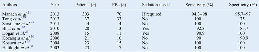
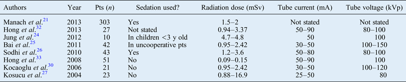
Sedation was used for CT scanning in 6 of the 14 studies;Reference Manach, Pierrot, Couloigner, Ayari-Khalfallah, Nicollas and Venail21–Reference Sodhi, Aiyappan, Saxena, Singh, Rao and Khandelwal26 however, its use did not influence the sensitivity or specificity of the technique, or the associated radiation exposure (as seen in Tables I–III). Thus, sedation does not appear to be a necessity for a procedure that lasts between 9.5 and 55 seconds.Reference Bhat, Hedge, Nagalotimath and Patil22, Reference Sodhi, Aiyappan, Saxena, Singh, Rao and Khandelwal26, Reference Kosucu, Ahmetoglu, Koramaz, Orhan, Ozdemir and Dinc27 Therefore, the safety of CT scanning in the diagnosis of foreign body aspiration may be determined by analysing its sensitivity and specificity, and calculating its radiation burden.
Sensitivity and specificity of CT in diagnosing foreign body aspiration

CT = computed tomography; FBs = foreign bodies
Radiation exposure during CT evaluation of suspected foreign body aspiration
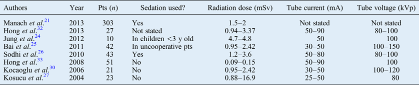
Four studiesReference Manach, Pierrot, Couloigner, Ayari-Khalfallah, Nicollas and Venail21, Reference Bai, Zhou, Gao, Shao, Califano and Ha25, Reference Kosucu, Ahmetoglu, Koramaz, Orhan, Ozdemir and Dinc27, Reference Kocaoglu, Bulakbasi, Soylu, Demirbag, Tayfun and Somuncu30 provided information regarding radiation exposure to patients during CT evaluation of suspected foreign body aspiration. The radiation dose ranged from 1.5 mSv to 16.9 mSv. An average radiation dose per patient across all studies was calculated as 2.16 mSv. The average background radiation dose in Australia is 1.5 mSv.Reference Webb, Solomon and Thomson34 CT = computed tomography; pts = patients; y = years
Studies with verification bias
In these studies, rigid bronchoscopy was not routinely performed in every patient. Whilst sensitivity and specificity could not be calculated, the studies demonstrate how computed tomography could be used in clinical practice. Pts = patients
Sensitivity
For CT scanning to be a safe alternative to rigid bronchoscopy in the diagnosis of foreign body aspiration, the sensitivity must be high so that positive cases are not missed. The sensitivity of CT ranged from 90 to 100 per cent in the studies reviewed, with four of the eight studies exhibiting perfect sensitivity (Table I).Reference Kosucu, Ahmetoglu, Koramaz, Orhan, Ozdemir and Dinc27–Reference Sarsilamz, Boyraz and Gelal29, Reference Haliloglu, Ciftci, Oto, Gumus, Tanyel and Senocak31 The lower sensitivity values were the result of false negative results; however, in these studies, pathology indicative of foreign body aspiration was evident on the scans even when no foreign body was seen.Reference Doğan, Coskun, Yikilmaz, Hasdiraz and Tahan23, Reference Kocaoglu, Bulakbasi, Soylu, Demirbag, Tayfun and Somuncu30 Inadequate slice thickness of the CT images can also contribute to low sensitivity.Reference Bhat, Hedge, Nagalotimath and Patil22 Sensitivity improves when the CT scans are reviewed by a second radiologist.Reference Manach, Pierrot, Couloigner, Ayari-Khalfallah, Nicollas and Venail21 Variation in sensitivity values will be analysed in the context of the relevant studies.
Computed tomography and rigid bronchoscopy had identical sensitivity results in the evaluation of suspected foreign body aspiration in four studies.Reference Kosucu, Ahmetoglu, Koramaz, Orhan, Ozdemir and Dinc27–Reference Sarsilamz, Boyraz and Gelal29, Reference Haliloglu, Ciftci, Oto, Gumus, Tanyel and Senocak31 Tong et al.Reference Tong, Zhang, Fang, Sha and Chi28 conducted a study of 37 patients where all negative CT results were confirmed with rigid bronchoscopy, with a sensitivity of 100 per cent. The remaining three studies also showed 100 per cent sensitivity; however, the sample sizes in those studies were small. There were only 4 patients in the Sarsilamz et al. study,Reference Sarsilamz, Boyraz and Gelal29 whilst the studies by Kosucu et al.Reference Kosucu, Ahmetoglu, Koramaz, Orhan, Ozdemir and Dinc27 and Haliloglu et al.Reference Haliloglu, Ciftci, Oto, Gumus, Tanyel and Senocak31 each comprised 23 patients. Although perfect sensitivities were demonstrated in these four studies, only Tong et al.Reference Tong, Zhang, Fang, Sha and Chi28 recruited sufficient patients for the result to be considered valid. For CT to be objectively evaluated as having 100 per cent sensitivity, the results will need to be replicated in larger scale studies.
Computed tomography has a high sensitivity provided the scan is interpreted accurately. Manach et al.Reference Manach, Pierrot, Couloigner, Ayari-Khalfallah, Nicollas and Venail21 conducted the largest prospective study in this review, comprising 303 cases, and initially demonstrated a sensitivity of 94.3 per cent. However, following a second interpretation of the CT images by a senior radiologist prior to endoscopy, the sensitivity improved to 98 per cent. Furthermore, foreign bodies missed with CT were identified in the images after the results of rigid bronchoscopy were known. This demonstrates that the diagnosis of foreign body is operator-dependent. Specific training in interpreting CT in the context of foreign body aspiration would increase the sensitivity of CT as a diagnostic tool. Sensitivity could also be improved by adopting a practice where two independent readers are required to review the CT images.
If radiologists are provided with a thorough clinical history of the patient, foreign bodies that are not visible on CT images can be detected by looking for other pathological indicators. The lowest sensitivity score of 90 per cent was reported by Kocaoglu et al.;Reference Kocaoglu, Bulakbasi, Soylu, Demirbag, Tayfun and Somuncu30 this was the result of one case of foreign body aspiration going undetected on CT. The CT images for this case showed right upper lobe collapse and an occluded segment, with no visible foreign body, the latter of which was a result of dense infiltrate. This patient was being investigated for chronic foreign body aspiration. In a chronic case, a dense inflammatory response to the foreign body is expected, which can potentially obscure the foreign body from view on CT imaging. Similarly, Dogan et al.Reference Doğan, Coskun, Yikilmaz, Hasdiraz and Tahan23 reported a false negative result in which dense infiltrates were identified on CT at the level where the foreign body was found at removal. A history of chronic aspiration should alert the radiologist to the potential for foreign bodies to be obscured by inflammation. Provided the doctor supplies the radiologist with a detailed history of the patient, and the radiologist takes this into consideration whilst reviewing the CT images, the threshold for diagnosis can be lowered.
The sensitivity of CT scans varies as a result of the slice thickness of the images; the accuracy is increased with a reduction in slice thickness. The final false negative result was described by Bhat et al.,Reference Bhat, Hedge, Nagalotimath and Patil22 and was reportedly due to a minute, longstanding foreign body of vegetative origin. Unlike the previous two examples, there was no evidence on CT to indicate the presence of a foreign body. The authors explained that in this circumstance the false negative result was most likely due to the inadequate slice thickness of 2 mm of the CT images. This value is large when compared to the slice thicknesses of the CT images used in the three studies with 100 per cent sensitivity: in the studies by Tong et al.,Reference Tong, Zhang, Fang, Sha and Chi28 Kosucu et al.Reference Kosucu, Ahmetoglu, Koramaz, Orhan, Ozdemir and Dinc27 and Haliloglu et al.Reference Haliloglu, Ciftci, Oto, Gumus, Tanyel and Senocak31 the slice thicknesses used were 1 mm, 1.25 mm and 1 mm, respectively.
Computed tomography scanning has only recently been used in diagnosing foreign body aspiration. At this stage, there are no standard parameters for using CT optimally in the diagnosis of foreign body aspiration. Imaging parameters should be standardised in future studies to enable increased sensitivity of CT, gained by using enhanced settings.
Specificity
A high specificity of CT is desired in the diagnosis of suspected foreign body aspiration, as false positive results will lead to unnecessary rigid bronchoscopy. Specificity values were given for eight of the studies reviewed;Reference Manach, Pierrot, Couloigner, Ayari-Khalfallah, Nicollas and Venail21–Reference Doğan, Coskun, Yikilmaz, Hasdiraz and Tahan23, Reference Kosucu, Ahmetoglu, Koramaz, Orhan, Ozdemir and Dinc27–Reference Haliloglu, Ciftci, Oto, Gumus, Tanyel and Senocak31 these ranged from 75 to 100 per cent (Table I). The selection criteria for patients enrolled in studies can affect the specificity.Reference Ransohoff and Feinstein38 Likewise, low sample sizes can give misleading results. As with sensitivity, specificity improves when the CT results are analysed by two independent readers. The specificity values of these trials are discussed in more depth below.
With regard to the influence of selection criteria on the results of specificity, the study by Tong et al.Reference Tong, Zhang, Fang, Sha and Chi28 only included patients with a clear history of foreign body aspiration, and therefore an abnormally low number of true negative results were seen. There was only one false positive result in that study; however, with only three true negative results, the specificity was calculated at 75 per cent. Thus, the specificity was artificially lowered by the study design. Trials should include all patients suspected of suffering from foreign body aspiration, rather than only selecting those with a clear history.
The study by Bhat et al.Reference Bhat, Hedge, Nagalotimath and Patil22 also demonstrated a low specificity, of 85.7 per cent. Once again, there was only 1 false positive result, but this study comprised only 20 participants. As discussed previously in relation to sensitivity, small numbers of participants in trials can lead to misleading results.Reference Ransohoff and Feinstein38 A further illustration of the effect of sample size can be observed in the four trials with perfect specificity.Reference Doğan, Coskun, Yikilmaz, Hasdiraz and Tahan23, Reference Kosucu, Ahmetoglu, Koramaz, Orhan, Ozdemir and Dinc27, Reference Sarsilamz, Boyraz and Gelal29, Reference Haliloglu, Ciftci, Oto, Gumus, Tanyel and Senocak31 Whilst a specificity of 100 per cent is encouraging, a study design which included a greater number of patients would provide a better representation of specificity.
As with sensitivity, specificity can be improved with a second analysis of the CT scans, as demonstrated by the Manach et al. study.Reference Manach, Pierrot, Couloigner, Ayari-Khalfallah, Nicollas and Venail21 The initial specificity in that study was calculated as 95.7 per cent, with a new specificity of 97 per cent demonstrated after the second analysis. This result once again highlights that the accuracy of CT in the diagnosis of foreign body aspiration can be improved with the implementation of a ‘two-read protocol’. If this protocol is followed, the specificity of CT scanning would be great enough to warrant its use as a first-line imaging modality in the diagnosis of foreign body aspiration.
Radiation exposure
The radiation exposure incurred by a patient when CT is used to determine foreign body aspiration needs to be analysed with respect to doses that are known to increase the risk of malignancy to a significant level. Whilst CT is non-invasive, it subjects patients to radiation and therefore an increased mortality risk. The paediatric population is subject to a higher lifetime mortality risk of cancer due to radiation exposure than adults.Reference Brenner, Elliston, Hall and Berdon39
When considering foreign body aspiration, it is important to note that the patient is no longer part of the ‘healthy population’. Therefore, in diagnosis of the condition, it is justifiable for the patient to be exposed to some radiation.Reference Stender40 This is different to a diagnostic screening test, in which a healthy individual is screened via radiation for a certain condition. The small potential risk of malignant transformation is arguably lower than the risks associated with undergoing rigid bronchoscopy under general anaesthesia.
A recent Australian study provides strong evidence for an increased cancer risk following exposure to CT scans in childhood or adolescence.Reference Mathews, Forsythe, Brady, Butler, Goergen and Byrnes41 It compared 680 000 people who had been exposed to CT scans with those who had not been exposed, in a population of 11 million. The study determined that each sievert of effective dose of CT caused 0.125 cancers.Reference Mathews, Forsythe, Brady, Butler, Goergen and Byrnes41 Using the average radiation dose calculated from the analysed studies of 2.16 mSv, 3703 CT scans used in the diagnosis of foreign body aspiration will result in 1 excess cancer. Whilst the risk of cancer is undeniably present, it is low, and must be weighed against that of undergoing rigid bronchoscopy.
The largest dose of radiation exposure was reported in the Kosucu et al.Reference Kosucu, Ahmetoglu, Koramaz, Orhan, Ozdemir and Dinc27 study, with a radiation dose range calculated between 0.88 and 16.99 mSv (Table II). The study described using a low tube voltage, and a tube current of 25–50 mA, which is equal to the lowest parameters used in the other studies. This variation could be explained by the fact that the authors used different methods to calculate the mean effective dose. Methods for calculating the mean effective dose include the Huba method, the Alessio method, and the Deak and Shrimpton dose–length product methods. Results derived using these methods may vary by as much as 30 per cent.Reference Newman, Ganguly, Kim and Robinson42 It was not specified in the studies reviewed how the exposure doses were calculated, which makes interpretation of the results difficult. Future studies on this topic should include the radiation dose incurred by the use CT (in the diagnosis of suspected foreign body aspiration), and specify how the dose was calculated, so that the safety of CT in this setting may be determined more accurately.
Radiation exposure in the paediatric population is of concern, as each dose of radiation is cumulative over an individual's lifetime.Reference Frush, Donelly and Rosen43 The majority of the studies reviewed demonstrated that CT used for foreign body aspiration diagnosis entailed only a low dose of radiation exposure. Dose reduction can be achieved by following a low-dose protocol,Reference Brisse and Aubert44 which will further minimise the radiation risks.
Studies with verification bias
Five studies included in this review were subject to verification bias;Reference Cevizci, Dokucu, Baskin, Karadag, Sever and Yalcin20, Reference Bai, Zhou, Gao, Shao, Califano and Ha25, Reference Sattar, Javed, Bodla, Anjum and Ch35–Reference Adeletli, Kurugoglu, Ulus, Ozer, Elicevik and Kantarci37 these are represented in Table III. In these studies, rigid bronchoscopy was not routinely performed in every patient; thus, a true sensitivity and specificity could not be determined for CT. Negative results on CT were interpreted as proof that no foreign body was present; these patients were treated medically, without being subjected to rigid bronchoscopy. The studies deserve mention as this situation would be indicative of how patients would be managed in clinical practice if CT were to be used as the standard in diagnosis of foreign body aspiration.
In instances where no foreign body is found by CT scans, patients can safely be managed medically, with good clinical outcome, thereby negating the need for rigid bronchoscopy. Bai et al.Reference Bai, Zhou, Gao, Shao, Califano and Ha25 conducted a prospective study in 2011 on 45 patients. All patients were required to have a CT scan prior to rigid bronchoscopy. Of the 45 patients, 3 had no CT findings indicative of a foreign body, and were treated conservatively with antibiotics. All of these patients improved clinically. The remaining 42 patients with positive CT findings underwent rigid bronchoscopy. The results were in exact concordance with the CT images. This study perfectly illustrates that CT is sufficiently accurate and safe for use in diagnosing foreign body aspiration, and can reduce the number of unnecessary rigid bronchoscopies performed with no apparent adverse implications for negative diagnosis.
Similarly, in studies by Satar et al.Reference Sattar, Javed, Bodla, Anjum and Ch35 and Adeletli et al.,Reference Adeletli, Kurugoglu, Ulus, Ozer, Elicevik and Kantarci37 patients that had no evidence of a foreign body on CT were followed up with medical management. The authors of both papers reported that all medically managed patients improved clinically, with no invasive intervention needed. A similar approach was taken by Cevizci et al.Reference Cevizci, Dokucu, Baskin, Karadag, Sever and Yalcin20 However, of the 20 patients with no suspected foreign body on CT, 7 continued to have prolonged symptoms exceeding 1 month. These cases were followed up with rigid bronchoscopy, but no foreign bodies were found. It could be concluded from these studies that medical follow up of a negative CT scan is appropriate management for suspected foreign body aspiration if it is not detrimental to patient well-being.
Sodhi et al.Reference Sodhi, Aiyappan, Saxena, Singh, Rao and Khandelwal26 took a similar approach by medically managing 13 patients with negative CT results. However, 4 of these 13 patients subsequently underwent fibre-optic bronchoscopy for further evaluation of their symptoms, and 1 patient was found to have a foreign body. The foreign body in this case was obscured on CT examination because of airway inflammation. As argued previously, whilst a foreign body could not be seen in this patient, the combination of severe airway inflammation obscuring the lumen and a positive history should be an indication for operative management. This further illustrates that CT results need to be interpreted in the context of patient history.
Whilst the five studies included in this section possess flawed research methodology for determining the accuracy of CT in the diagnosis of foreign body aspiration, they do demonstrate that medical management of most patients who have negative CT results produces good outcomes. The single case of a missed foreign body may have been avoided if the CT results were interpreted in the context of the patient's medical history. These studies illustrate that CT used in the diagnosis of foreign body aspiration is sufficiently safe and accurate, and can result in a reduction in the number of unnecessary rigid bronchoscopies.
Implications for clinical practice
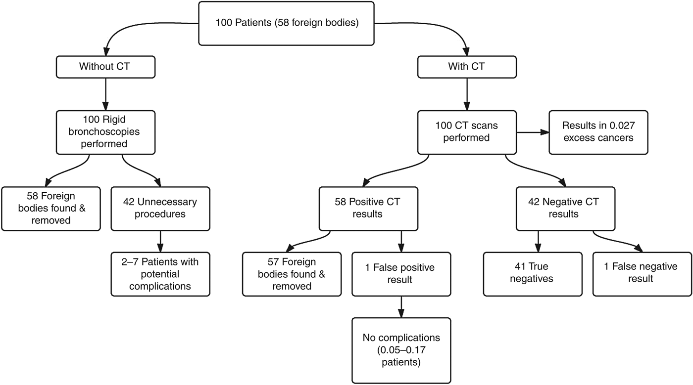
In Australia, 42 per cent of rigid bronchoscopies are performed on patients without a foreign body. The largest study in this review gave a sensitivity of 98 per cent and specificity of 97 per cent when reviewed by two readers.Reference Manach, Pierrot, Couloigner, Ayari-Khalfallah, Nicollas and Venail21 Based on the current data, if 100 patients are investigated for foreign body aspiration without CT, 42 patients will not have a foreign body and will have undergone rigid bronchoscopy unnecessarily. If every patient undergoes a CT prior to rigid bronchoscopy, there will be 58 positive results found. Of these 58 patients, only 1 would undergo rigid bronchoscopy unnecessarily, resulting in a reduction in unnecessary procedures of 97 per cent, as illustrated by Figure 1.
Potential outcomes for 100 patients evaluated for foreign body aspiration with and without computed tomography (CT) (implemented in the diagnostic process), based on current data.
According to the above scenario, there will be one false negative result. Whilst this would be detrimental to that patient, it has to be compared to the potential complications in the 42 patients that would have undergone rigid bronchoscopy unnecessarily. The rate of rigid bronchoscopy complications is reported as between 5 and 17 per cent.Reference Tomaske, Gerber and Weiss6, Reference Ciftci, Bingol-Kologlu, Senocak, Tanyel and Buyukpamukcu9, Reference Cavel, Bergeron, Garel, Arcand and Froehlich45 Hence, in the scenario described, there would be complications in 2–7 of the 42 patients who would have unnecessarily undergone the procedure, as opposed to only 1 patient affected by the false negative result (the foreign body goes undetected by CT scanning). According to the radiation data, the use of 100 CT scans would result in 0.027 cancers. In the hypothetical scenario described, it can be concluded that the use of CT significantly reduces the number of unnecessary rigid bronchoscopies, and results in an overall improvement in patient outcomes.
Whilst there is no denying the utility of rigid bronchoscopy in the management of foreign body aspiration, patients without a foreign body are exposed to a number of unnecessary risks. Rigid bronchoscopy necessitates general anaesthesia and carries the complications of an invasive procedure, which could be avoided by the use of non-invasive CT scanning.
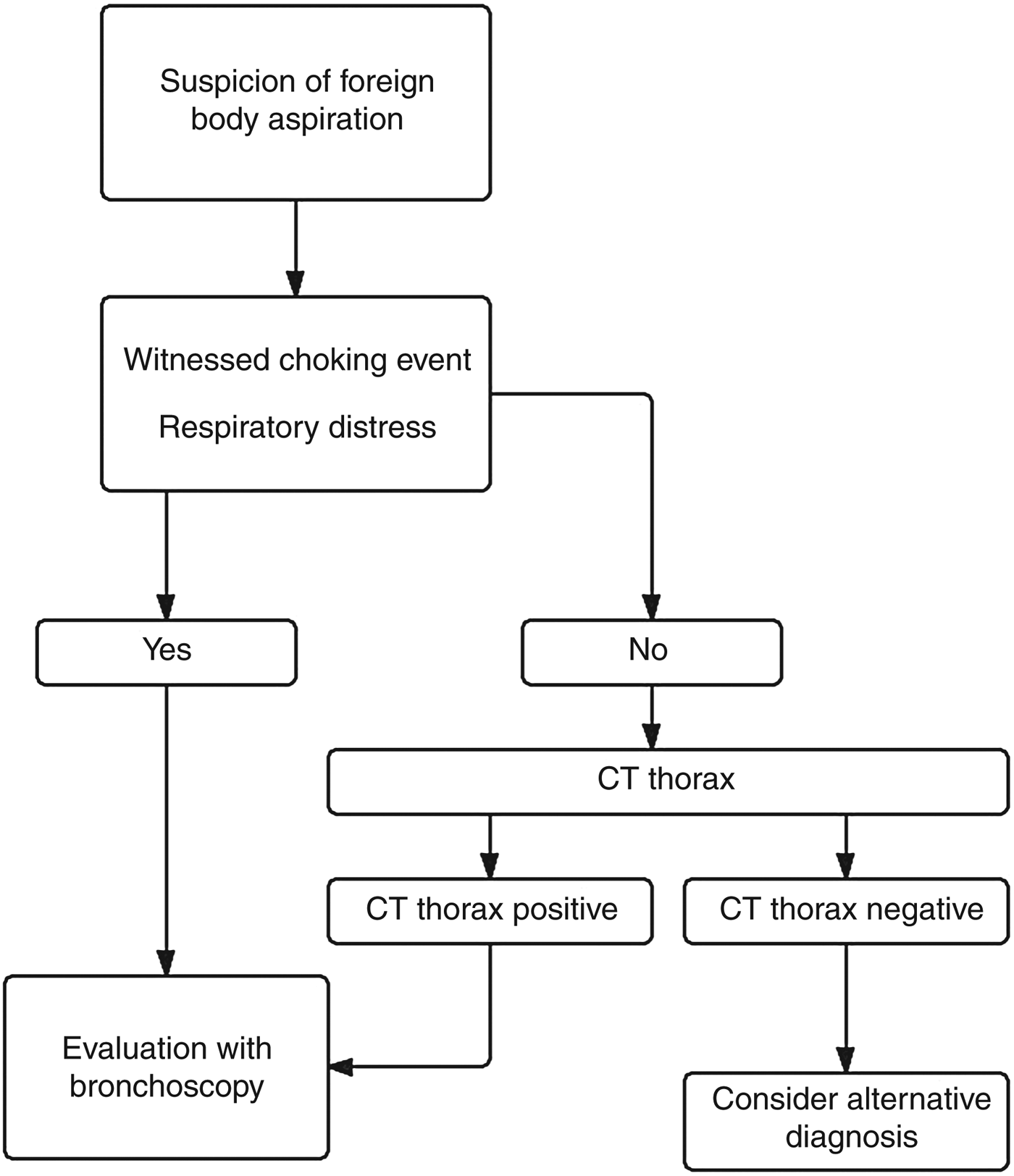
The authors propose an algorithm for the diagnosis of foreign body aspiration based on the conclusions of this review, as shown in Figure 2. Any patient in respiratory distress with a positive history for foreign body aspiration should immediately be taken for surgical management. Those patients not in respiratory distress are to be analysed via a CT scan. Patients with a negative thorax CT scan should be managed medically and followed up. If there is no clinical improvement, they may be evaluated with bronchoscopy.
A proposed algorithm for diagnosis of foreign body aspiration based on the conclusions of this review. CT = computed tomography
Conclusion
From the available literature, it is concluded that CT scans used in the diagnosis of foreign body aspiration are safer, less invasive and have comparable accuracy to rigid bronchoscopy. The accuracy of CT scanning with a two-read protocol, using thinner scan slice thickness and interpreting negative CT results in the context of patient history, has been proven to be comparable to the gold standard. If low-dose protocols are followed, radiation exposure levels are acceptable. Studies subject to verification bias have demonstrated the practical application of CT used in the diagnosis of foreign body aspiration; CT is associated with good clinical outcomes for patients. Given the available data in the literature, this review concludes that CT is a sensitive, specific, safe and non-invasive modality for the diagnosis of foreign body aspiration. Moreover, its future use will decrease the number of unnecessary rigid bronchoscopies.
Acknowledgement
We would like to thank Dr Karin Steinke of the Royal Brisbane and Women's Hospital for her radiology advice.