I. INTRODUCTION
Micrometer- and submicrometer-sized copper particles are used in electromagnetic interference packaging,Reference Brusic, Frankel, Roldan and Saraf1 in electronically conductive pastes,Reference Ohmi2, Reference Songping and Shuyuan3 and in hybrid integrated circuits.Reference Songping and Shuyuan3–Reference Hosokura and Maeda6 In addition to applications in the electronic industry, potential applications of copper particles in powder metallurgy are also attracting more attention.Reference Hao, Xu, Han, Li and Bai7–Reference Chung9 Porous bulk materials generated from copper particles by powder metallurgy techniques have adjustable damping capacity, thermal conductivity, and mechanical strength, and can be used as impact energy absorbers, silencers, heat exchangers, and structural materials.Reference Hao, Xu, Han, Li and Bai7, Reference Chung9
Compared with particle generation methods reported in the literature, such as chemical reduction,Reference Tsai and Dye10, Reference Hirai, Nakao and Toshima11 polyol method,Reference Kurihara, Chow and Schoen12, Reference Fievet, Fievetvincent, Lagier, Dumont and Figlarz13 and vaporization techniques,Reference Cardenas-Trivino, Klabunde and Dale14, Reference Saton and Kimura15 spray pyrolysis has advantages of controllable particle size, simple experiment setup, and large scale production ability.Reference Jain, Skamser and Kodas16–Reference Messing, Zhang and Jayanthi20 Spray pyrolysis devices are typically composed of a precursor reservoir, droplet generator, furnace reactor, and particle collection equipment. Various metal salt precursors can be selected depending on the desired product particle composition. Particle diameter is tunable from tens of nanometers to micrometers by adjusting the salt concentration in the precursor. In addition, temperature and carrier gas flow rate are two important operating parameters for the control of particle morphology.Reference Messing, Zhang and Jayanthi20
To achieve the desired conductivity and product reliability, copper particles free of copper oxide are required. To produce oxide-free particles using spray pyrolysis, a reducing environment is required to prevent the formation of a stable oxide.Reference Stopic, Ilic and Uskokovic21–Reference Biskos, Vons, Yurteri and Schmidt-Ott24 Use of 10 vol% hydrogen was reported for copper nanoparticle generation, but hydrogen with a concentration from 4 to 75 vol% (in air at 1 atm) leads to a risk of explosion.Reference Gurav, Kodas, Pluym and Xiong25 In 2003, Kim et al. reported the generation of copper nanoparticles using N2 as a carrier gas and ethanol (ET) as a cosolvent.Reference Kim, Babushok, Germer, Mulholland and Ehrman26 Under the reported conditions, the ET decomposes providing a reducing environment.Reference Firmansyah, Kim and Kim27–Reference Okuyama and Lenggoro30 The flash point of ET is 14 °C, however, which makes ET a National Fire Protection Association (NFPA) class 3 flammable material. For industrial applications, use of materials that have a flash point above 100 °C is preferred. Ethylene glycol (EG) has a flash point of 111 °C,Reference Speight31 and is NFPA class 1 flammable material requiring preheating before ignition.32 In this work, we report generation of micrometer-sized copper particles with either EG or ET at different temperatures. The effects of the cosolvent and temperature on particle morphology and composition were monitored, and the reaction process was investigated.
II. EXPERIMENT
A. Particle generation
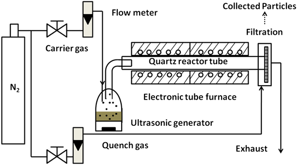
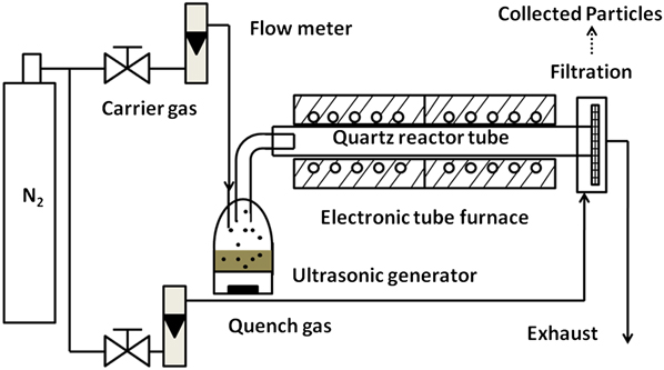
A spray pyrolysis reactor system, illustrated in Fig. 1, was developed which can produce several grams per hour of micrometer-sized powder. The system is composed of a precursor solution reservoir, an ultrasonic droplet generator, tube furnaces, a quartz tube reactor, and a filtration system for particle collection. 1.2 M copper nitrate aqueous solution diluted with 40 vol% cosolvent, either ET or EG, was used as a precursor. 40 vol% was selected because less cosolvent is known to lead to the formation of oxide particles,Reference Kim, Babushok, Germer, Mulholland and Ehrman26 and a ratio higher than 40% reduces the atomization efficiency of the ultrasonic system.Reference Zhon, Peabody, Glicksman, Marshall and Ehrman33 Copper nitrate salt was selected because it is inexpensive, commercially available and highly soluble [145 g Cu(NO3)2 in 100 g water at room temperature].Reference Speight31 The ultrasonic generator operates at a frequency of 1.7 MHz, and atomizes the precursor solution to droplets with volume average diameters of around 5 micrometers. Two tube furnaces, with a total length of 81 cm were combined in series. A quartz tube reactor with inner diameter of 2.5 cm was placed in the center of the furnaces. Temperatures in the furnaces ranged from 400 to 1000 °C. Particles could not be collected because of the residual EG when the furnace temperature was lower than 400 °C, and 1000 °C is the highest operating temperature our furnaces and fittings can reach. A polytetrafluoroethylene filter was used to collect the particles. Industrial grade nitrogen (N2) with a minimum 95% purity was used as carrier gas. For regular copper particle generation, the carrier gas flow rate was 2.5 L/min leading to a residence time around 2 s with two furnaces used. A high flow rate of 10 L/min and a single furnace was used to decrease the residence time to less than 0.4 s for the investigation of the reaction process. Particles were quenched with additional 10 L/min nitrogen before collection.
Illustration of the co-solvent spray pyrolysis process.
B. Particle characterization
Scanning electron microscopy (SEM; Hitachi SU-70, Hitachi High-Technologies Europe GmbH, Krefeld, Germany) was used to image the particles. Particle diameter was manually measured from SEM images. For each particle, the diameter was determined by averaging the value of the vertical distance from the bottom to the top of the particle and the horizontal distance from the left to the right, and a minimum of 200 particle diameters were recorded for each sample. Cross section images of the particles were achieved by ion beam milling pretreatment (Kratos Minibeam operated at 3.5 kv, Kratos Analytical Ltd., Manchester, UK) and SEM. The particle sample was attached to the conductive paste, and an argon ion beam was directed to the paste with a contact angle of 60° and an interaction area of around 1 cm2. The intensity of the ion beam was approximately 26 μA/cm. This system was able to remove about a half micrometer of material in 2 h. Transmission electron microscopy (TEM; JEOL JEM 2100 LaB6, JEOL Ltd., Pleasanton, CA) was also used to examine the hollow structure of the particles. Particles were analyzed by an x-ray diffractometer (XRD; Bruker Smart 1000, Bruker AXS, Madison, WI) with the diffraction angle ranging from 20° to 90°. Detailed information about crystal structure and phase abundance was obtained from Rietveld refinement, which is based on a least squares method. Parameters of crystal structure and abundance are adjusted to match the theoretical diffraction profile with the measured profile.Reference McCusker, Von Dreele, Cox, Louer and Scardi34 Particle density was measured by the pycnometry method (Micromeritics helium pycnometer, Micrometrics Corporate, Norcross, GA). Particle composition was monitored by Fourier transform infrared (FTIR) spectroscopy. For FTIR, the sample and KBr were mixed in a 1:5 mass ratio and then pressed to form a pellet for the analysis. 24 scans were run for each sample with resolution of 4 cm−1. The spectra were collected from 400 to 4000 cm−1.
III. DISCUSSION
A. Copper powder generated with ET
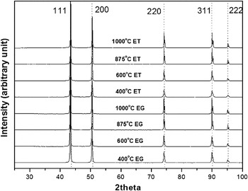
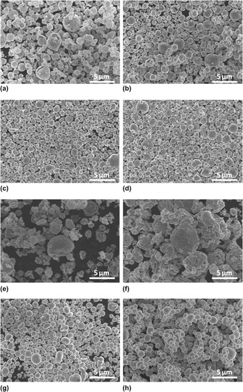
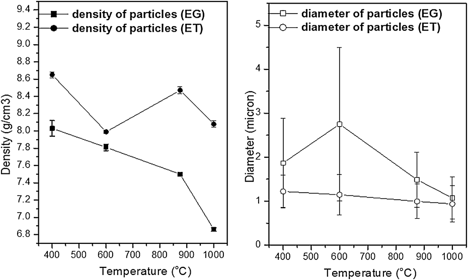
Particles were generated from 1.2 M copper nitrate solution at temperatures of 400, 600, 875, and 1000 °C 40 vol% ET was added into the solution as the cosolvent, and a low carrier gas flow rate of 2.5 L/min was used, giving a residence time from 1.5 to 3.3 s depending on the temperature. XRD of the particles generated at different temperatures (Fig. 2) showed diffraction peaks attributed to a high-symmetry face-centered cubic (FCC) structure of copper [International Center for Diffraction Data (ICDD) PDF No. 01-071–3761]. No copper oxide peaks or any other impurity peaks were detected suggesting that oxide-free copper particles were successfully generated at temperatures ranging from 400 to 1000 °C. Increasing the temperature did not change the composition of the particles. However, different particle morphologies were detected as shown in Figs. 3(a)–3(d). At 400 °C, mainly hollow particles were generated [Fig. 3(a)]. At higher temperatures, particles had smoother surfaces, and hollow particles were less prevalent but still always observed at 1000 °C. When increasing temperature from 400 to 1000 °C, the average diameter of the particles slightly decreased from 1.2 ± 0.4 μm to 0.9 ± 0.4 μm.
XRD of copper particles generated at different temperatures with ethanol (ET) or ethylene glycol (EG).

SEM images of copper particles generated at (a) 400 °C with ethanol (ET) (b) 600 °C with ET (c) 875 °C with ET (d) 1000 °C with ET (e) 400 °C with ethylene glycol (EG) (f) 600 °C with EG (g) 875 °C with EG (h) 1000 °C with EG.

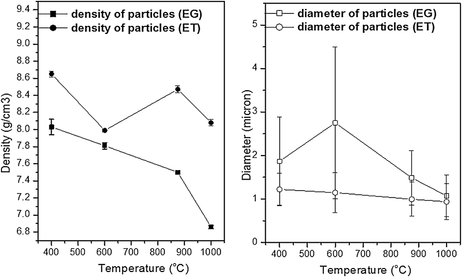
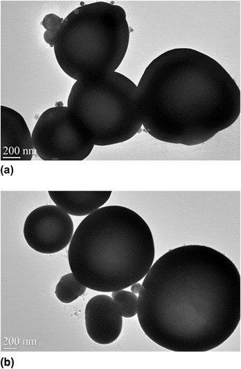
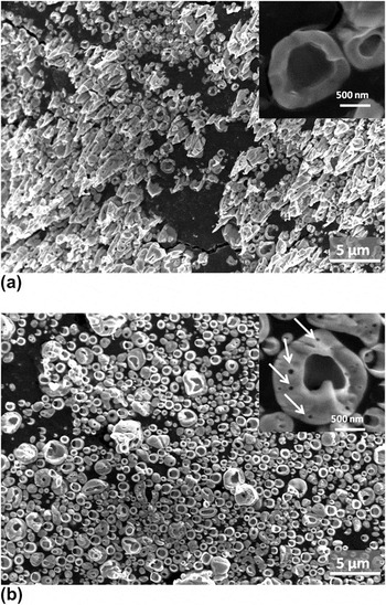
Higher temperatures allow the metal atoms to have a greater mobility promoting densification. However, according to the helium pycnometry result, (Fig. 4, red line and circle symbols) the particle apparent densities were significantly lower than the pure copper density of 8.9 g/cm3 and the density decreased with increasing temperature. The greatest density was achieved at 400 °C rather than at 1000 °C, and this counterintuitive result may be explained by considering how the density measurement is made. The helium pycnometry test measures the volume of a known mass of particles enabling one to calculate density. At 400 °C, the particles were hollow with lots of cracks allowing for better permeation of the gas into the particles such that the particles have a smaller volume and therefore a larger density closer to the theoretical value. By increasing the furnace temperature to 1000 °C, a smoother shell was formed and the gas could not penetrate into the core of the particles causing the volume of the particles to be larger and the density smaller. This was confirmed by TEM images that show the existence of less electron dense regions inside the particles, indicated by the light area in the center of each particle [Fig. 5(a)]. Cross section images of copper particles generated at 1000 °C were obtained by ion beam milling and shown in Fig. 6(a). Hollow structured particles with solid shells were observed.
Density and number average diameter of copper particles as a function of furnace operating temperature and cosolvent, ethanol (ET) or ethylene glycol (EG). The error bars represent measurement uncertainty in the case of density and the standard deviation of the particle size distribution in the case of diameter.
TEM pictures of copper particles generated at 1000 °C with (a) ethanol and (b) ethylene glycol.

(a) SEM pictures of copper particles generated at 1000 °C with cosolvent of 40 vol% ethanol. Hollow shell structures were observed. The inset image shows representative cross section morphology. (b) SEM pictures of copper particles generated at 1000 °C with cosolvent of 40 vol% ethylene glycol. Particles have been pretreated by ion bombardment for two hours. When ethylene glycol was used as cosolvent, small pores in the shell wall formed, indicated by white arrows in the inset image.

B. Copper powder generated with EG
Copper particles were generated using conditions similar to the previous experiments, but with ethanol replaced by ethylene glycol. XRD, SEM images and the density of the particles generated at different temperatures are shown in Figs. 2, 3(e)–3(h), and 4 (black line with square symbols) respectively. XRD indicates that all the particles were composed of pure copper regardless of the temperature used. SEM images in Fig. 3(e) show that, at 400 °C, porous particles were generated with pore diameters of less than one hundred nanometers. The average diameter of the generated particles was 1.9 ± 1.0 μm. In Fig. 3(a), we show the images of particles generated with ET. Most of the particles were hollow, and big cracks could be observed on the surface of the particles. Based on the analysis of the images, we can find the particle morphology exhibited differences because of the change of the cosolvent. The effect of cosolvent on particle morphology became more obvious with a generation temperature of 600 °C. With ET, each particle had a primary hollow region [Fig. 3(b)], but multiple small pores could be detected on the surface of each particle generated with EG as shown in Fig. 3(f). Further increasing temperature to 875 and 1000 °C, particle average diameter decreased to 1.5 ± 0.6 μm and 1.1 ± 0.5 μm, respectively. At temperatures higher than 875 °C, the surface of the particles became smooth, and few hollow particles were observed [Figs. 3(g) and 3(h)]. Increasing temperature from 400 to 600, 875, and 1000 °C, as shown in Fig. 4, resulted in a density decrease from 8.0 to 7.8, 7.5, and 6.9 g/cm3. The apparent densities of the particles generated with EG also decreased with increasing temperature. The low density of particles generated at high temperature may also be explained by the hollow shell structure of the particle and poor permeability of helium. From TEM pictures of particles generated at 1000 °C [Fig. 5(b)], a significant light area in the center of each particle was observed, suggesting the existence of a shell structure. All particle samples made with EG have relatively lower apparent densities and greater particle size than those made with ET, suggesting that voids inside the particles were more easily formed when EG was used.
Cross section images of copper particles generated with EG at 1000 °C were obtained by ion beam milling and shown in Fig. 6(b). Most of the particles show a shell structure with shell thickness of around 100 nm. Small pores were observed in the shell wall, indicated by white arrows. In prior literature,Reference Jain, Skamser and Kodas16, Reference Widiyastuti, Wang, Lenggoro, Iskandar and Okuyama35 formation of the shell structure was explained by the temperature and concentration gradient along the radial direction of the droplets that formed during the heating stage of the process. Higher temperatures on the surface of the droplets result in the evaporation of the solvent and the increasing of the precursor concentration within the droplet. Once the concentration is higher than the saturation concentration, the precursor precipitates at the droplet surface. It is believed that the small pores in the wall result from the initial lamellar structure of copper hydroxy nitrate [Cu2(OH)3NO3] formed during the precipitation process, which will be discussed later.
C. Reaction process investigation
When the cosolvent was changed from ethanol to ethylene glycol, a change in particle morphology was observed. For better understanding the processes occurring during particle formation, short residence time experiments were performed. In these experiments, incomplete reaction occurred and intermediate products and morphologies were observed allowing for a more detailed analysis of the particle formation process. A larger carrier gas flow rate of 10 L/min was used with only one furnace. Residence time estimates are listed in Table I. At 400 °C with 10 L/min carrier gas flow rate, it takes 0.4 s to pass through one furnace, and 3.3 s is needed if 2.5 L/min flow rate and two furnaces are used. All residence times were estimated based on the temperature measured along the furnace center line.Reference Kim, Babushok, Germer, Mulholland and Ehrman26 For the calculation, furnace was separated to n = 16 infinitesimal elements, and in each element the temperature T i was considered to be uniform. Considering the expansion effect of the gas, the time required for the carrier gas to pass through each volume element can be described by:
 $$t_{\rm{i}} = F/A \times T_{\rm{r}} /T_{\rm{i}} ,$$
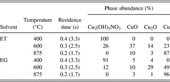
Composition of samples generated under short residence time conditions at various temperatures. Short residence times were achieved when a single furnace and 10 L/min carrier gas flow rate were used. The residence times listed in brackets were estimated based on regular experimental conditions, where two furnaces and 2.5 L/min carrier gas flow rate were used.

where F is carrier gas flow rate, A is cross section area and T r is room temperature. Residence time t was estimated by:
 $$t = \mathop\sum\limits_1^n {t_{\rm{i}} } .$$
SEM, XRD, and FTIR were used for the analysis of the particles.
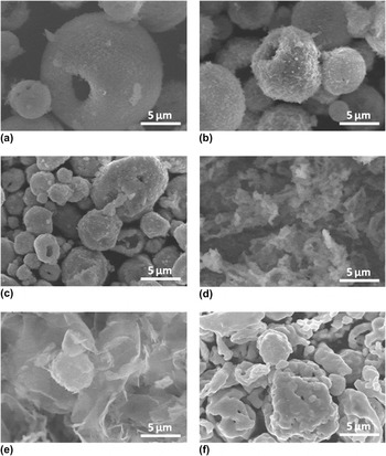
The typical morphology of particles generated at 400 °C with ET and 0.4 s residence times is shown in Figure 7(a). Under these conditions, spherical solid particles with rough surfaces and hollow structures were observed. Similar hollow structures were observed when increasing the furnace temperature to 600 and 875 °C [Figs. 7(b) and 7(c)]. The residence times were 0.3 and 0.2 s correspondingly. According to previous work, a rapid increase in temperature, resulting from high furnace temperatures, will lead to rapid evaporation of solvent and the development of a salt concentration gradient in the droplet.Reference Jain, Skamser and Kodas16, Reference Gurav, Kodas, Pluym and Xiong25 Once the salt concentration on the surface exceeds the saturation concentration, salt will precipitate out at the surface of the droplet and form a shell around the remaining liquid leading to the formation of hollow particles.
SEM images of particles generated under short residence time conditions at (a) 400 °C with ethanol (ET) (b) 600 °C with ET (c) 875 °C with ET (d) 400 °C with ethylene glycol (EG) (e) 600 °C with EG (f) 875 °C with EG.

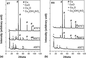
In addition to the morphology changes, the chemical reaction process was also monitored. In the article by Kim et al., they proposed a reaction mechanism for the conversion of a solution of copper nitrate in water to copper particles,Reference Kim, Babushok, Germer, Mulholland and Ehrman26 It was believed that copper nitrate [Cu(NO3)2]decomposed to CuO, which further reduced to pure copper under the highly reducing atmosphere created from ET decomposition at temperatures above 600 °C. However, our XRD results indicated that Cu(NO3)2 first reacts to give Cu2(OH)3NO3. XRD of particles generated at 400, 600, and 875 °C with 10 L/min N2 are shown in Fig. 8(a), and phase abundances of each composition in the particles were calculated based on Rietveld refinement (Table I). For the 400 °C sample, the XRD was composed of primary peaks at 12.8°, 25.7°, and 33.5° and a series of small peaks ranging from 20° to 70°, belonging to Cu2(OH)3NO3 with both monoclinic and orthorhombic crystal structures (ICDD PDF Nos. 00-015-0014 and 00-014–0687). Similar decomposition behavior for copper nitrate was also reported by LvovReference Lvov and Novichikhin36 and Jackson.Reference Jackson, Fonseca and Holcombe37 Particles generated at 600 °C were composed of Cu2(OH)3NO3, Cu, CuO, and Cu2O. Signals from CuO (ICDD PDF No. 03-065–2309) and Cu2O (ICDD PDF No. 01-077–0199) were relatively weak with primary peaks at 39° and 36.5°, respectively, overlapping with the peaks from Cu2(OH)3NO3. At 875 °C, particles were mainly Cu with 10% CuO and 3% Cu2O.
XRD of copper particles generated in short residence time at different temperatures with cosolvent of ethanol (a) and ethylene glycol (b).

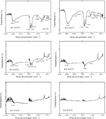
FTIR results are shown in Fig. 9 (left). For the 400 °C sample, the existence of Cu2(OH)3NO3, was supported by FTIR, which had a strong absorption from 3500 to 3200 cm−1, attributed to the stretching of O–H bonds in Cu2(OH)3NO3. O–N bonds resulted in the absorption at 1420 cm−1. Our IR data is consistent with Park’s report of Cu2(OH)3NO3,Reference Park and Lee38 except a sharp peak at 1380 cm−1, which can be explained as the bending vibration of C–H in methyl from unreacted ET. Particles at 600 °C had similar infrared (IR) absorption behavior as the 400 °C sample, but with lower intensity because most of the Cu2(OH)3NO3 has decomposed to CuO and Cu2O. Samples at 875 °C are mainly composed of Cu, and there was no IR absorption except background from potassium bromide (KBr).
FTIR of particles generated in short residence times at different temperatures with cosolvents of ET (a)-(c) and EG (d)-(f).

For checking whether use of EG results in a similar decomposition process, short residence time experiments were run using 40 vol% EG as the cosolvent. The typical morphology of particles at 400 °C is shown in Fig. 7(d). Flake and fragment-like structures were observed, indicating a different precipitation behavior during the evaporation of solvent when ET was replaced with EG. During the experiment at 400 °C and 0.4 s residence time, most of the sample on the filter was visibly wet, suggesting that the solvent had not completely evaporated. This is likely because EG is less volatile (vapor pressure of 0.06 mmHg at 20 °C) compared with water (18 mmHg at 20 °C) and ET (43 mmHg at 20 °C),Reference Speight31 and the evaporation rate is dominated by the vapor pressure if droplets are the same size and experience the same temperature history.Reference Pruppacher and Klett39, Reference Friedlander40 At 600 °C, we can observe the outline of the particles, and lamellar structures were detected. Upon increasing temperature to 875 °C, porous particles were generated as shown in Fig. 7(f).
The composition of each sample was calculated based on Rietveld refinement of XRD patterns [Fig. 8(b)] and listed in Table I. 400 °C samples were composed of 91 mol% Cu2(OH)3NO3, with the remainder consisting of 5 mol% CuO and 4 mol% Cu2O. The diffraction peaks were wider compared with the sample in ET, because EG was not totally evaporated and Cu2(OH)3NO3 did not crystallize well. At 600 °C, strong and sharp diffraction peaks resulting from pure Cu were observed, with small diffraction peaks at 36.5° and 12.8° indicating the existence of Cu2O and Cu2(OH)3NO3. Refinement showed that particles consisted of 49 mol% Cu, 29 mol% Cu2O, 10 mol% CuO, and 12 mol% Cu2(OH)3NO3. Particles generated at 875 °C contained 96 mol% copper, 1 mol% Cu2O, and 3 mol% CuO. The results indicated that EG promotes the formation of copper compared with ET at the same temperature. When ET was used as cosolvent, 23 and 87 mol% Cu were detected in the particles generated at 600 and 875 °C separately.
FTIR results are shown in Fig. 9 (right). The IR of samples at 400 °C in EG is similar to the sample in ET, suggesting that both are composed of Cu2(OH)3NO3. The detection of the absorption of O–H from 3500 to 3200 cm−1, O–N bond around 1420 cm−1 and CuO bond vibration at 511 and 430 cm−1 confirms this. However, it is important to note that the sample in EG showed a big broad peak at 3420 cm−1 rather than two separated ones which we observed in the ET sample from the Cu2(OH)3NO3. This is because some EG was still in the sample as indicated by the stretching vibration of OH, which has a broad absorption peak at 3370 cm−1. Therefore the peak detected at 3420 cm−1 in the EG generated sample is a combination of three peaks at 3450, 3430, and 3370 cm−1. The existence of EG in the sample was also supported by the signals at 2950 and 2280 cm−1 from C–C bond vibration and 1080 cm−1 from stretching vibration of C–O bond. Samples generated at 600 °C had weak FTIR signal with peaks at 3420, 1380, and 1080 cm−1. These peaks indicated that even at 600 °C, there was still some EG in the particles. The 875 °C sample was pure copper and led to no IR absorption except the background of KBr.
D. Proposed process mechanism
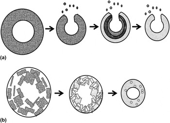
Considering all of the information from SEM, XRD, and FTIR, we propose a copper particle formation process with ET as illustrated in Fig. 10(a). After the evaporation of ET and water, copper nitrate hydrolyzes to Cu2(OH)3NO3 which then decomposes to CuO and Cu2O. Rapid evaporation of water and ET promotes the formation of a concentration gradient in the droplet and the formation of hollow structures. The final step is the reduction of copper oxide in the reducing atmosphere created by ET decomposition. When particles are generated in EG, a different precipitation process takes place as shown in Fig. 10(b). As droplet temperature increases, water evaporates, and Cu(NO3)2 hydrolyzes to Cu2(OH)3NO3 with most of EG still in a liquid state resulting in the formation of a Cu2(OH)3NO3 layer on the surface. Then these Cu2(OH)3NO3 precipitates combine to make porous particles, which have a structure as observed in Figs. 3(e) and 3(f). Higher temperatures and longer residence times allow the melting of the lamellar structure, resulting in the shell. It is hypothesized that incomplete fusing leaves small pores in the wall as shown in Fig. 6. Copper oxide could be reduced either by the CO and H2 resulting from the decomposition of EG, or by reaction with EG when both were present.
Illustration of possible copper particle formation process with cosolvents of (a) ethanol (b) ethylene glycol. Supporting information: High resolution SEM images of copper particles generated at (a) 400 °C with ethanol (ET) and (b) 400 °C with ethylene glycol (EG).

IV. SUMMARY AND CONCLUSIONS
Copper particles were generated by a cosolvent spray pyrolysis process at furnace temperatures ranging from 400 to 1000 °C with 40% ethanol (ET) or ethylene glycol (EG) used as a cosolvent with nitrogen gas as the carrier gas. We found that using ET as the cosolvent resulted in copper particles with a significant fraction of hollow structures, and big cracks were observed on the particle surface, whereas the use of EG resulted in porous structures at 400 and 600 °C, and hollow shell particles with smooth surfaces at 875 and 1000 °C. Because of the better gas permeation into hollow structure compared with porous and shell structures, particles generated with ET had an apparent density greater than those produced with EG. With either ET or EG, particle density was lower than the theoretical value, and decreased with increasing temperature. The decrease in density with increasing temperature indicates that formation of hollow shell-like particles is promoted by high furnace temperatures.
The reaction process was then investigated by short residence time experiments. Results indicated that copper nitrate first hydrolyzed to Cu2(OH)3NO3, and then decomposed to CuO and Cu2O, which was then reduced to pure copper. Differences in the evaporation rate of ET, EG and water resulted in the different precipitation behavior of Cu2(OH)3NO3, which affected the morphology of the product particles.
Results from both standard and short residence time experiments indicated that the formation process and resulting particle morphology were strongly dependent on the cosolvent used. ET leads to denser particles, but hollow structured particles could not be completely eliminated even at 1000 °C. EG promotes the formation of copper at lower temperatures compared with ET. In our experiments, micrometer-sized spherical oxide-free copper particles with smooth surfaces were generated with a cosolvent of EG at lowest temperature of 875 °C in 1.7 s, but void spaces inside the EG-generated particles were observed.
ACKNOWLEDGMENTS
This work was supported by the National Science Foundation (CBET-0755703) and by the DuPont Company. We also acknowledge the support of the Maryland NanoCenter and its NispLab. The NispLab is supported in part by the National Science Foundation as a Materials Research Science and Engineering Center Shared Experimental Facility.