Non-technical summary
Dysnoetoporidae is a family of cheilostome bryozoans including only the genus Dysnoetopora, with three recognized species from the Late Cretaceous of the United States, Crimea, and Germany. The aim of this study was to record for the first time the presence of a fossil dysnoetoporid bryozoan in South America by describing the new genus and new species Chenquepora miocenica. This taxon was found in Miocene deposits of the Chenque Formation (early Langhian, ~15.37 Ma), on the Atlantic coast of Argentine Patagonia. Chenquepora differs from Dysnoetopora in its encrusting colony and, consequently, in the absence of an endozone with long zooids arranged parallel to the direction of growth. This new record extends its paleobiogeographic distribution to the Southern Hemisphere and its stratigraphic range from the Campanian (Late Cretaceous) to the Miocene, showing that this extinct family survived the Cretaceous-Paleogene (K-Pg) mass-extinction event. Furthermore, the description of C. miocenica n. gen. n. sp., based on material from the Chenque Formation, increases the known taxonomic diversity of the phylum Bryozoa in the Neogene of Argentine Patagonia.
UUID: https://zoobank.org/c07713bd-1d9b-4c41-986d-5e4bd781d7e7
Introduction
Dysnoetoporidae Voigt, Reference Voigt1971 is a family of exclusively fossil cheilostome bryozoans that includes only the genus Dysnoetopora Canu and Bassler, Reference Canu, Bassler and Wade1926, with three species: D. celleporoides Canu and Bassler, Reference Canu, Bassler and Wade1926, D. dimissa (White, Reference White1879), and D. confusa Voigt, Reference Voigt1951. Canu and Bassler (Reference Canu, Bassler and Wade1926) initially regarded this genus as belonging to the order Cyclostomatida, without assigning it to a particular family, mainly because of the presence of long tubes with diaphragms in the central portions of the branches, and the absence of ovicells. Dysnoetopora was later transferred to the cyclostome family Heteroporidae by Bassler (Reference Bassler and Moore1953) without discussing the reasons for this decision, and maintained in this family by Toots and Cutler (Reference Toots and Cutler1962). Voigt (Reference Voigt1971) was the first to realize that this peculiar bryozoan was actually a cheilostome due to the occurrence of avicularia, D-shaped orifices suggesting the presence of opercula, communication between zooids by septula, ascopore proximal to the orifice, and absence of the typical interzooidal pores characteristic of the Cyclostomatida. Although Dysnoetopora shows some affinities with the cheilostome family Celleporidae, Voigt (Reference Voigt1971) erected the family Dysnoetoporidae because the celleporids lack an ascopore and always brood their larvae in ovicells.
The species of Dysnoetopora have erect colonies and autozooids characterized by the presence of an ascopore proximal to the orifice, an endozone with long tubes of axial zooids, and the absence of ovicells and spines. Moreover, in the type species, D. celleporoides from the Maastrichtian Ripley Formation of Coon Creek, Tennessee, USA, the proximal frontal shield of the vicarious avicularia is gymnocystal and has an ascopore (Taylor and McKinney, Reference Taylor and McKinney2006). The oldest species of the genus, D. dimissa, was found in the Mesaverde Formation, Campanian of Wyoming, USA. The status of a third species, D. confusa from the Upper Maastrichtian near Hannover, Germany, was re-evaluated by Voigt (Reference Voigt1971) who doubtfully referred this species to Dysnoetopora. Martha et al. (Reference Martha, Matsuyama, Scholz, Taylor and Hillmer2019) even doubted that the type material of D. confusa belonged to the Cheilostomatida due to its poor state of preservation.
The aim of the present study was to describe a new genus and species of the family Dysnoetoporidae based on colonies found in the Miocene of the Chenque Formation on the Atlantic coast of Argentine Patagonia. In addition, we discuss the paleobiogeographic relationships of this Gondwanan taxon in South America.
Geological setting
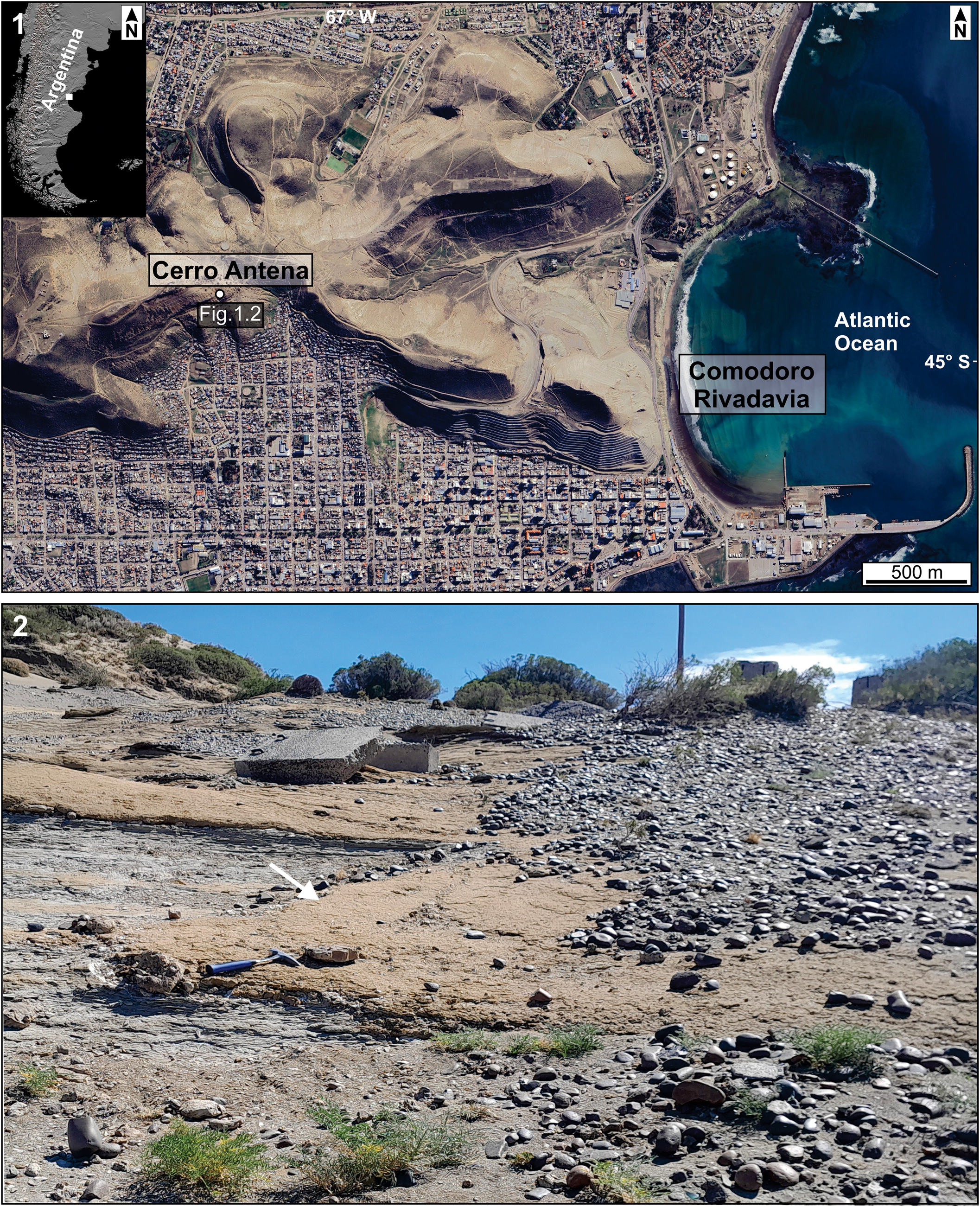
The Chenque Formation crops out near the city of Comodoro Rivadavia, Chubut, Argentina (Fig. 1.1). It has a thickness of ~350 m and consists mainly of siliciclastic deposits accumulated in shallow marine environments (Bellosi, Reference Bellosi1995; Paredes, Reference Paredes2002; Buatois et al., Reference Buatois, Bromley, Mángano, Bellosi and Carmona2003; Carmona et al., Reference Carmona, Buatois, Mángano and Bromley2008, Reference Carmona, Ponce, Buatois and Mángano2022). These clastic deposits mainly consist of psammopelitic sediments (Paredes, Reference Paredes2002), associated to a lesser extent with isolated fragments of marine organisms, mostly macroinvertebrates, and with in situ biohermal ostreid levels carrying a diverse epibiont assemblage (Del Río, Reference Del Río2004; Mooi et al., Reference Mooi, Martínez and Del Río2016; Fig. 1.2). The colonies analyzed in the present study were encrusting the inner and outer surfaces of barnacle plates detached from oysters (Fig. 2.1, 2.2).
Study area: (1) location of Cerro Antena (Chenque Formation) in Comodoro Rivadavia, Chubut, Argentina; (2) outcrop from which Chenquepora miocenica n. gen. n. sp. was recovered (arrow). Note 30 cm long rock hammer in (2) for scale.

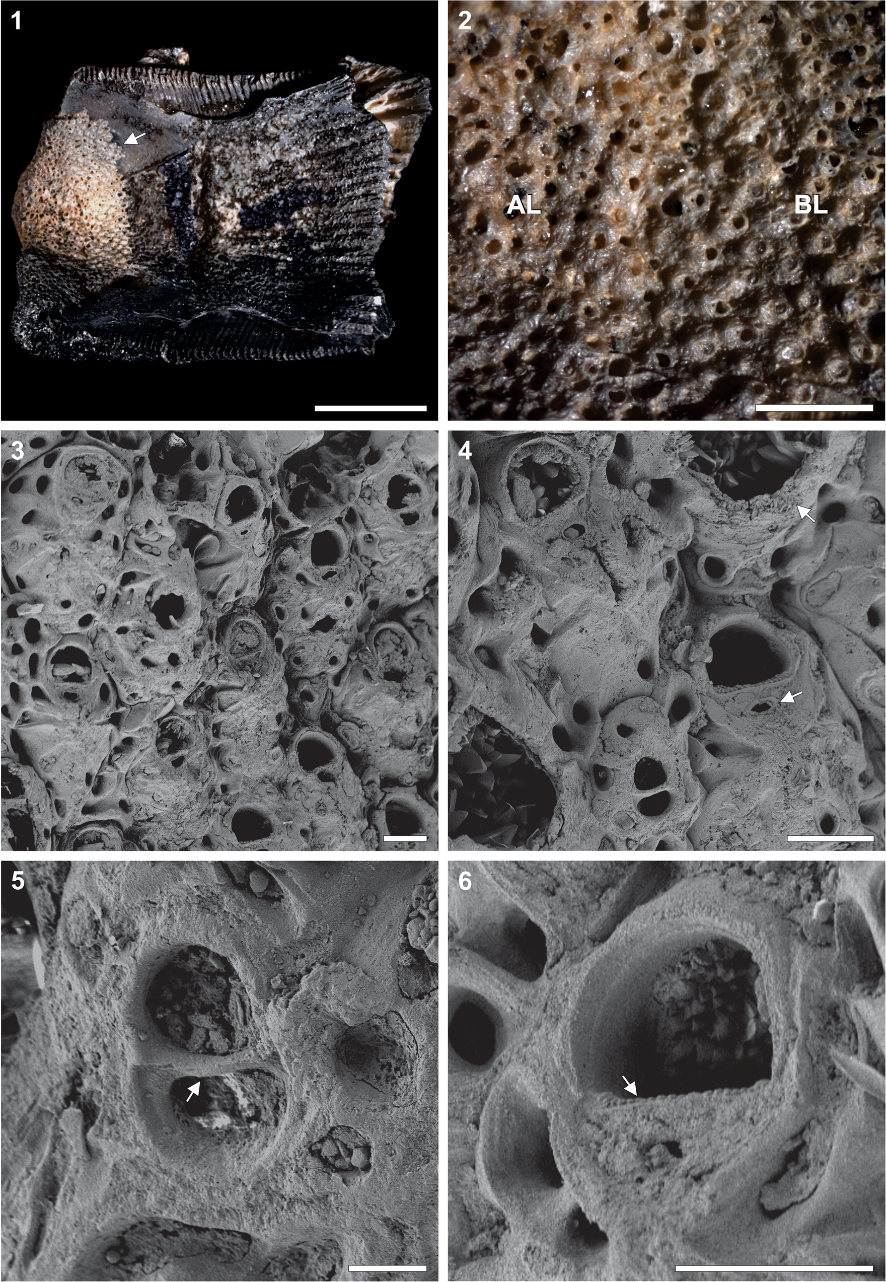
Chenquepora miocenica n. gen. n. sp.: (1) holotype, MEF 7194, colony (arrow) encrusting a barnacle plate; (2) basal layer (BL) and additional layer (AL); (3) general aspect of autozooids in the basal layer; (4) autozooids and interzooidal avicularium, with lower arrow showing the ascopore and upper arrow pointing to a growing autozooid; (5) interzooidal avicularium with complete crossbar (arrow); (6) D-shaped orifice with serrated proximal margin (arrow). Scale bars = 5 mm (1), 1 mm (2), 100 μm (3, 4, 6), 40 μm (5).

Based on 87Sr/86Sr analysis of well-preserved oyster valves, Cuitiño et al. (Reference Cuitiño, Scasso, Santos and Mancini2015) estimated the age of the fossiliferous level bearing the new taxon as 15.37 ± 0.2 Ma (early Langhian–Miocene).
Material and methods
The colonies examined in this study were extracted from a sandy/bioclastic concentration. The material was later cleaned in an ultrasonic bath and immersed in a solution of 30% hydrogen peroxide for 3 min.
The holotype and paratypes were sputter coated with gold/palladium and photographed with a Zeiss Gemini 360 scanning electron microscope (SEM) in the Museo Argentino de Ciencias Naturales (MACN). The type material was also photographed with an AxioCam MRc camera mounted on a Zeiss Stemi 2000-C stereomicroscope using the software AxioVision SE64 (https://carl-zeiss-axiovision-se64-rel.software.informer.com/4.9/). Measurements were taken from the SEM images with ImageJ 1.54 g (Schneider et al., Reference Schneider, Rasband and Eliceiri2012).
Repository and institutional abbreviation
The type material was deposited in the Egidio Feruglio Museum, Trelew, Chubut, Argentina (MEF).
Systematic paleontology
Order Cheilostomatida Busk, Reference Busk and MacGillivray1852
Infraorder Hippothoomorpha Gordon, Reference Gordon1989
Superfamily Hippothooidea Busk, Reference Busk1859
Family Dysnoetoporidae Voigt, Reference Voigt1971
Revised diagnosis
Colony erect or encrusting multilaminar, celleporiform. Frontal budding present. Frontal shield of autozooids gymnocystal, with marginal septular pores. Orifice D-shaped, proximal edge straight. Ascopore present. Orientation of autozooidal orifices variable. Avicularia interzooidal or vicarious. Spines absent. Ovicells unknown, probably absent.
Genus Chenquepora new genus
Type species
Chenquepora miocenica new genus new species.
Diagnosis
Colony encrusting, with basal layer of fully developed autozooids and overlapping layer of incomplete zooids produced by frontal budding. Autozooids with gymnocystal frontal shield and ascopore; proximal margin of orifice straight, lacking sinus. Frontal shield convex, with central umbo and unevenly distributed frontal septular pores along zooidal margins. Avicularia interzooidal, without ascopore; crossbar complete. Ovicells, spines, and axial zooids absent.
Etymology
The generic name refers to the Chenque Formation, the stratigraphic unit in which this taxon was found. Tchenque is the name that the aboriginal people of this Patagonian region gave to pre-Columbian indigenous tombs (Guichón et al., Reference Guichón, Barberena and Borrero2001).
Remarks
The differences between Chenquepora n. gen. and Dysnoetopora are summarized in Table 1. The two genera differ in the growth habit of the colony (encrusting in Chenquepora, erect and dendroid in Dysnoetopora; Canu and Bassler, Reference Canu, Bassler and Wade1926; Taylor and McKinney, Reference Taylor and McKinney2006). Consequently, the endozone with long tubes of axial zooids of Dysnoetopora is absent in Chenquepora. Moreover, the interzooidal avicularia of Chenquepora have the morphology found in most ascophoran bryozoans, whereas the peculiar vicarious avicularia of D. celleporoides have a gymnocystal proximal frontal shield and an ascopore (Taylor and McKinney, Reference Taylor and McKinney2006).
Morphological comparison between Dysnoetopora Canu and Bassler, Reference Canu, Bassler and Wade1926 and Chenquepora n. gen., mainly based on their type species.

Type specimens
Holotype, MEF 7194, one encrusting colony on the internal and external surfaces of a barnacle plate (Fig. 2.1), Cerro Antena, Chenque Formation (45°51′6.91″S, 67°30′10.88″W), near the city of Comodoro Rivadavia, Chubut Province, Argentina (Fig. 1.1). Paratypes, MEF 7195, 7196, one colony and one fragment, same data and substratum as the holotype.
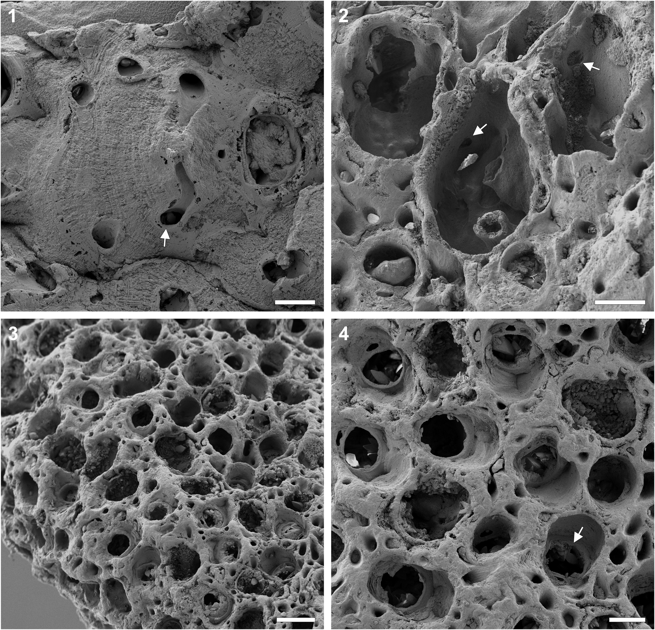
Chenquepora miocenica n. gen. n. sp.: (1) paratypes, MEF 7195, 7196, detail of one autozooid, with arrow showing a marginal septular pore; (2) internal view of the autozooids showing pore plates (arrows); (3) additional layer grown on top of the basal zooids; (4) detail of the additional layer with arrow showing the orifice and ascopore of an autozooid from the basal layer. Scale bars = 200 μm (3), 100 μm (2, 4), 50 μm (1).

Measurements (μm) of Chenquepora miocenica n. gen. n. sp., holotype, MEF 7194.

Diagnosis
Colony encrusting, with two layers of zooids. Frontal shield gymnocystal, convex, with central umbo and frontally facing septular pores along margins. Orifice D-shaped, proximal margin straight, serrated. Ascopore of the autozooids transversely elliptical. Avicularia interzooidal, ovoidal or pear-shaped, without ascopore; crossbar complete. Ovicells and spines absent.
Occurrence
Colonies of this new species were only found at the locality ‘Cerro Antena’ in the middle section of the Chenque Formation near the city of Comodoro Rivadavia, Chubut Province, Argentina (Fig. 1.1), early-middle Miocene. Despite the large amount of bryozoan material that we examined, it is worth noting that this taxon was absent at all other localities of the Chenque Formation.
Description
Colony encrusting. Basal layer with subhexagonal autozooids separated by grooves, 0.28–0.43 mm × 0.17–0.31 mm, arranged in longitudinal series (Fig. 2.3). In older parts of the colony at some distance from the growing margin, the basal layer becomes covered by an additional layer generated by frontal budding and composed of zooids without calcareous frontal shields (Fig. 3.3, 3.4). Frontal shield of basal autozooids gymnocystal, convex, with central umbo and series of ~7 or 8 marginal septular pores sunk into thickened calcification of older autozooids and simulating areolar pores within areolae; the intervening ridges converging toward umbo (Figs. 2.3, 2.4, 2.6, 3.1). Orifice D-shaped, 0.071–0.091 mm long × 0.081–0.1 mm wide; distal margin curved; proximal margin straight (i.e., lacking sinus), serrated (Fig. 2.6). Communication between zooids by pore plates (Fig. 3.2). Ascopore small, transversely elliptical, very close to proximal margin of orifice, flanked by V-shaped ridges (Fig. 2.3, 2.4, 2.6). Avicularia interzooidal, ovoidal or pear-shaped, uncommon, 0.07–0.14 mm × 0.04–0.07 mm; rostrum rounded; crossbar complete, robust, slightly curved, without ligula (Fig. 2.4, 2.5). Ovicells and spines absent. Ancestrula and early astogeny unknown.
Etymology
The specific name refers to the Miocene age of the Chenque Formation.
Remarks
One of the main differences between Chenquepora and Dysnoetopora is the encrusting colony growth habit (Table 1). Although the colonies of D. celleporoides and D. dimissa have encrusting colony bases during their early growth stages (Toots and Cutler, Reference Toots and Cutler1962; Taylor and McKinney, Reference Taylor and McKinney2006), those of Chenquepora n. gen. are completely encrusting. Another important difference is the morphology of the avicularia. In Chenquepora, these polymorphs are interzooidal, whereas in D. celleporoides, they are vicarious, spatulate, and usually larger than the autozooids. In addition, the avicularia of Chenquepora are of the type seen in most ascophorans. In D. celleporoides, however, there is a proximal gymnocystal shield pierced by a pore that might be regarded as an ascopore (Taylor and McKinney, Reference Taylor and McKinney2006).
The colonies of Chenquepora miocenica n. gen. n. sp. were always found encrusting the internal and external surfaces of barnacle plates (Fig. 2.1), implying postmortem colonization of dead and disarticulated biological substrata by the settling bryozoan larvae.
Perhaps the most striking feature of Chenquepora miocenica n. gen. n. sp. is that its colonies show two areas with markedly contrasting morphology. Near the growing margin, the colony is unilaminar, with the zooids showing a regular arrangement (Fig. 2.1). In older areas, however, a second layer of incomplete, frontally open zooids is developed, giving the colony an extraordinarily confusing exterior appearance (Fig. 3.3). Voigt (Reference Voigt1971) described a similar phenomenon in Dysnoetopora celleporoides, in which the basal zooids become deeply immersed and surrounded by immature open zooids, the frontal walls of which were not yet calcified.
Discussion
Some cheilostome families, e.g., Microporellidae, Fenestrulinidae, Haplopomidae, Calwelliidae, and Inversiulidae, share with Dysnoetoporidae the presence of an ascopore. None of these families could accommodate Chenquepora for the following differences (see also Taylor and McKinney, Reference Taylor and McKinney2006): Microporellidae and Fenestrulinidae have articulated spines (absent in Chenquepora) and lack frontal budding (present in Chenquepora); Haplopomidae and Inversiulidae have pores evenly distributed throughout the frontal shield (only marginal septular pores in Chenquepora); and Calwelliidae has erect colonies with zooids paired back-to-back (encrusting colonies in Chenquepora). Chenquepora miocenica n. gen. n. sp. is here included in the Dysnoetoporidae because of the presence of a gymnocystal frontal shield with a suboral ascopore, and the absence of ovicells and spines.
Paleobiogeographic relationships
The paleobiogeographical distribution of Dysnoetopora is limited to the Mesozoic of the Northern Hemisphere. Two species, D. celleporoides and D. dimissa, have been recorded in the Late Cretaceous (Campanian–Maastrichtian) of the United States (Canu and Bassler, Reference Canu, Bassler and Wade1926; Toots and Cutler, Reference Toots and Cutler1962; Voigt, Reference Voigt1971; Taylor and McKinney, Reference Taylor and McKinney2006). Dysnoetopora celleporoides has also been found in the Upper Maastrichtian of Crimea (Voigt, Reference Voigt1962, Reference Voigt1964, Reference Voigt1971; Taylor and McKinney, Reference Taylor and McKinney2006).
The Cretaceous Period was a time of elevated tectonic activity during which North America and Eurasia were separated from Gondwana, and the Neo-Tethys evolved into a global east-west oriented seaway (Abed, Reference Abed2013). The circumglobal Tethyan current flowed westward driven by paleowinds, thus connecting the Neo-Tethys with world oceanic basins (Abed, Reference Abed2013).
In the Southern Hemisphere, the breakup of the Atlantic margins of South America and Africa began during the Early Cretaceous (Aptian) from south to north (Lawver and Gahagan, Reference Lawver and Gahagan2003). The activation of the rift known as the Mid-Atlantic Ridge resulted in the total separation of the Brazilian northeast from the Guinean Gulf during the Paleogene. As a consequence, the early Atlantic Ocean flooded this narrow passage, generating a shallow marine connection between the central and southern oceans. Toward the Late Cretaceous (Maastrichtian), the connection between the North and South Atlantic was already fully developed, and relatively deeper areas were already present (Ladant et al., Reference Ladant, Poulsen, Fluteau, Tabor, MacLeod, Martin, Haynes and Rostami2020). Thus, the ancestors of Chenquepora n. gen. might have entered the South Atlantic from the Northern Hemisphere before the Cretaceous-Paleogene (K-Pg) extinction event. This hypothesis implies a late arrival of the Dysnoetoporidae to the Atlantic margin of Patagonia.
The presence of Chenquepora n. gen. in the Chenque Formation might alternatively be explained as the survival of a more extended Mesozoic distribution of the Dysnoetoporidae encompassing the Atlantic Ocean, much greater than the Laurasian distribution proposed for the Cretaceous. Thus, Dysnoetopora might have disappeared during the K-Pg extinction event, with Chenquepora n. gen. remaining as a relict taxon in the Southern Hemisphere until the Miocene. This last hypothesis, however, is difficult to evaluate, because there are no Cretaceous records of Dysnoetoporidae in Patagonia. Based on the available evidence, we regard the first of the two hypotheses as more plausible.
The presence of Chenquepora n. gen. in the early Miocene of Patagonia is an infrequent example of an extinct Northern Hemisphere family having a related lineage in southern South America. In contrast, the Neogene bryozoan assemblages in Atlantic Patagonia show close relationships with the Australasian bryozoan fauna (Casadío et al., Reference Casadío, Campbell, Taylor, Griffin and Gordon2010; Pérez et al., Reference Pérez, López-Gappa and Griffin2015, Reference Pérez, López-Gappa, Vieira and Gordon2020; López-Gappa et al., Reference López-Gappa, Pérez and Griffin2017, Reference López-Gappa, Ezcurra, Martha and Pérez2023). Both the Chenque Formation and the early Miocene Monte León Formation in Santa Cruz Province share several genera with similar stratigraphic units in Australia, New Zealand, and neighboring islands (Pérez et al., Reference Pérez, López-Gappa and Griffin2015; López-Gappa et al., Reference López-Gappa, Pérez and Griffin2017; López-Gappa and Pérez, Reference López-Gappa and Pérez2023).
During the early Oligocene, the complete opening of the Drake Passage caused a change in the evolution of the bryozoan assemblages of the southern Southwest Atlantic. Subsequently, the clockwise circulation of the Antarctic Circumpolar Current, driven by westerly winds, allowed entrance to the coast and shelf of Atlantic Patagonia of larvae from species inhabiting the Southeast Pacific archipelagos (Hedgpeth, Reference Hedgpeth1969; Moyano, Reference Moyano1997). In this way, the Cenozoic bryozoan assemblages of Atlantic Patagonia became more closely related to the extant bryozoan fauna of the Southeast Pacific.
Conclusions
Chenquepora miocenica n. gen. n. sp. is described based on material from the Chenque Formation, Argentine Patagonia. This new record extends the stratigraphic range of the extinct family Dysnoetoporidae from the Late Cretaceous (Campanian) to the early-middle Miocene (Langhian), thus surviving the K-Pg mass extinction event. The species of Dysnoetopora are distributed in the Northern Hemisphere, whereas C. miocenica n. gen. n. sp., the only species of Chenquepora n. gen., is the first member of the family known in the Gondwanan region. The presence of this species in Patagonia suggests a migration of representatives of this family through a north-south marine corridor during the opening of the Atlantic Ocean toward the end of the Cretaceous. The description of C. miocenica n. gen. n. sp., based on material from the Chenque Formation, increases the known taxonomic diversity of the phylum Bryozoa in the Neogene of Argentine Patagonia.
Acknowledgments
F. Tricárico operated the SEM at MACN. We are indebted to F. Oporto Romero for his help during fieldwork and to M.G. Liuzzi for her advice with the stereomicroscope photographs. M. Key Jr. and two anonymous reviewers provided helpful comments that improved an earlier version of the manuscript. This research was possible thanks to a Doctoral Fellowship of CONICET to D. Iturra.
Competing interests
No potential conflict of interest is reported by the authors.