Introduction
The Chinese alligator, Alligator sinensis Fauvel, 1879, is listed as a critically endangered species and is exclusively native to eastern China and primarily distributed in the middle and lower reaches of the Yangtze River and Taihu Lake. Crocodiles, in common with other reptiles, usually harbour a wide variety of protozoan and metazoan external and internal parasites (Tellez, Reference Tellez2013). In other parts of the world, information on parasitic infections of crocodiles is richly available (Paperna & Lainson, Reference Paperna and Lainson2003; Enabulele et al., Reference Enabulele, Ozemoka, Awharitoma and Aisien2013). One species of pentastome (Sebekia oxycephala), two species of nematodes (Dujardinascaris waltoni and Multicaecum tenuicolle), four species of trematodes (Polycotyle ornata, Acanthostomum coronarium, Archaeodiplostomum acetabulatum and Pseudocrocodilicola americaniense) and one species of hemogregarine (Haemogregarina crocodilnorum) were recovered from American alligators from South Carolina (Hazen et al., Reference Hazen, Aho, Murphy, Esch and Schmidt1978). The presence of eight helminths (Acanthostomum americanum, Pelaezia loossi, Telorchis sp. juvenile, Pseudoneodiplostomum groschafti sp. n., Dujardinascaris helicina, Contracaecum sp. Type 2 larvae, Micropleura sp. and Paratrichosoma recurvum) was revealed in the Morelet's crocodile from the Lagoon of Celestún, Yucatan, Mexico (Moravec, Reference Moravec2001). Nevertheless, little is known about the parasite fauna of Chinese alligators, although Wang et al. (Reference Wang, Nie and Wang1995) showed that nematodes in the alimentary tract of the Chinese alligator induced loss of appetite and mortality. Wang et al. (Reference Wang, Huang, Yang, Jiang, Niu, Zhao, Yu, Deng and Chen2007) collected faecal samples of crocodiles from Chengdu Zoo, Sichuan, China, and incidentally identified cysts of Amoeba in one faecal sample of captive Chinese alligators. Zhao et al. (Reference Zhao, Zhou, Wang, Tu, Tang and Wu2014) found some cysts of protozoa and eggs of nematodes and trematodes in faecal samples of A. sinensis from the National Nature Reserve of Chinese Alligator.
In order to contribute further to this list of parasites, the present study records the occurrence of Ortleppascaris sinensis sp. nov. collected from the stomach and upper intestine of A. sinensis during recent investigations of Chinese alligators in the National Nature Reserve of Chinese Alligator, Xuanzhou, southern China. The specimens were identified using both morphological and molecular approaches.
Materials and methods
Collection and examination of nematodes
In July and August 2014, two adult Chinese alligators, A. sinensis Fauvel, 1879 (total body length 162 cm and 197 cm), were collected by wardens of the National Nature Reserve of Chinese Alligator in Anhui (30°94′N, 118°79′E), namely Chinese Crocodile Lake, an important breeding centre in southern China that plays a vital role in the conservation of A. sinensis (Thorbjarnarson et al., Reference Thorbjarnarson, Wang, Ming, He, Ding, Wu and McMurry2002). The alligators were immediately electrically shocked and subsequently killed for examination of parasite prevalence. Nematodes were recovered and washed in physiological saline and then fixed in hot 70% ethanol. For light microscopy examination, nematodes were cleared with lacto-phenol. Drawings were made with the aid of an Olympus BX51 drawing attachment (Olympus, Japan). For scanning electron microscopy (SEM), specimens were fixed in 2.5% glutaraldehyde, post-fixed in 1% osmium tetroxide, dehydrated through a graded ethanol series, critical-point dried and sputter coated with gold. Specimens were examined using an S-4800 field emission scanning electron microscope at an accelerating voltage of 15.0 kV. All measurements are given in millimetres unless otherwise indicated.
Molecular analysis
Three nematodes were used for molecular analysis, which was based on the sequences of the complete internal transcribed spacers, ITS (ITS1, 5.8S ribosomal rDNA and ITS2), partial small subunit DNA segments (18S) and the cytochrome oxidase I (COI) gene. Tissue for DNA extraction was taken from the middle part of the body while taxonomically important anterior and posterior parts were preserved for morphological identification. Genomic DNA was obtained using traditional phenol–chloroform extraction. The 18S rDNA, ITS and COI segments were amplified according to the original descriptions, using the primers Nem_18S_F (5'-CGCGAATRGCTCATTACAACAGC-3') and Nem_18S_R (5'-GGGCGGTATCTGATCGCC-3') (Floyd et al., Reference Floyd, Rogers, Lambshead and Smith2005), NC5 (5'-GTAGGTGAACCTGCGGAAGGATCATT-3') and NC2 (5'-TTAGTTTCTTTTCCTCCGCT-3') (Gasser et al., Reference Gasser, Rossi and Zhu1999), and LCO1490 (5'-GGTCAACAAATCATAAAGATATTGG-3') and HCO2198 (5'-TAAACTTCAGGGTGACCAAAAAATCA-3') (Folmer et al., Reference Folmer, Black, Hoeh, Lutz and Vrijenhoek1994), respectively. Polymerase chain reaction (PCR) products were electrophoresed in 1% agarose gels, excised and purified with a column DNA gel extraction kit (Sangon Biotech, Shanghai, China), and then automatically sequenced (ABI 3730, USA). The nucleotide sequences obtained were compared using BLAST against known sequences in GenBank.
Results
Morphology of Ortleppascaris sinensis sp. nov.
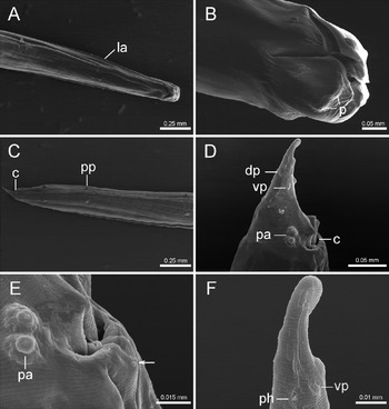
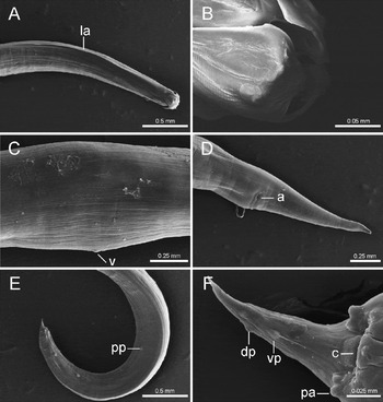
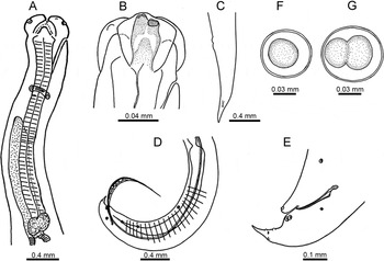
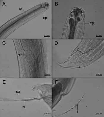
General. Live worms move rapidly forward by twisting the body with active movement of the anterior end, whereas the body in situ makes a slow curving movement. The colour of living specimens is oyster white. The body appears slender without being coiled, with males being smaller than females. There are three well-developed lips, two ventral and one dorsal; the three lips are almost equal in size. Lips are longer than they are wide, with thin delicate margins, and without denticles or interlocking processes. Each lip has a prominent unpaired median lobe of pulp. Interlabia are well developed, triangular, rounded anteriorly, reaching anteriorly more than half the length of the lips (figs 1A and B, 2B). Cervical alae and lateral thickenings of the cuticle are present in the oesophageal region (figs 1A, 2A). The excretory pore is nearer to the lips than to the nerve ring, opening just behind the ventral interlabium (figs 3A and B, 4A and B). Cervical papillae are present behind the level of the nerve ring. The oesophagus is muscular, relatively stout and terminates in an irregularly lobed ventriculus containing nuclei of the oesophageal glands (figs 3A, 4C). There is a long anterior dorsal intestinal caecum (figs 3A, 4A). The male has a slender, sharp gubernaculum (Reference Enabulele, Ozemoka, Awharitoma and Aisienfigs 3E, Reference Floyd, Rogers, Lambshead and Smith4D) and alate spicules (figs 3D, 4E). The male tail is slender, pointed, slightly curved ventrally, and bears on each side four widely spaced precloacal papillae, two narrow spaced paracloacal, one subdorsal and two subventral postcloacal papillae, and one lateral papilla at the level of the centre of the gubernaculum (Reference Baylisfigs 1C and Reference Enabulele, Ozemoka, Awharitoma and Aisien3D). The mature female is wide in the posterior third of the body, with a long, tapering tail (figs 2D, 3C). The vulva is smooth, without lip or papilla (fig. 2C), anterior to the mid-region of the body. The vagina is relatively long, extending posteriorly to form the undivided part of the uterus.
Ortleppascaris sinensis sp. nov., male. (A) Ventral view of the anterior end. (B) Sublateral view of the cephalic end; lips have thin, delicate margins, each lip with a prominent unpaired median lobe of pulp; interlabia reach anteriorly more than half the length of the lips. (C) Subventral view of the posterior end, in which four pairs of precloacal papillae are easily seen. (D) Detail of the tail. (E) Amplification of the region of the cloacal opening, ventral view; arrow indicates median small pre-anal papilla. (F) Posterior part of the tail, lateral view. la, Lateral alae; p, pulp; pp, precloacal papillae; c, cloacal aperture; pa, paracloacal papillae; vp, subventral postcloacal papillae; dp; subdorsal postcloacal papillae; ph, phasmid.

Ortleppascaris sinensis sp. nov.: (A) subdorsal view of the anterior end of the female; (B) sublateral view of the dorsal lip of the male, lips have thin delicate margins; (C) lateral view of the vulva of the female; (D) subventral view of the tail of the female; (E) lateral view of the posterior end of the male showing the four precloacal papillae; (F) detail of the posterior part of the male tail, lateral view. la, Lateral alae; v, vulva; a, anus; pp, precloacal papillae; c, cloacal aperture; pa, paracloacal papillae; vp, subventral postcloacal papillae; dp; subdorsal postcloacal papillae.

Ortleppascaris sinensis sp. nov.: (A) anterior end of the male, lateral view, showing the oesophagus, nerve ring, excretory pore, ventriculus and intestinal caecum; (B) right subventral lip of the female, showing the position of the excretory pore; (C) tail of female, lateral view; (D) posterior end of the male, lateral view, showing spicules and the ejaculatory duct; (E) tail of male, lateral view, showing gubernaculum; (F) eggs, content of eggs uncleaved; (G) eggs, content of eggs cleaved into two blastomeres.

Ortleppascaris sinensis sp. nov.: (A) anterior end of male, lateral view; (B) cephalic end of the female; (C) anterior end of male; (D) tail of male, lateral view; (E) posterior part of the male tail; (F) end of spicule. nr, Nerve ring; ep, excretory pore; v, ventriculus; g, gubernaculum; sa, alae of spicule; s, spicule.

Males (based on nine specimens; measurements of holotype in parentheses, units mm unless stated): smaller than females; body length 26.85–38.62 (33.82), maximum width 0.58–0.92 (0.58). Lips/labia 0.14–0.19 (0.16), and interlabia 0.08–0.11 (0.10) long. Behind the lips, the cuticle is thickened in the lateral region (figs 1A, 4A and B), 1.77–2.04 (1.94) long, thickening to its widest behind the nerve ring, ending about halfway along the oesophagus (fig. 1A). The excretory pore is situated anteriorly, just behind the lips in a mid-ventral position (figs 3A, 4A). The length of the oesophagus is 2.80–3.90 (3.16), with maximum width 0.14–0.28 (0.19), representing 8.08–12.52% (9.34%) of the body length. The ventriculus is 0.16–0.30 (0.23) long and 0.17–0.30 (0.23) wide. The nerve ring is 0.66–0.95 (0.77) from the anterior extremity. The intestinal caecum is 1.90–2.93 (2.50) long. The tail is 0.17–0.25 (0.17) long, slender, pointed and slightly curved ventrally (figs 2E and 4D). On each side of the tail are four precloacal papillae (figs 1C, 2E, 3D), and one lateral (fig. 1C), two paracloacal, one subdorsal and two subventral postcloacal papillae (figs 1D, ). There are two median small pre-anal papillae (fig. 1E). Lateral phasmids are found near the subdorsal papilla (fig. 1F). Spicules are long, slender and equal in length, 6.44–13.49 (10.27), representing 24.00–34.93% (30.38%) of the body length, with delicate alae along the middle part (figs 3D, 4E), and a gradually tapering sharp point (figs 3D, 4F); the anterior end is slightly enlarged in the form of a cylinder (fig. 3D). The gubernaculum is 0.15–0.17 (0.17) long, and sharply pointed with a slender shaft and rounded proximal expansion (figs 3E, 4D). The ejaculatory duct is 1.72–2.78 (2.69) long, and slender, with a slightly expanded anterior end (Fig. 3D). The seminal vesicle is wide. No caudal alae or ridges are present in the region of the cloacal aperture.
Female (based on two specimens; measurements of allotype in parentheses): bodies are more robust than those of males; the body length is 44.77–49.53 (44.77) with maximum width 1.04–1.20 (1.04). Lips are 0.19–0.27 (0.19) long, and interlabia are 0.07–0.14 (0.07). Distinct lateral alae (fig. 2A) are 2.31–2.65 (2.31) long, extending about 55.78–56.21% (56.21%) the length of the oesophagus. The excretory pore is hole-like, opening in the ventral interlabium between subventral lips (figs 3B, 4B). The oesophagus is 4.11–4.75 (4.11) long, with maximum width 0.25–0.26 (0.25), representing 9.18–9.60% (9.18%) of the body length. The ventriculus is 0.26–0.36 (0.26) long and 0.26–0.36 (0.26) wide. The nerve ring is 0.97–1.10 (0.97) from the anterior extremity. The intestinal caecum is 3.42–3.78 (3.42) long. The vulva is 19.57–22.13 (19.57) from the anterior end, situated at 43.71–44.68% (43.71%) of the body length. The vulva is smooth, without lip or papilla (fig. 2C). The vagina is 1.45–1.56 (1.45) long, extending posteriorly to form the undivided part of the uterus. Two uterine branches extend in opposite directions. There is a lengthy tapering tail, 1.21–1.27 (1.21) long, with a sharply pointed tip (fig. 2D). Phasmids are indistinct, and about three-quarters of the distance from the anus to the tail (figs 2D, 3C).
Eggs: vary from almost spherical to oval, are thin-walled with a smooth surface, and measured 75.12–98.80 × 60.63–79.68 μm. The content of the eggs was mostly uncleaved or cleaved into more than two blastomeres (figs 3F and G).
Molecular evidence
Sequences from three different O. sinensis sp. nov. specimens were identical (100% pairwise identity) for the ITS, 18S rDNA and COI gene amplified fragments. Excluding primers, 859, 891 and 645 base pairs were obtained for the ITS (GenBank KM891739), 18S rDNA (GenBank KM891740) and the COI gene (GenBank KM891741), respectively. The sequences obtained in this study were used in GenBank for BLAST search, but, unfortunately, there is only one sequence of Ortleppascaris sp. registered in GenBank for ITS (JQ654456), 18S (JQ654455) and COI gene (JQ654457) among the genus Ortleppascaris. These sequences are for Ortleppascaris sp. reported from the livers of cane toads, Rhinella marina (Linnaeus, 1758), captured in eastern Brazilian Amazonia (Silva et al., Reference Silva, Melo, Silva, Goncalves, Giese, Furtado and Santos2013). The maximum score (query coverage, maximum identity) in BLAST analysis was 239 (18%, 93%), 750 (47%, 98%) and 682 (100%, 86%) for ITS, 18S and COI gene sequences, respectively. The nematodes O. sinensis sp. nov. and Ortleppascaris sp. are from different hosts and territories. BLAST analyses supported the morphological identification that O. sinensis sp. nov. is a new biological species.
Taxonomic summary
-
Type host. Chinese alligator Alligator sinensis Fauvel, 1879 (Crocodilian: Alligatoridae).
-
Site in host. Stomach and upper small intestine.
-
Type locality. The National Nature Reserve of Chinese Alligator (30°94′N, 118°79′E), located in the middle and lower reaches of the Yangtze River, Xuanzhou, Anhui Province, the People's Republic of China (collected in July and August 2014).
-
Prevalence and intensity. Two male Chinese alligators examined were both infected; seven specimens in one Chinese alligator and four specimens in the other alligator.
-
Type material. Holotype, allotype and paratype are all deposited in the Department of Parasitology, Wannan Medical College, Anhui Province, China (nos: WNMC-N-100 to WNMC-N-106).
-
Etymology. The specific name of this nematode relates to the genitive form of the species name of the host.
Remarks
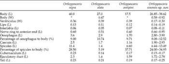
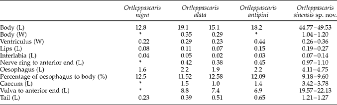
Nematodes O. sinensis sp. nov. observed in the present study are different in size and morphology from those recorded in other crocodiles (tables 1 and 2). Male O. sinensis sp. nov. in the Chinese alligator were found to be longer than O. antipini and O. alata, but shorter than O. nigra (table 1). Ortleppascaris sinensis sp. nov. have the largest lips and interlabia, the smallest ventriculus, and the longest distances from the nerve ring to the anterior end among these species. The caecum and ejaculatory duct in the present specimens are longer than those in O. antipini and O. alata, but shorter than those in O. nigra. However, among the specimens obtained in our study, the tail length (0.173–0.247) is similar to that observed in the three other species of Ortleppascaris (table 1). Among females, specimens of O. sinensis sp. nov. are the longest (table 2). Correspondingly, O. sinensis sp. nov. have the largest lips, interlabia, oesophagus, caecum, tail and the longest distances from the nerve ring to the anterior end, and from the vulva to the anterior end (table 2).
Comparative morphometrics (measurements in mm) of male Ortleppascaris nigra, O. alata and O. antipini in crocodile hosts after Sprent (Reference Sprent1978) with O. sinensis in the present study; L= length, W= width, * measurement not given.

Comparative morphometrics (measurements in mm) of female Ortleppascaris nigra, O. alata and O. antipini in crocodile hosts after Sprent (Reference Sprent1978) with O. sinensis in the present study; L= length, W= width, * measurement not given.
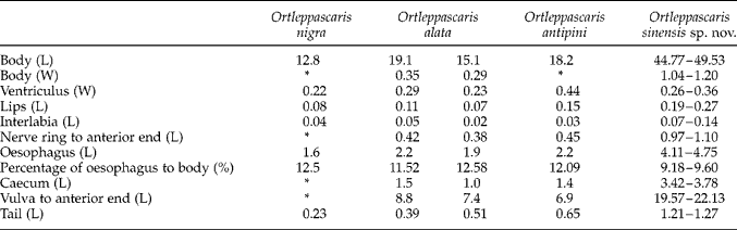
Until the present study, SEM has been used for the description of the Ortleppascaris nematode parasites O. nigra, O. alata and O. antipini in crocodiles and alligators by Sprent (Reference Sprent1978), and for the larvae of Ortleppascaris sp. parasites in cane toads by Silva et al. (Reference Silva, Melo, Silva, Goncalves, Giese, Furtado and Santos2013). According to the observations of structures by SEM and light microscopy, including the position of the excretory pore, the shape of the lips, gubernaculum and tail, and the spicules with delicate alae along their middle part, specimens observed in the present study are similar to O. antipini recorded in Alligator mississippiensis (Sprent, Reference Sprent1978). However, the two median, small pre-anal papillae observed in male O. sinensis sp. nov. have not been described in O. antipini. The position of phasmids observed in the new species are also different (3/4 vs. 2/3 from anus to tail), and the end point of the lateral alae along the oesophagus is also different (1/2 vs. 3/4). Moreover, morphological measurements are also different, as described above (tables 1 and 2). Otherwise, compared with O. antipini males recorded by Sprent (Reference Sprent1978), males of the present material are distinctly larger (26.851–38.622 vs. 15–27), but the percentage of spicules to body length was smaller (24.00–34.93% vs. 38–49%). Compared with O. antipini females, our specimens were longer (44.774–49.529 vs. 29-39), and the vulva was situated further from the anterior end (43.71–44.68% vs. 35–37%).
There is only one previously reported study of the genetic characteristics of the genus Ortleppascaris sp. (Silva et al., Reference Silva, Melo, Silva, Goncalves, Giese, Furtado and Santos2013), and there is a great discrepancy in the genetic characteristics of the ITS (0.7%) and 18S (0.2%) and COI (14.0%) genes between Ortleppascaris sp. and O. sinensis sp. nov. Today, mitochondrial DNA (mtDNA) is most effective for identifying nematodes, especially for closely related species, and for researching genetic variations, etc. The COI gene is a good genetic marker within the mitochondrial DNA. In this study, the COI gene showed the greatest difference (14%) compared with Ortleppascaris sp. The molecular data presented here represent an important advance in understanding the phylogenetics of O. sinensis sp. nov. and will contribute to future studies in population genetics.
Discussion
Ortleppascaris was removed from the genus Dujardinascaris (Sprent, Reference Sprent1977) by Sprent (Reference Sprent1978) and placed in a new genus Ortleppascaris according to differentiated characteristics of the lips, ventriculus, cuticle and excretory system. Sprent (Reference Sprent1978) pointed out that the species of Ortleppascaris have delicate wing-like margins, weakly developed interlabia, an irregularly shaped ventriculus, long cervical alae and an excretory pore near the subventral lips. In his article, Sprent described three species of the genus, O. nigra (Geodelst, Reference Geodelst1916) Sprent, Reference Sprent1978, O. alata (Baylis, Reference Baylis1947) Sprent, Reference Sprent1978 and O. antipini (Mozgovoy, Reference Mozgovoy1950) Sprent, Reference Sprent1978. As of today, O. nigra, O. alata, O. antipini and O. sinensis sp. nov. seem to be the only four species of this genus parasitizing crocodiles and alligators as adults. Some larvae of Ortleppascaris have also been observed in toads and frogs (Moravec & Kaiser, Reference Moravce and Kaiser1995; González & Hamann, Reference González and Hamann2007; Silva et al., Reference Silva, Melo, Silva, Goncalves, Giese, Furtado and Santos2013). Adults of these Ortleppascaris species have been observed in the gastrointestinal tracts of A. mississippiensis, Crocodylus moreletii, C. rhombifer, C. niloticus, C. cataphractus, Osteolaemus tetraspis, Caiman crocodilus, C. yacare, C. intermedius, Melanosuchus niger and Crocodylus sp. (Sprent, Reference Sprent1978; Goldberg et al., Reference Goldberg, Bursey and Aquino-Shuster1991; Catto & Amato, Reference Catto and Amato1994; Moravec & Kaiser, Reference Moravce and Kaiser1995; Scott et al., Reference Scott, Simcik and Craig1999; Huchzermeyer, Reference Huchzermeyer2003; Waddle et al., Reference Waddle, Kinsella, Ross, Rojas-Flores, Percival and Forrester2009; Tellez, Reference Tellez2013); the biogeography of Ortleppascaris was recorded in Florida, Louisiana, Texas, Mexico, Cuba, Russia, Congo, Zimbabwe, Trinidad, Venezuela, Brazil, Paraguay and Eastern India (Sprent, Reference Sprent1978; Goldberg et al., Reference Goldberg, Bursey and Aquino-Shuster1991; Catto & Amato, Reference Catto and Amato1994; Moravec & Kaiser, Reference Moravce and Kaiser1995; Scott et al., Reference Scott, Simcik and Craig1999; Huchzermeyer, Reference Huchzermeyer2003; Waddle et al., Reference Waddle, Kinsella, Ross, Rojas-Flores, Percival and Forrester2009; Tellez, Reference Tellez2013). However, this is the first report of Ortleppascaris infection in China; it is also the first record of the parasite in A. sinensis. The recovery of O. sinensis sp. nov. from Chinese alligators in China represents a new geographic record for these parasites and adds to the number of helminths known to infect this crocodile.
Acknowledgements
We greatly appreciate authorization from the National Nature Reserve of Chinese Alligator, Xuanzhou, Anhui Province, China for sample collection. We are also greatly indebted to Professor Chaopin Li (Wannan Medical University) for assistance in identifying the nematode and for encouragement during this study. The authors’ thanks are also due to the staff of the Laboratory of Electron Microscopy, Testing Centre of Yangzhou University, for their SEM technical assistance.
Financial support
This study was supported by the National Natural Science Foundation of China (nos: 31272337 and 31472019) and Key Project of Wannan Medical University of China (nos: WK2013Z04).
Conflict of interest
None.