

INTRODUCTION
Delirium, an acute confusional state, is a difficult diagnostic concept. It is a syndrome lying on a continuum between full consciousness and deep coma. The symptoms of delirium are protean. They range from frenetic hyperactivity to a withdrawn, hypoactive state and result from biotoxic stressors overwhelming the internal resources and defenses of the brain. There may be considerable fluctuations of symptoms. Periods of relative lucidity occur as may sundowning (the exacerbation of symptoms as night falls). This flux lessens as further organic damage occurs and coma ensues.
Delirium is an inevitable consequence of the dying body and brain. It is common during the final 24–48 h of life, occurring in up to 88% of terminally ill cancer patients (Lawler et al., 2000). Delirium, particularly with hypoactive features, is underrecognized. In the elderly, up to 70% of all deliria are missed by the clinician (Rockwood et al., 1994). Diagnosing delirium is clearly problematic to clinicians. Such are the uncertainties about the concept that Rockwood and Bhat (2004) suggested abandoning the notion of the brain being a specific victim in delirium in favor of the brain being an innocent bystander. They proposed that delirium should be considered as a physical and not a “mental disorder.” Not being able to clinically describe brain failure should not, however, lead to abandoning the disorder “delirium,” and to focus only on the primary causes. Modern psychiatric diagnostic classification systems are mostly descriptive rather than etiologically based. There is however a psychopathologic logic to the symptoms of delirium. They are not just a cluster of assorted symptoms; they emerge from specific pathology in the brain. A better understanding of the concept of delirium might improve diagnostic acumen. And nowhere is this more important than in palliative medicine. Palliating confusion in those precious last days of life should be addressed with the same rigor as are pain, nausea, and emotional distress. The fluctuations of the mental status are both the key to understanding and to managing the syndrome. There are not definitive, evidence-based delirium management guidelines (Britton & Russell, 2000), despite four millennia of anecdotal descriptions and clinical observations. The personal distress and behavioral disruptions created by delirium in the dying may be able to be palliated and the quality of remaining life improved by careful assessment and management.
Several terms are used to describe the deficits of delirium; rarely are they defined, and often they are used loosely and inconsistently. The intent of this article is to review the terminology, clarify that which is most appropriate based on our understanding of the pathophysiology of delirium, and to present a simplified clinical concept of the syndrome.
DEFINING DELIRIUM
The Diagnostic and Statistical Manual of Mental Disorders, fourth edition (DSM-IV) criteria defines delirium as a disturbance of consciousness associated with a change of cognition, a rapid onset, fluctuation of symptoms, and evidence of organic etiology (American Psychiatric Association, 1994). Clinically determining “consciousness” is difficult. Much of the confusion and lack of clinical clarity concerning delirium is accounted for by this conceptual problem. The poorly defined terminology used, the use of the term “consciousness” to refer both to clinical and philosophical concepts, and the genuine difficulty in delineating an “interstate” clinical phenomenon compound the clinician's uncertainties.
In neurology, the terms “consciousness” and “awareness” are often used synonymously, and being conscious presupposes that the subject is “awake” and can be “aroused.” Ross (1991) and Breitbart and Sparrow (1998) suggested that disorders of the “arousal” system and of “cognition” are the primary abnormalities of delirium. The terms “attention” and “awareness” are referred to in DSM-IV, but not “arousal.” Trzepacz and Meagher (2005) suggested that the two main components of consciousness are “wakefulness” (“alertness”) and “awareness.” Burns et al. (2004) suggested that in delirium tremens a “hyperalert” state ensues and the patient responds to all external stimuli indiscriminately. These authors also interchange the terms “hyperalert” and “hyperactive.” Breitbart and Sparrow (1998) added “hyperarousal” to describe this subtype of delirium. “Hypervigilance” may be used to describe frenetic delirium. The Delirium Symptom Interview, crucial to the evolution of DSM-IV criteria for delirium, associated “hyperactivity” with “hypervigilance,” and “hypoactivity” with “unawareness” and “decreased alertness” (Liptzin, 1999). Geschwind (1982) considered delirium to be a global impairment of “attention.” Lipowski (1980, p. 7) stated that “it appears clinicians, by and large, excel in observing and describing natural phenomena, but often display deplorable looseness in the use of words that are indispensable for labeling, classifying, and explaining what is observed and recorded.” Consciousness, awareness, arousal, alertness, vigilance, awakeness, motoric activity, and attention are states requiring precise definition, despite being strongly interconnected (Young & Pigott, 1999).
Consciousness
Greiner (1817) pioneered the concept of “clouding” of consciousness as the main pathogenetic feature of delirium, and this influence prevailed up to DSM-III in 1980 (Lindesay, 1999). Clouding of consciousness was related to awareness and attention. However DSM-IIIR in 1987 dropped “clouding of consciousness,” for it considered it difficult to measure, and used reduced level of consciousness in its place. Difficulty keeping awake during examination was said to be an indicator of “reduced” level of consciousness rather than “clouding.” Differentiating “clouding” from “reduced” consciousness is surely clinically uncertain. DSM-IV in 1994 reintroduced “disturbance of consciousness” as the primary feature of delirium, and DSM-IVTR retained this emphasis (American Psychiatric Association, 2000). ICD 10 (World Health Organization, 1992) considers “impairment of consciousness” as the major diagnostic criterion and “attention” as being the indicator of this but does not refer to “awareness.” James (1890) defined consciousness as the awareness of oneself (or one's cognitive experience) and the environment. For the clinician, consciousness is that state that allows cognition to occur (Lipowski, 1985). Cognition refers to all the processes involving perception, remembering, creating imagery, and thinking (Lipowski, 1980). Being conscious allows us an appreciation, an awareness, of our “self” and its relationship with our environment. The level of consciousness is the essence of delirium. However there is not a simple or easily clinically applicable tool with which to measure this. The EEG is impossible to perform in disturbed patients and a numerical scale for delirium equivalent to coma scores such as the Glasgow Coma Scale has yet to be designed.
Moruzzi and Magoun (1949) implied that the capacity for consciousness depends on activity in the brain stem and diencephalon, which exert a powerful influence on the cerebral hemispheres. Jackson conceptualized consciousness within the hierarchy of the nervous system, and suggested that damage to lower levels of organization would result in global and protean mental symptoms (Ey, 1962). Damage to the lowest levels, such as the brain stem, disrupts consciousness and thus awareness (Dietrich, 2003). Full consciousness is dependent on intact (evolutionary) old and new brain functioning. Consciousness is an amalgam of awareness, alertness, awakeness, arousal, attention, and vigilance. These features are dependent to a degree on separate physiological, psychological, and anatomical systems, yet are strongly interconnected. Consciousness does not have a specific anatomy or biochemistry. The prefrontal cortex is not the “seat” of consciousness (Dietrich, 2003), but it is “en route” and on top of the hierarchy. Consciousness is the product of a functionally complete nervous system. Determining consciousness at the bedside is just too difficult.
Awareness
Awareness is the content of consciousness and is dependent on an intact cerebral cortex (Bateman, 2000). Awareness is the content of higher mental functioning (Trzepacz, 1999). Being unaware, and alert, is to be in a persistent vegetative state. Awareness is typically very disturbed in delirious states and characterized by impairments of global cerebral functioning, including perceptual, motor, emotional, and thinking abnormalities. To use the radio as an analogy, awareness is equivalent to being “tuned in.”
In delirium, awareness is relatively easily determined to be abnormal because of the vast array of symptoms in multiple domains of cerebral functioning. Any and all mental symptoms may occur in delirium. Most commonly patients have compilations of psychotic, affective, motor, and cognitive symptoms. Hallucinations (particularly visual, occasionally tactile), delusions (usually paranoid), altered mood (depressed, manic, labile), thought disorders, word-finding difficulties, disinhibited behaviors (anger, inappropriate) and a fragmented or reversed sleep–wake cycle all may occur. Awareness involves cortical thalamic structures but is reliant on intact subcortical functioning. Awareness is intricately related to the lower level function of alertness, which is moderated by diencephalic structures.
Awareness is influenced by an electrophysiological phenomenon known as the gamma or “40-Hz rhythm.” This electrical oscillation is produced by thalamocortical circuits that synchronize or link the various regions of the brain to foster functional integration. Episodic bursts of enhanced energy assist the complex task of the central nervous system (CNS) in organizing and formatting the information it has available in order to coalesce it into concerted function (Young and Pigott, 1999). There is an inherent pulsatile activity of the brain, which is likely to be expressed as fluctuating performance.
Alertness
Alertness refers to a state of enhanced readiness to receive and process information and to respond (Brown & Bowman, 2002). It is a general state of cognitive readiness. Alertness refers to the level of consciousness (Bateman, 2000). When alert, stimuli are processed more efficiently and responses are initiated more rapidly. Alertness enhances the rapidity of orienting responses, focusing the head and eyes to the target of interest, thus enhancing information processing (Brown & Bowman, 2002). Alertness is the key that activates awareness. It is a prerequisite for awareness to occur. To use the radio analogy, alertness is equivalent to “loudness.” High alertness is an attentive state. To be alert one needs an intact Ascending Reticular Activating System (ARAS) with intact connections to diencephalic structures (Bateman, 2000). The ARAS gates sensory input and modulates cortical excitability and motor output by its connections to thalamic and limbic structures. The visual system and the orienting responses are essential for alertness and are probably impaired early in delirium. Alertness is intimately related to awakeness, for, when one is asleep, the ability to respond to the environment is lessened. It is not possible to determine a single neurochemical system for alertness (Brown & Bowman, 2002).
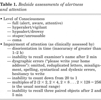
The level of consciousness, the position on the continuum between full consciousness and deep coma, is determined by the alertness of the individual. At the bedside, evaluating alertness is a key task. Descriptors such as a hyperalert (vigilant, frenetic) state, obtundation (lethargus, torpor, hypoalertness), stupor (unrousability), or coma are crude indicators of the level of alertness. These terms, although by no means particularly objective, do convey a differential response to the environment. They provide useful crude clinical information.
Alertness shows a diurnal pattern. The most alert periods are during the morning, there tends to be a low point in the midafternoon, and then an early evening rise. The most difficult time to maintain alertness, if awake, is between 2 a.m. and 6 a.m., when the body temperature is lowest. An increasing sleep deficit decreases alertness. Fatigue associated with the pathology causing a delirium may well be so profound that it overrides the ability to stay alert. Foods (such as warm milk and bananas) may moderate alertness and facilitate sleep; psychostimulant medications enhance alertness, as does mild exercise. Stimulus load governs, to some extent, alertness. If this is very low, then the organism reacts sluggishly. If it is very high, a catastrophic-type response may occur, the brain is overwhelmed and is transiently functionally paralyzed by the bombardment. Alertness is in a state of constant flux.
Vigilance, the focused readiness to detect and respond to a given stimulus, is a state of directed alertness. It can be distinguished from alertness, which is a more generalized state. A given stimulus may be processed by a vigilant brain yet it may not react to less specific stimuli because of its state of impaired alertness. With vigilance, one is alert to particular possibilities, threats, and dangers. With enhanced vigilance, general performance tends to suffer, attention scatters, and ultimately paranoia may intervene (Brown & Bowman, 2002).
Arousal
Traditionally arousal, rather than alertness, has been the term to describe fundamental brain activation. Arousal is a more general term referring to activation of the peripheral physiological state as well as forebrain cortical activation (Brown & Bowman, 2002). The Yerkes–Dodson law states that in very high states of arousal, mental performance falls off and alertness fades. Alertness and arousal are similar but distinct states. The relationship between arousal and performance is not necessarily linear (Brown & Bowman, 2002). Factors that increase alertness may also (but not always) increase arousal, but very high arousal creates physiological effects such as tachycardia and sweating, and these may be distracting. Thus cognitive impairment can be associated with high arousal. Arousal and alertness may conflict. Too little arousal, or mental lethargy, as may occur in severe illness or fatigue, may not be sufficient to stimulate alertness. Alertness and arousal are both mildly enhanced in hypomanic states and superior cognitive performance can result; however, in mania, mental performance can fall off dramatically. Arousal refers not only to the associated involvement of the autonomic nervous system but can also refer to motoric activity, which in delirium may be hyperactive, hypoactive, or, most commonly, a mixture of activity.
Awakeness
Since the early 17th century, a relationship between sleep, dreams, and delirium has been considered. Hunter described delirium as a “diseased dream arising from a diseased sleep” (Lipowski, 1980, p. 16), and Heinroth suggested that delirium was a state intermediate between sleep and wakefulness (Lindesay, 1999). The similarities between delirium and dreaming are obvious. The often disrupted sleep–wake cycle of delirium and the environmental unawareness associated with deep sleep, which is not total, make the relationship between sleep and delirium a close one. Awakeness is a predominantly voluntarily controlled off–on switch for “consciousness,” in the sense that at bedtime we are able to intentionally induce this change. Zeman (2001) considered one meaning of consciousness to be that of the waking state, and indeed this has been the classical viewpoint. One needs to be awake to be conscious. Yet agitated delirious patients are not asleep in the usual sense of the word. The persistent vegetative patient is “awake but not aware” (Zeman, 2001). Patients in “minimally conscious states” are awake but have only partial preservation of awareness and responsiveness.
The ARAS is the most important activating system in the brain linking the brain stem and the thalamus. Romano and Engel (1944) demonstrated generalized slowing of the EEG patterns in delirious subjects. Diffuse theta and delta waves and poor organization of the background rhythm are the features. These features are different from those of sleep. Awake subjects have alpha and beta waves and, if alert, low-amplitude, high-frequency beta waves dominate. The presence and proportion of beta activity is correlated with a subjective sense and behaviors of alertness. Theta waves emerge with drowsiness; delta waves arise in “slow-wave” sleep. The neurotransmitters involved in the maintenance of wakefulness include cholinergic pathways from the brain stem to the basal forebrain, noradrenergic pathways from the locus coereleus, and dopaminergic and serotonergic pathways from the brain stem. Consciousness is not merely being awake, and delirium is not being asleep.
Attention
For attention to occur alertness and awakeness must be present. James (1890, p. 381) considered attention “the taking possession by the mind, in clear and vivid form, of one of what seem simultaneous possible objects or trains of thought.” Moscovitch (1979, p. 379) defined attention as “a control process that enables the individual to select, from a number of alternatives, the task he will perform or the stimulus he will process, and the cognitive strategy he will adopt to carry out these operations.” Because we can't process all sensory inputs simultaneously, there must exist processes that select some inputs and filters out others (Vecera & Luck, 2002). These processes collectively are referred to as “attention.” Improving organization of internal and external sensations by attending to them provides the template of primitive cognition. The ability to pay attention and select relevant stimuli, register and monitor extraneous stimuli, and coherently organize those of importance are lost in delirium (Geschwind, 1982). Unfocused and distracted selectivity results in chaotic cognitions and the typical symptoms of delirium. Visuospatial attention is also impaired in delirium (Mach et al., 1996). This process provides higher level cerebral structures with the raw material onto which can be attached and fixed cerebral function. Several brain regions are involved in attention—frontal, area 46 of the prefrontal cortex, the right hemisphere, and the thalamus (Trzepacz, 1999). Cholinergic neurotransmitters (and reciprocally dopamine) are particularly involved with attention, especially in the thalamus (Trzepacz, 1999). Attention is the sentry gate of consciousness (Zeman, 2001); however it alone may not be sufficient for consciousness. Blindsight refers to the ability to register spatial cues even though these cues and stimulus are unseen. Attentiveness does not invariably lead to consciousness, though the association is generally a close one.
Attention can be objectively measured at the bedside. Objective measures of awareness, alertness, arousal, vigilance, and awakeness are most difficult. Inattention, distractabilty (the failure to select), and perseveration (the inability to shift attention) are objective, though secondary or indirect indicators of alertness and awakeness. They can serve as clinical barometers of consciousness. Though epiphenomenon, inattention is a valuable clinical index of delirium.
DIAGNOSING DELIRIUM
The most clinically overt sign of delirium is that of brain stem dysfunction, or alertness. Delirium is a primarily a disorder of alertness, and the disorders of arousal, awareness, and attention are consequential. Bedside assessment of alertness is coarse. Deficits in arousal and awareness, although apparent in delirium, are difficult to quantify. In determining attention, the clinician has a tool to better differentiate the normal state and the pathological state of brain failure, and subsequently gauge progression. Attentional deficits, which incorporate memory and language impairment, can be objectively quantified.
Attention serves as an indirect indicator of alertness and awakeness. Inattention can be assessed. Inability to direct, focus, and sustain attention represents inattention. Many bedside tests, such as 100–7 s, are not only most difficult to explain to delirious persons, they are genuinely difficult for most. Counting down from 20 to 1 and reversed spelling of the patient's name or of a five-letter word are simple tests of inattention and also involve aspects of concentration and recall. Multiples of two is a useful bedside test of attention and arithmetic dexterity. It is easy to explain, commences very gently, quickly becomes challenging and there is an approximate usual or normal range.
If attention is impaired, the registration of new information does not occur and immediate and short-term memory loss results. Geschwind (1982) considered paramnesia, the distortion rather than the loss of memory, as more typical in delirium than actual forgetfulness. Disorientation has been considered to be a relevant clinical sign of delirium. Orientation to time of the day is the most valid clinical test of disorientation. Day, date, month, and year are of lesser relevance as a personalized watch or phone supplies this information, supermarkets are open every day of the week, and sports are played most days of the year. It is surprising how often at the bedside delirious patients muddle mornings and afternoons. Uncertainty regarding time of day is frequent in delirium. Misidentification of other people (usually mistaking the unfamiliar for the familiar), a positive symptom, is a consequence of poor attention to detail or compromised registration.
Assessing language function, although dismissed by some authors such as Trzepacz and Meagher (2005), can be rewarding and acceptable to the patient. Language requires accurate attentiveness to learned and remembered cognitive skills. Simply asking a patient to write his or her address on a piece of blank paper is not difficult even for the physically frail, provided, of course, that they are not blind or hemiplegic. Complex tasks are the first to falter with compromised cerebral function. Written language is symbolic representation of symbols (words); thus even with mild impairment of cerebral activity it is affected. Dysgraphia is not only a reasonably sensitive diagnostic aid in delirium, it is an easily applicable serial assessment of clinical progress (Macleod & Whitehead, 1997; Baranowski & Patten, 2000).
Of the core symptoms of delirium, disorganized thinking, or loss of coherence (Geschwind, 1982), is most difficult to quantify. This lack of clarity of thinking, literally confusion, is not, however, exclusive to delirium. Delusions, illusions, visual hallucinations, disinhibition, moodiness, and fear are diagnostically nonspecific and “downstream” (in terms of CNS hierarchy). Psychomotor behavior may be an exception and a gauge of level of consciousness. Hippocrates considered that “silent” delirium had a grave prognosis (Lipowski, 1980). Hypoactive delirium is closer to the state of unresponsiveness or coma than is hyperactive delirium. Positive symptoms, the hyperactivity, are no longer possible in reaction to the declining level of consciousness. Hyperactive delirium, not surprisingly, has the best treatment response (Stagno et al., 2004).
Validated delirium rating scales, of which there are at least 13, have been strongly advocated by the literature (Leentjens & van der Mast, 2005). A study by Zou et al. (1998) showed that psychiatrists had lower sensitivity and specificity in diagnosing delirium than the Confusion Assessment Method (CAM; Inouye et al., 1990). The CAM is generally considered to be the best validated diagnostic tool and is the most widely used and best studied. It does not attempt to quantify mental status aberrations. “Disorganized thinking,” a major feature in the CAM, is not specific to organic dysfunction. The key advantage of the CAM algorithm is that it is brief, nonintrusive, and able to be used by nonpsychiatric clinicians. The Mini-Mental State Exam (MMSE) is designed for the assessment of cognitive impairment, but is not specific for delirium. For a frail patient it is a grueling task to complete. Fayers et al. (2005) found that delirium could be fairly accurately screened for by using just 4 of the 20 items on the MMSE. These are tests of orientation (current year and date), spelling a 5-letter word backwards, and copy design. There are many other psychometric instruments for evaluating delirium. The core issues for clinicians with respect to using such tools is whether or not they are easy to administer, able to be completed by the patient, and clinically valid. Otherwise they tend not to be used. Probing for symptoms and pleading for cooperation, in an attempt to complete an examination, enhances anxiety, unnecessarily advertises to the patient their cognitive inadequacies, and invokes anger and sometimes aggression (Stedeford, 1978). If a person can conform to the usual clinical practice of participating in the acquisition of a history and a mental status examination they are unlikely to be delirious. Patient characteristics, symptom characteristics, and clinician preferences are so varied that such tests tend not to be used as they might be. Although they are invaluable for research, these methods are not user friendly for palliative care patients (and doctors). Relying on the basic medical assessment format of history and examination, at least for physicians, allows for accommodation to the patient's limitations, yet the gathering of adequate clinical information. A history from others, a crude estimate of alertness, and a couple of measures of attention may be as much as can be attained from a frenetic patient (Table 1). Clinically this is usually sufficient. Dying is not an appropriate occasion to burden patients with unnecessary questioning and examination.
Bedside assessments of alertness and attention

THE DELIRIANT THRESHOLD
The existence of delirium is determined by the characteristics of the organic insult and those of the host. The absolute strength and activity of the noxious agent(s), together with the vulnerability of the person to such an insult are the critical factors. Environmental influences may moderate these risks by muting the toxin or strengthening the host (Lipowski, 1980). A clinical “point” or threshold is reached at which the brain fails and a disturbance of consciousness occurs. Conceptualizing delirium in terms of a “threshold” model, a model well established for epilepsy, is particularly useful in understanding the various risks factors and the interrelationship between predisposing and precipitating factors (Sherrington, 1906; Leentjens and van der Mast, 2005). If vulnerability at baseline is low, patients may be resistant to delirium despite exposure to significant precipitating factors, but if vulnerability at baseline is high, delirium is likely to occur with exposure to only minor precipitating factors (Inouye & Charpentier, 1996). The balance between predisposing (or vulnerability) and precipitating factors sets the “deliriant” threshold. Each individual has his or her own unique threshold, which may change over time and in response to internal and external demands.
The risk factors for delirium are multiplicative rather than additive (Inouye, 1998). Age (the extremes of age), preexisting dementia, medical illness, alcohol abuse, depression, diminished activities of daily living, abnormal serum sodium, and learning and visual impairments (Elie et al., 1998; Inouye, 1998) all lower the deliriant threshold. A previous delirium is a risk factor for postoperative delirium, with an odds ratio of 4.08, which is greater than the odds ratio for age over 70 (3.2) and that of preexisting cognitive impairment (2.16; Litaker et al., 2001). This suggests that a kindling type phenomenon may occur for delirium as for epilepsy. Impaired brain reserve lowers the baseline deliriant threshold and allows weaker precipitating insults to result in dysfunction. The lucid interval between a delirogenic insult and the presentation of delirious symptoms, sometimes hours to days later, is likely to be an index of the deliriant threshold. The preventative and protective influences of environmental and psychosocial factors are apparent to clinicians and do influence the deliriant threshold (Meagher, 2001).
Sherrington (1906) applied the threshold concept to a single neuron; yet it is as relevant to the brain in its entirety. In epilepsy, the therapeutic goal is to reduce the risk of seizure activity by raising the convulsive threshold. This is achieved by moderating the environment and by chemically raising the biological point of catastrophe. For a very few epileptics, surgery can remove the cause, but for most the cause is irreversible. Irreversible delirium should be managed with a similar philosophy of care. The deliriant threshold needs to be raised by biopsychosocial (or multicomponent) interventions.
THE FLUCTUATIONS OF CONSCIOUSNESS
Trzepacz and Meagher (2005) stated that the underlying reason for fluctuating symptoms in delirium is poorly understood. The combined influences of normal biological rhythm and the organism's reaction to disease provide some explanation. Alertness, awakeness, and awareness have a degree of inherent variability. In the healthy brain, consciousness waxes and wanes. In the failing brain, a more dramatic influence on stability occurs.
John Hughlings Jackson (1835–1911), the English conceptual neurologist and founder of “the modern discipline,” based his theoretical understanding of the function of the nervous system on the philosophy of Herbert Spenser. The CNS is evolved on evolutionary principles, and, upon dissolution, its function reverts to that of an earlier stage of development. A loss of structure in the nervous system results in a compensatory increase of functioning of lower order structures. This gives rise to new forms of behavior that may be interpreted as “symptoms.” J. R. Reynolds (1828–1896) referred to negative and positive symptoms as the negation of vital properties and excess or alterations of vital function respectively (Berrios, 1985). Jackson used these terms in his description of a functional hierarchy of nervous activity (Power, 1965). Damage to the nervous system “releases” the inhibition over the remaining healthy tissue. The Doctrine of Dissolution is the reverse of the process of evolution (Berrios, 1985). Using epilepsy as the clinical model, Jackson suggested that “a loss or defect of consciousness” permits increased automatic behavior. Under certain circumstances, the organism partially or wholly reverts to an earlier stage of functional organization (bodily or mentally), thus providing it the opportunity to subsequently repair and regain the former more sophisticated functioning (Power, 1965). A reduction from the voluntary to the automatic, and from the complex to the simple, occurs in response to physical (and psychological) damage to the CNS. There are similarities to Freud's theory of psychological regression, for Freud was influenced by Jackson. Disease does not create, it sets free (Ey, 1962). In delirium, as the level of alertness diminishes “positive” symptoms are released. Thoughts and memories are expressed uncensored, and uninhibited emotions spill out. But retained are also facets of clarity of idea and feeling. Lucid periods eventuate, though just how clear mental functioning is during these periods has not been investigated. The biological rhythm of consciousness and the deteriorating or ongoing disease determines that these phases of clarity are transient. The pulsatile nature of cerebral functioning is amplified by the ebb and flow of neurological regression and compensatory reaction.
THE PHILOSOPHY OF DELIRIUM MANAGEMENT
Comprehension of the core deficit of delirium, alertness, not only accounts for the huge variety of symptoms but it also directs the clinician to identify the gross clinical signs associated with the disorder. From the perspective of the clinician, the critical assessment features are those of the history and estimates of alertness and attention. Appreciating the fluctuations of symptoms and a threshold for symptom expression provides the philosophy of management. Reversing causation may not be possible in terminal delirium, but raising the deliriant threshold by multicomponent interventions can result in palliation of symptoms and the improvement of the quality of remaining life. The threshold model encourages an appreciation of the individualistic risk factors and the usual multiplicity of etiology, particularly in palliative care patients.