Introduction
Rhabdomyoma is an extremely rare condition originating from striated muscle, mostly from the heart. These tumours commonly occur in the head and neck, especially in the upper aerodigestive tract.Reference Carron, Darrow and Karakla1 There are two tissue types of rhabdomyoma in the head and neck: fetal and adult. Rhabdomyomas are benign and extremely slow-growing. Patients usually present with hoarseness, dysphagia and globus-like symptoms. Treatment involves excision of the offending lesion, thereby limiting growth and preserving surrounding tissues. This treatment is usually curative. However, recurrence can occur, and can indicate sarcomatous change or incomplete excision.
Laryngeal rhabdomyomas are extremely rare, with only approximately 40 cases being reported worldwide. They usually affect men over the age of 40 years. Here, we present a male patient whose diagnosis had been missed on several occasions, and whose subsequent surgical management led to the correct diagnosis and treatment.
Clinical record
A 76-year-old man was seen in the head and neck clinic following years of dyspnoea and noisy breathing. On this occasion, he presented with progressive, severe hoarseness and dysphagia.
He had an Epworth score of 19 out of 24, and was being treated by the respiratory physicians for obstructive sleep apnoea (OSA) with continuous positive airway pressure (CPAP) at night. He had been diagnosed as having OSA following an overnight sleep study, the results of which suggested severe OSA (with an apnoea–hypopnoea index of 35 and three desaturations to a maximum of 72 per cent).
On examination, the patient had vesicular breathing.
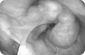
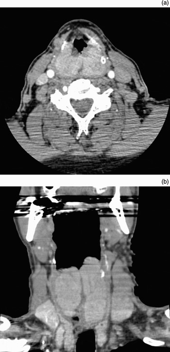
Transnasal laryngoscopy showed massively enlarged arytenoid tissue bilaterally, partially obstructing the patient's airway and both piriform fossae (Figure 1). A contrast-enhanced computed tomography (CT) scan showed lobulated masses 3.5 cm in diameter (Figure 2). Neck examination was normal. Magnetic resonance imaging (MRI) of the neck indicated symmetrical, lobular thickening of both aryepiglottic folds extending into the post-cricoid region. The laryngeal airway was clearly compromised.
Transnasal laryngoscopic view of the arytenoid cartilage. Bilaterally enlarged arytenoid cartilages are clearly seen encroaching on the airway.

(a) Axial computed tomography (CT) scan at the level of the glottis, showing bilaterally enlarged arytenoids. (b) Coronal CT scan of the larynx showing bilateral globular enlargement of the arytenoids.

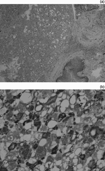
Following discussion with the head and neck multidisciplinary team, incisional, laser-assisted biopsies were undertaken on both sides to establish a definitive diagnosis. Histopathological evaluation revealed the lesions to be synchronous, bilateral rhabdomyomas of the laryngeal arytenoids (Figure 3).
(a) Photomicrograph of mucosa with adherent epithelium (right) and rhabdomyoma (left). (b) Photomicrograph with immunostain showing uniform positivity for desmin, a muscle marker. The cells were also positive for myoglobulin, another muscle marker, but negative for keratins, macrophage marker and S100 protein.

Post-operatively, the patient's breathing and snoring improved significantly. Further resections were planned in order to debulk the same area, in an attempt to fully remove the lesions and further improve the patient's symptoms.
The first laser-assisted debulking procedure was uneventful.
However, five months later, endoscopic examination revealed that the lesions had returned. A second debulking procedure was conducted, with moderately severe bleeding due to removal of the tumour capsules in an attempt to prevent recurrence. At this point, a tracheostomy was deemed necessary to protect the patient's airway.
The patient went on to make a good recovery, and was subsequently decannulated with a good swallow and patent airway. He no longer required CPAP, and his snoring had settled.
Unfortunately, the patient's symptoms deteriorated over the next four months. Examination revealed that the lesions had once again returned. A third resection was performed and the tracheostomy re-sited. Further histopathological examination again confirmed the diagnosis and its benign nature.
At the time of writing, the patient was six months post-resection and living with a tracheostomy in situ; however, both he and his wife were unhappy about the amount of care required. There were early signs of tumour recurrence in the same locations.
A dilemma therefore existed: if the patient's tumours progress, should local resections be continued, or should a laryngectomy be performed for symptom control (a controversial step in light of his benign condition)?
Discussion
Rhabdomyomas of the larynx are extremely rare, benign tumours of striated muscle. They are found in adults and children and are categorised separately from cardiac rhabdomyomas. Extracardiac rhabdomyomas are classified as fetal, genital or adult types.Reference Willis, Abdul-Karim and Di Sant'Agnese2
Genital rhabdomyomas, as the name suggests, are usually found in the vulva or vagina in women aged 20–45 years.
Fetal rhabdomyomas are either myxoid or cellular. Myxoid fetal rhabdomyomas are usually found in the subcutaneous tissue of the head and neck, commonly in the postauricular region in children younger than three years of age.Reference Granich, Pilch, Nadol and Dickersin3 Their histological appearance is of elongated, spindle or oval-shaped cells with a vague cytoplasm and striated muscle similar in appearance to that seen in uterine development in the first trimester (weeks seven to 12). Immunohistochemical staining is positive for desmin and vimentin, with less uptake of myoglobin than in adult rhabdomyoma. Cellular fetal rhabdomyomas occur in the head and neck region of adult men.Reference Di Sant'Agnese and Knowles4 Their histological appearance is more of a cellular than a myxoid type, containing elongated, spindly, immature skeletal muscle cells with a lack of stroma.
Adult type rhabdomyomas (nearly always) occur three times more commonly in men than women over the age of 40 years.Reference Kapadia, Meis, Frisman, Ellis, Heffner and Hyams5 Common sites include the base of the tongue, floor of the mouth, pharynx and larynx. Uncommon sites include the orbit, lip, cheek, soft palate, uvula, oesophagus, mediastinum and stomach.Reference Bastian and Brocker6–Reference Box, Newman and Anastasiades10 The clinical history is of a progressive worsening of symptoms due to slow tumour growth and benign pathology. Previous case reports have reported progressive hoarseness, dysphagia, airway obstruction, a palpable mass and serous otitis media from eustachian tube obstruction.
Macroscopically, adult type rhabdomyomas are red-brown or grey in colour, lobulated, and quite soft.Reference Bastian and Brocker6 Their histological appearance is characterised by large ovoid or polygonal cells with eosinophilic cytoplasm.Reference Willis, Abdul-Karim and Di Sant'Agnese2 The nucleus is usually large and situated peripherally, and the nucleoli are small. The cytoplasm is granulated due to the glycogen present in the cell. Ultrastructural features include hypertrophic z bands, intranuclear inclusions and mitochondria with linear intracristal inclusions.
The radiological features are usually those typical of benign neoplasms. Extracardiac rhabdomyomas have well circumscribed margins, with no invasion of surrounding tissue or the submucosa.Reference Liang, Loevner and Kumar11 Computed tomography findings may mimic the appearance of a malignant lesion, with indistinct borders and blending with adjacent isodense muscle tissue. On the other hand, MRI (especially T1- and T2-weighted images) is much better at delineating rhabdomyomas from surrounding structures, as they appear isointense or hyperintense to muscle and are homogeneously enhancing without necrosis or haemorrhage.
The diagnosis of laryngeal rhabdomyoma is made using a combination of clinical and histopathological findings. The differential diagnosis for such patients includes cysts, laryngocoeles, squamous cell carcinoma, amyloidoma, neurofibroma, granular cell tumours, leiomyoma, rhabdomyoma and rhabdomyosarcoma. In our patient, all of these were considered in the first instance, but inconclusive evidence from the early tissue samples meant that the final diagnosis was not made until the patient's disease had progressed significantly.
Treatment of extracardiac rhabdomyoma, as in our patient, consists of complete excision whilst attempting to maintain the integrity of surrounding tissue structures such as vocal folds and swallowing apparatus.
Recurrence commonly occurs in more than one-third of cases, due to incomplete excision.Reference Kapadia, Meis, Frisman, Ellis, Heffner and Hyams5 In elderly patients, such recurrence does not appear to be a problem as the lesion is slow-growing. Rhabdomyomas, although benign, must be followed up over a long period of time to monitor their slow growth. Recurrence should alert the clinician to the possibility of sarcomatous change. Rhabdomyosarcomas grow faster, and have the histopathological features of atypical mitotic figures, nuclear pleomorphism and foci of invasion.Reference Canalis, Platz and Cohn12
We recommend endoscopic local resection for the symptomatic patient fit enough to tolerate the procedure. Laryngectomy should be reserved for patients with severe or progressive symptoms not responsive to local resection, or for those with sarcomatous change.