Introduction
Aspergillus fumigatus is a filamentous fungus which is considered a major respiratory pathogen in birds. This filamentous fungus was first found in the lungs of a bustard (Otis tarda) in 1863 by Fresenius (Arné et al., Reference ARNÉ, THIERRY, WANG, DEVILLE, LE LOC'H, DESOUTTER, FEMENIA, NIEGUITSILA, HUANG, CHERMETTE and GUILLOT2011). Active proliferation and sporulation of this common soil saprophyte on organic material produces large amounts of airborne small-sized asexual spores (conidia) that are easily dispersed in air, and can be inhaled and deposited deep in the respiratory tract (Girma et al., Reference GIRMA, ABEBAW, ZEMENE, MAMUYE and GETANEH2016). As with other opportunistic pathogens, the consequences of inhalation of A. fumigatus conidia ultimately depend on the interaction between host (e.g. the integrity of the innate immune system) and microbial factors (virulence traits). Moreover, it has become clear that the virulence of A. fumigatus is multifactorial, and that the expression of specific virulence traits is often determined by the type of underlying immunosuppression of the host. Current evidence suggests that A. fumigatus has evolved mechanisms that aid in its survival in the environment by conferring a competitive advantage against microorganisms that share the same ecological niche. These mechanisms include thermotolerance, the secretion of extracellular proteases, nutritional scavenging, and extensive secondary metabolism.
Susceptible hosts develop polymorphic clinical forms in relation to either localised or disseminated lesions. Acute aspergillosis generally occurs in young birds resulting in high morbidity and mortality. The chronic form is sporadic, causes less mortality and generally affects older birds, especially breeders which can have a compromised immune system due to poor husbandry conditions.
A. fumigatus has been isolated from lesions in wild birds since the early 1800s. Major die-offs of free-ranging wild and captive birds have been reported following the dumping of mouldy grain in areas where birds feed (Arné et al., Reference ARNÉ, THIERRY, WANG, DEVILLE, LE LOC'H, DESOUTTER, FEMENIA, NIEGUITSILA, HUANG, CHERMETTE and GUILLOT2011). Infection by Aspergillus spp. has been reported in almost all domesticated avian species and production types. In these birds, the spontaneous outbreaks may cause mortality ranging from 4.5% to 90%, whereas the age of diseased birds varied from three days to 20 weeks (Arné et al., Reference ARNÉ, THIERRY, WANG, DEVILLE, LE LOC'H, DESOUTTER, FEMENIA, NIEGUITSILA, HUANG, CHERMETTE and GUILLOT2011). Apart from direct losses related to mortality, FCR and growth rate in recovering birds remain poor. Indeed, airsacculitis is a major reason for carcass condemnation at slaughter inspection. The economic significance of aspergillosis is most readily apparent in turkey production where the disease occurs late in the growing cycle or primarily affects costly breeder toms.
Poultry exposure and pathogenesis
Aspergillus species are ubiquitous saprophytes that inhabit poultry environments, in particular soil and organic debris (compost). Initial contamination of poultry farms may occur through the use of a mouldy litter or introduction of one-day-old birds where their down has retained conidia in hatchery facilities. Further contamination may involve inappropriate bedding management, poor quality feedstuffs, or admission of outside air loaded in conidia (Girma et al., Reference GIRMA, ABEBAW, ZEMENE, MAMUYE and GETANEH2016). Organic substrates such as litter, feed, and even feathers can easily fulfil the colonisation requirements of A. fumigatus (Tekaia and Latgé, Reference TEKAIA and LATGÉ2005). Humidity and temperature conditions encountered in poultry farms promote the rapid growth of hyphae and efficient asexual multiplication resulting in a copious production of easily airborne hydrophobic conidia, which are subsequently dispersed and inhaled by the birds (Girma et al., Reference GIRMA, ABEBAW, ZEMENE, MAMUYE and GETANEH2016). Constant animal movements under high stocking densities, litter refreshment or deficient ventilation may contribute to generate a conidial aerosol (Arné et al., Reference ARNÉ, THIERRY, WANG, DEVILLE, LE LOC'H, DESOUTTER, FEMENIA, NIEGUITSILA, HUANG, CHERMETTE and GUILLOT2011).
Due to inhalation of the air and contact with the litter, birds have continual exposure to the conidia. Therefore, a comprehensive understanding of contributory factors leading to productive infection requires precise information on instantaneous conidia concentration on one hand and fluctuations in aerosol composition on the other, prevailing in production facilities. Numerous longitudinal surveys have been conducted in different production systems in order to characterise the mycoflora of litter, feedstuffs and air inside buildings.
Virulence represents the ability of a pathogen to invade the host, overcome its natural defences, and proliferate subsequently in the organism ( Figure 1 ). When in a favourable environment, A. fumigates conidia germinate and concurrently produce enzymes that degrade organic materials into nutrients for further assimilation. The fungus secretes various enzymes such as proteases and toxic secondary metabolites (Tekaia and Latgé, Reference TEKAIA and LATGÉ2005). Gliotoxin is an immunosuppressive mycotoxin produced by various isolates of A. fumigates, and concentrations exceeding 20 μg/g and 70 μg/g have been detected in poultry feedstuffs and in tissues obtained from turkeys with airsacculitis. Exposure to high levels of gliotoxin in turkeys induces either death of lymphocytes or a lymphoblastogenic response. Additionally, a considerable amount of this toxin can be found in the lungs of turkeys just four days after experimental inoculation of A. fumigates (Richard et al., Reference RICHARD, DVORAK and ROSS1996). However, the distinction of true virulence factors remains uncertain because either environmental or clinical isolates seem to be able to induce aspergillosis in susceptible hosts (Tekaia and Latgé, Reference TEKAIA and LATGÉ2005). In experimental conditions, intra-air sac inoculation of turkeys with mammalian, avian, or environmental A. fumigatus isolates induced mortality and lesions in all groups. A. fumigatus can express virulence without requiring specific and unique fungal determinants. Indeed, the analysis of the genome of A. fumigatus suggested that its primary ecological niche is in plants and that opportunistic infections of animal hosts are a dead end for this fungal species (Tekaia and Latgé, Reference TEKAIA and LATGÉ2005).
Pathogenesis of aspergillosis in poultry.

Both host and fungus characteristics explain the particular susceptibility of birds to A. fumigatus infection. Aspergillosis is primarily an infection of the respiratory tract (Fedde, Reference FEDDE1998). Birds placed in environments contaminated with aerosolised conidia show significant pathology after only a short duration of exposure. Anatomy and physiology of the avian lung-air sac system are strikingly different from that of the broncho-alveolar lung of mammals. Nine air sacs function as bellows to move air through the lungs’ gas-exchange surface (Fedde, Reference FEDDE1998). Upper respiratory clearance mechanisms rely on mucous-covered epithelial cells possessing cilia and lining the trachea, the primary bronchi and the roots of the secondary bronchi (Fedde, Reference FEDDE1998). The epithelium of the upper airway presents a highly lytic activity. When unanaesthetised chickens or anesthetised pigeons were exposed to aerosolised populations of various size fluorescent microspheres, particles with a diameter of 3 μm or less were found throughout the respiratory tract. Conversely, bigger spheres were confined to upper airways where mucociliary-dependent clearance might occur. Therefore, A. fumigatus conidia are small enough, being 2-3 μm in diameter, to bypass initial physical barriers and disseminate deeply in the respiratory system. The gas pathway through lungs accounts for the susceptibility of the caudal air sacs to pathogen infections including mycosis, compared to the cranial air sacs (Fedde, Reference FEDDE1998). The larger diameter of A. flavus conidia (3.5-4.5 μm) may explain their lower pathogenicity when compared to A. fumigatus in experimental infections (Richard et al., Reference RICHARD, DVORAK and ROSS1996).
In 2009 de Féménia et al. evaluated the effects of Aspergillus conidia on human respiratory cell apoptosis. A. fumigatus and A. flavus conidia inhibited cellular apoptosis, while A. nidulans, A. niger, and A. oryzae conidia did not. However, there were no differences in the inhibition of apoptosis by A. fumigatus conidia from either human, avian, or environmental isolates (de Féménia et al., Reference DE FÉMÉNIA, HUET, LAIR-FULLERINGER, WAGNER, SARFATI, SHINGAROVA, GUILLOT, BOIREAU, CHERMETTE and BERKOVA2009). The avian lung-associated immune system includes a bronchus-associated lymphoid tissue (BALT) localised at the junctions of primary and secondary bronchi and at the ostia to the air sacs, an interstitial immune system combining lymphocytes and macrophages and a phagocyte system (Fedde, Reference FEDDE1998). The latter should provide an immediate front line defence of the extensive gas-exchange surface area as observed in mammals. Lavages of the normal steady-state avian respiratory system yield very few resident phagocytes that gather in clusters at the entrance to air capillaries. Avian air sacs are particularly prone to contamination because they are submitted to an airflow that favours particle deposition. They have no available macrophages to remove foreign items and have an epithelial surface nearly devoid of a mucociliary transport mechanism. In contrast, access to blood-gas barrier tissue is protected by an extensive phagocytic epithelium. Furthermore, although rather refractory to elicitation by inert stimulants like nonviable A. fumigatus conidia, the avian respiratory system responds efficiently to invasion by pathogens with a rapid influx of heterophils and macrophages from the subepithelial compartment and pulmonary vasculature. During the acute phase response, kinetics of migration suggests that heterophils egress earlier and in larger number from the tissues to the lumen of the lungs.
Recognition of A. fumigatus by innate immune cells occurs via cellular receptors known as pattern recognition receptors (PRRs), which detect pathogen-associated molecular patterns, molecular motifs common to multiple pathogenic microorganisms, such as Toll-like receptors (TLRs), and lectins. Those primary polymorphonuclear leukocytes are vital cellular components of innate immunity and function by killing the pathogens following phagocytosis (Kunkle and Sacco, Reference KUNKLE and SACCO1998). Classical avian macrophage properties include chemotaxis, phagocytosis, pathogen elimination, and cytokine production. Many macrophages of turkeys exposed to A. fumigatus by aerosol after 45 minutes had conidia attached to them or had ingested one or more conidia. The ability of birds to respond to fungal antigens by organising an appropriate and lasting cell-mediated response could be a determinant in infection resolution which means that one of the key events in the establishment of aspergillosis may be the resistance to phagocytosis and its slow killing in vivo (Tekaia and Latgé, Reference TEKAIA and LATGÉ2005).
Finally, if the avian respiratory system appears to lack any of the clearance mechanisms found in mammals, an effective resolution of infection largely depends on a precocious and strong recruitment of activated phagocytes. A. fumigatus down-regulates the TLR response and signalling which could represent a mechanism of fungal immune evasion. Melanin synthesis has been identified as an additional mechanism involving in immune evasions. Conidial melanin is a polyketide polymer that forms a dense layer on the conidial surface which protects it from oxidative damage. A. fumigatus is well equipped for the breakdown of extracellular protein and the scavenging of nutrients. The array of proteinases produced by A. fumigatus includes a metalloproteinase, alkaline proteinase and elastase. These enzymes have double roles, acting both as chemical drills that clear a way for invading hyphae through host tissues, and mediating digestion and assimilation of proteinaceous substrates. Penetration of the respiratory mucosa by A. fumigatus occurs by extension of hyphae through intact epithelial cells of respiratory system. Aspergillus species produce a remarkable number of highly diverse secondary metabolites, which facilitate adaptation to diverse ecological niches. Secondary metabolites have garnered intense interest, as many of these products have beneficial pharmaceutical properties. However, some secondary metabolites such as gliotoxin are potent mycotoxins that damage avian cells.
Pathology associated with natural aspergillosis
Acute aspergillosis may include a variety of nonspecific clinical signs; anorexia, lethargy, ruffled feathers, respiratory signs, polydipsia, polyuria, stunting, or sudden death. In chicks, contaminated in ovo or during hatching, the disease, commonly known as brooder pneumonia, is usually fatal in the first ten days of life and results in a major respiratory distress (Girma et al., Reference GIRMA, ABEBAW, ZEMENE, MAMUYE and GETANEH2016). Two outbreaks of omphalitis where the primary cause was A. fumigates have been investigated in young turkeys (Cortes et al., Reference CORTES, SHIVAPRASAD, KIUPEL and SENTIES-CUEM2005). In poultry farms, mortality rates may rise slightly (Throne Steinlage et al., Reference THRONE STEINLAGE, SANDER, BROWN, LOBSINGER, THAYER and MARTINEZ2003) or increases suddenly, peak for a few days, and then returns to the initial level (Akan et al., Reference AKAN, HAZROĞLU, ILHAN, SAREYYÜPOĞLU and TUNCA2002).
Respiratory signs include dyspnoea, gasping, hyperpnoea with panting, non-productive coughing, wheezing, cyanosis and sometimes nasal discharge (Akan et al., Reference AKAN, HAZROĞLU, ILHAN, SAREYYÜPOĞLU and TUNCA2002; Throne Steinlage et al., Reference THRONE STEINLAGE, SANDER, BROWN, LOBSINGER, THAYER and MARTINEZ2003). In the chronic form, dyspnoea, depression, dehydration and emaciation are described. Nervous system involvement causes ataxia, tremor, opisthotonos, lateral recumbency, torticollis, seizures, convulsions, lameness and hind limb paresis (Akan et al., Reference AKAN, HAZROĞLU, ILHAN, SAREYYÜPOĞLU and TUNCA2002; Throne Steinlage et al., Reference THRONE STEINLAGE, SANDER, BROWN, LOBSINGER, THAYER and MARTINEZ2003). Cloudiness of the eye with severe conjunctivitis and turbid discharge are associated with paralysis in broiler breeders (Akan et al., Reference AKAN, HAZROĞLU, ILHAN, SAREYYÜPOĞLU and TUNCA2002). A. fumigatus can colonise skin and surgical wounds and induce necrotic granulomatous dermatitis or even systemic aspergillosis (Arné et al., Reference ARNÉ, THIERRY, WANG, DEVILLE, LE LOC'H, DESOUTTER, FEMENIA, NIEGUITSILA, HUANG, CHERMETTE and GUILLOT2011).
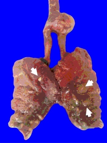
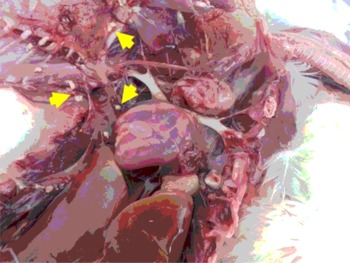
The severity and the degree of development of the disease determine both morphology and extension of macroscopic lesions. Large scale involvement of the respiratory tract can occur before clinical signs are apparent. Typically, lesions consist of white to yellowish granulomas ranging from miliary (<1 mm in diameter) to large roughly spherical granulomatous nodules (>2 cm) involving serosae and parenchyma of one (Corkish, Reference CORKISH1982) or multiple organs ( Figure 2 ). Single or multiple necrotic areas are visible on cut surfaces. The primary location of lesions is the air sacs and lungs, although the oesophagus, proventriculus, gizzard, small intestine, liver, kidney, spleen, skin, trachea, peritoneum, brain, eye, muscle or heart may be involved (Akan et al., Reference AKAN, HAZROĞLU, ILHAN, SAREYYÜPOĞLU and TUNCA2002; Throne Steinlage et al., Reference THRONE STEINLAGE, SANDER, BROWN, LOBSINGER, THAYER and MARTINEZ2003; Cacciuttolo et al., Reference CACCIUTTOLO, ROSSI, NARDONI, LEGROTTAGLIE and MANI2009). Parenchyma is either consolidated or has focal granulomas of varied size (Beytut, Reference BEYTUT2007). When coalescing in air sacs, these masses form cheesy caseous plaques covering the thickened membranes and even obstructing the entire lumina where fungal sporulation may occur, as evidenced by a grey-greenish velvety surface (Akan et al., Reference AKAN, HAZROĞLU, ILHAN, SAREYYÜPOĞLU and TUNCA2002; Throne Steinlage et al., Reference THRONE STEINLAGE, SANDER, BROWN, LOBSINGER, THAYER and MARTINEZ2003; Cacciuttolo et al., Reference CACCIUTTOLO, ROSSI, NARDONI, LEGROTTAGLIE and MANI2009). Broiler pullets have reportedly presented periorbital and eyelid swelling with cheesy yellow exudates in the conjunctival sac (Akan et al., Reference AKAN, HAZROĞLU, ILHAN, SAREYYÜPOĞLU and TUNCA2002).
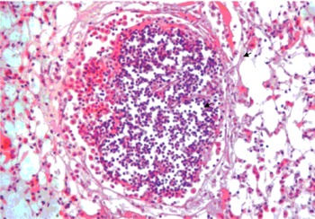
Cerebellum, ribs, sternum, yolk sacs, and hip joints constituted unusual locations of aspergillosis lesions (Olias et al., Reference OLIAS, HAUCK, WINDHAUS, VAN DER GRINTEN, GRUBER and HAFEZ2010). Haematoxylin-eosin stain is often augmented with periodic acid-Schiff, Grocott, and Gomori's methenamine silver stains in order to display fungal elements in embedded tissue sections (Cacciuttolo et al., Reference CACCIUTTOLO, ROSSI, NARDONI, LEGROTTAGLIE and MANI2009). The fluorescent optical brightener blankophor proves to be a valuable tool for detecting hyphae (Olias et al., Reference OLIAS, HAUCK, WINDHAUS, VAN DER GRINTEN, GRUBER and HAFEZ2010). Based on histopathological differences, Cacciuttolo et al. (Reference CACCIUTTOLO, ROSSI, NARDONI, LEGROTTAGLIE and MANI2009) distinguished a deep nodular and a superficial diffuse form of aspergillosis. A well-organised granulomatous reaction develops in non-aerated parenchyma whereas a superficial diffuse form, containing fungal elements and a non-encapsulated pyogranulomatous reaction, predominates in serosae and lungs (Figure 2a, b and c). Organised granulomas are clearly encapsulated by an outer thick fibrous layer (Beytut, Reference BEYTUT2007), whereas pyogranulomas lack clear borders. The first feature corresponding to late granuloma may be an important sign of the chronic form of aspergillosis, especially in adults (Olias et al., Reference OLIAS, HAUCK, WINDHAUS, VAN DER GRINTEN, GRUBER and HAFEZ2010). Pyogranulomas organisation presents a centre with variable amounts of septate, dichotomously branching hyphae surrounded by a palisade of radially arranged foreign body giant cells, macrophages, heterophils, and lymphocytes. Phagocytised fungal elements are regularly observed in the eosinophilic cytoplasm of multinucleated cells. Lymphocytes may infiltrate the margins of the granuloma (Cacciuttolo et al., Reference CACCIUTTOLO, ROSSI, NARDONI, LEGROTTAGLIE and MANI2009). Progressive inflammation in lungs resulting in small granuloma coalescence induces more extensive lesions and may lead to parabronchial obliteration with necrotic eosinophilic material containing erythrocytes, degenerated heterophils, and exfoliated epithelial cells (Beytut, Reference BEYTUT2007). Numerous conidiophores and free spores appear in granulomas that open to the air spaces of the respiratory tract (Beytut, Reference BEYTUT2007). Typical granulomatous reactions, which may or may not be associated with fungal elements, have been observed in the brain (Cortes et al., Reference CORTES, SHIVAPRASAD, KIUPEL and SENTIES-CUEM2005), conjunctiva (Beytut, Reference BEYTUT2007), liver, spleen, gizzard, small intestine (Beytut, Reference BEYTUT2007), hip joints (Olias et al., Reference OLIAS, HAUCK, WINDHAUS, VAN DER GRINTEN, GRUBER and HAFEZ2010) and trachea (Corkish, Reference CORKISH1982).
(a) Exudative pneumonia with yellow-grey nodules (1-3 mm) (arrows) in broiler chicken with aspergillosis.

(b) Yellow-grey plaques (arrows) in the air sacs of broiler chicken with aspergillosis.

(c) Lung showing granulomatous lesion with fungal hyphae (arrow) and cellular infiltration (arrow head).

Pathology associated with experimental aspergillosis
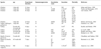
Animal models are established to improve our understanding of pathogen virulence, disease pathogenesis, and therapy feasibility. Because of the unique anatomical and physiological features of the avian respiratory system (Fedde, Reference FEDDE1998), the use of birds to study avian aspergillosis as a global entity is required. More restricted models, such as embryonated eggs, have been developed to investigate A. fumigatus development in ovo. Numerous experiments, which differed largely in both A. fumigatus conidial concentrations and inoculation routes, have been reported on varied avian species ( Table 1 ). In some surveys, birds were concurrently challenged with A. flavus conidia (Taylor and Burroughs, Reference TAYLOR and BURROUGHS1973; Richard et al., Reference RICHARD, CUTLIP, THURSTON and SONGER1981). The purpose of intratracheal, intrapulmonary, or intra-air sac injections was to induce a primary respiratory disease (Beernaert et al., Reference BEERNAERT, PASMANS, HAESEBROUCK and MARTEL2008). These inoculation routes allow the delivery of consistent numbers of conidia between individual birds (Arné et al., Reference ARNÉ, THIERRY, WANG, DEVILLE, LE LOC'H, DESOUTTER, FEMENIA, NIEGUITSILA, HUANG, CHERMETTE and GUILLOT2011). Nevertheless, all of these routes bypass the upper airways and their associated defence mechanisms, especially when thoracic or abdominal air sacs were chosen (Fedde, Reference FEDDE1998). The basic pattern of early pneumonia may be modified. Following a unique air sac inoculation, lesions are generally confined to the ipsilateral lung and air sacs. However, lesions are mostly compatible with those seen in field cases in which airsacculitis and pneumonia are the predominant problems (Beernaert et al., Reference BEERNAERT, PASMANS, HAESEBROUCK and MARTEL2008).
Models of avian aspergillosis (Arné et al., Reference ARNÉ, THIERRY, WANG, DEVILLE, LE LOC'H, DESOUTTER, FEMENIA, NIEGUITSILA, HUANG, CHERMETTE and GUILLOT2011).

aAt inoculation time; bdexamethasone injections (Y: yes, N: no); cwAER: wet aerosol; dAER: dry aerosol; L: contact with a contaminated litter; IT: intratracheal;IP: intrapulmonary; IAS: intra-air sac; dnumbers of conidia/bird except for aerosol (total dispersed inoculum); ND: nondetermined
Conidial nebulisation aims to mimic natural conditions of contamination but requires a strict standardisation of procedures. Variable parameters include time exposure (5 to 60 min), nebulisation chamber volume, conidial concentration and inoculum presentation (Faublee and Boller, Reference FAUBLEE and BOLLER1975; Richard et al., Reference RICHARD, CUTLIP, THURSTON and SONGER1981; van Cutsem, Reference VAN CUTSEM1983). Pulverised dry conidia compared to wet conidial aerosols yields better results in terms of mortality and morbidity suggesting that the size of the droplets containing conidia could be a drawback (Klimes and Severa, Reference KLIMES and SEVERA1964). However, the dosage of a dry inoculum lacks precision and its manipulation is delicate (Arné et al., Reference ARNÉ, THIERRY, WANG, DEVILLE, LE LOC'H, DESOUTTER, FEMENIA, NIEGUITSILA, HUANG, CHERMETTE and GUILLOT2011).
Aerosol infection allows a large number of conidia in the lung as measured by the number per gram of lung immediately after exposure. A loading charge of 5× 10⁵ viable conidia/g lung tissue killed 50% of three week-old turkeys (Richard et al., Reference RICHARD, CUTLIP, THURSTON and SONGER1981), whereas no mortality was reported in one-day-old chickens exhibiting 3×10⁴ spores/g after aerosolisation (Taylor and Burroughs, Reference TAYLOR and BURROUGHS1973). Experimental immunosuppression was used in order to induce aspergillosis in less susceptible species or to reduce the variability of diseased bird responses. The protocols consisted of repeated dexamethasone injections (2 to 5 mg/kg) in pigeons (Beernaert et al., Reference BEERNAERT, PASMANS, HAESEBROUCK and MARTEL2008), chickens (Corrier et al., Reference CORRIER, ELISSALDE, ZIPRIN and DELOACH1991) and turkeys (Le Loc'h et al., Reference LE LOC'H, ARNE and BOURGEROL2006). Genotyping of isolates recovered from internal organs allowed to verify that experimental infection resulted from the inoculated A. fumigatus strain (Beernaert et al., Reference BEERNAERT, PASMANS, HAESEBROUCK and MARTEL2008). In most cases, experimental aspergillosis is a hyperacute infection obtained by administering a high concentration of conidia at a single time point. When observed, deaths and morbidity occur generally between days one and 14 (Richard et al., Reference RICHARD, CUTLIP, THURSTON and SONGER1981; van Cutsem, Reference VAN CUTSEM1983; Chaudhary and Sadana, Reference CHAUDHARY and SADANA1988; Le Loc'h et al., Reference LE LOC'H, ARNE and BOURGEROL2006). From seven days after inoculation, surviving animals may develop chronic lesions (Faublee and Boller, Reference FAUBLEE and BOLLER1975). These models reproduce a pulmonary aspergillosis with clinical signs and internal lesions. In some cases, extensive intraocular lesions (Richard et al., Reference RICHARD, CUTLIP, THURSTON and SONGER1981) or neurological symptoms with associated brain granulomas (Faublee and Boller, Reference FAUBLEE and BOLLER1975) have been described after aerosol exposure.
Sequential necropsies at various times after inoculation allow observations on the pathogenesis of the disease and the kinetics of internal lesions development (Taylor and Burroughs, Reference TAYLOR and BURROUGHS1973; Richard et al., Reference RICHARD, CUTLIP, THURSTON and SONGER1981; Femenia et al., Reference FEMENIA, FONTAINE, LAIR-FULLERINGER, BERKOVA, HUET, TOWANOU, RAKOTOVAO, GRANET, LE LOC'H, ARNÉ and GUILLOT2007). Immediately after aerosol exposure and up to 24 h later, A. fumigatus conidia were detected in circulating blood of three week-old turkeys. In contrast with mycological cultures of lung tissue, the proportion of Aspergillus-positive cultures from liver and brain decreased with time. Numerous conidia could be observed in many macrophages harvested by lung lavages. Therefore, brain or eye lesions due to A. fumigatus may follow rapid haematogenous dissemination of conidia mediated by macrophage migration from the respiratory system (Faublee and Boller, Reference FAUBLEE and BOLLER1975).
Air sac membranes were slightly opaque 24 h after inoculation and scattered with miliary white foci. As fulminant airsacculitis evolved, the severity of macroscopic lesions increased rapidly, with progressive thickening, vascularisation, and opacification. Concurrently, 1-5 mm granulomas developed and tended to coalesce in plaques. Early lesions in lungs consisted of marginal oedema, progressive consolidation, and the formation of small white nodules (Kunkle and Rimler; 1996; Le Loc'h et al., Reference LE LOC'H, ARNE and BOURGEROL2006). In the first hours following infectious challenge, clear oedema, extensive epithelial alterations, and massive infiltration of heterophils, macrophages, and lesser lymphocytes occurred in both lungs and air sacs. Scarce swollen and germinating conidia were observed in inflammatory exudates, in areas of necrosis and aggregates of epithelial macrophages or multinucleated giant cells (Taylor and Burroughs, Reference TAYLOR and BURROUGHS1973; Richard et al., Reference RICHARD, CUTLIP, THURSTON and SONGER1981; Femenia et al., Reference FEMENIA, FONTAINE, LAIR-FULLERINGER, BERKOVA, HUET, TOWANOU, RAKOTOVAO, GRANET, LE LOC'H, ARNÉ and GUILLOT2007). A. fumigatus seemed to infect the air sac membrane interstitium rather than its surface leading to a severe airsacculitis within 24 h. Granulomas with heterophilic or necrotic centres and fungal elements were reported as soon as 24 h after air sac inoculation in nine and 19 week-old turkeys (Kunkle and Rimler, Reference KUNKLE and RIMLER1996), but not in one-day-old turkeys (Femenia et al., Reference FEMENIA, FONTAINE, LAIR-FULLERINGER, BERKOVA, HUET, TOWANOU, RAKOTOVAO, GRANET, LE LOC'H, ARNÉ and GUILLOT2007), where diffuse inflammation predominated till 48 h.
Chronologically, multifocal inflammatory response was gained in cellularity with marked mixed cellular infiltrates and granulomatous reaction in respiratory tissues. Circumscribed pyogranulomas developed with central eosinophilic necrosis restricting short hyphae and conidia surrounded by intact heterophils, epithelioid and foreign body giant cells (Richard et al., Reference RICHARD, CUTLIP, THURSTON and SONGER1981; Kunkle and Rimler, Reference KUNKLE and RIMLER1996; Femenia et al., Reference FEMENIA, FONTAINE, LAIR-FULLERINGER, BERKOVA, HUET, TOWANOU, RAKOTOVAO, GRANET, LE LOC'H, ARNÉ and GUILLOT2007). Beyond six days of exposure, inflammatory and necrotic foci seemed to regress in surviving birds exhibiting well-organised granulomas encapsulated by a thick layer of fibrous tissue (Faubl´ee and Boller, 1975; Richard et al., Reference RICHARD, CUTLIP, THURSTON and SONGER1981). Destruction of A. fumigatus occurred as attested by fungal elements observed in multinucleate giant cells and the regular diminution of positive cultures from tested organs (Taylor and Burroughs, Reference TAYLOR and BURROUGHS1973; Richard et al., Reference RICHARD, CUTLIP, THURSTON and SONGER1981; Kunkle and Rimler, Reference KUNKLE and RIMLER1996; Femenia et al., Reference FEMENIA, FONTAINE, LAIR-FULLERINGER, BERKOVA, HUET, TOWANOU, RAKOTOVAO, GRANET, LE LOC'H, ARNÉ and GUILLOT2007). In order to improve our current understanding of pathogenesis, crucial first steps of host-pathogen interaction should be carefully monitored.
Diagnosis
The signs of aspergillosis are non-specific, so early diagnosis is difficult and usually unreliable. Diagnosis usually relies upon an accumulation of evidence from the history, clinical presentation, haematology and biochemistry, serology, radiographic changes, endoscopy and culture of the fungus. Exotic pets may benefit from cumulative diagnostic tests including biochemistry, haematology, radiography, laparoscopy, or endoscopy that are not available in a poultry context.
Aspergillosis should be suspected when debilitated birds with respiratory distress do not respond to antibiotic treatment (Olias et al., Reference OLIAS, HAUCK, WINDHAUS, VAN DER GRINTEN, GRUBER and HAFEZ2010) and when careful history reveals the presence of underlying environmental or immunosuppressive factors. Definitive diagnosis is based on the isolation of A. fumigatus by culture or by the detection of the organism during histological examination. It is fundamental to keep in mind that many birds can host Aspergillus conidia in the respiratory system leading to a dormant infection without clinical symptoms or macroscopic lesions (Cacciuttolo et al., Reference CACCIUTTOLO, ROSSI, NARDONI, LEGROTTAGLIE and MANI2009). Furthermore, A. fumigatus may not be the primary cause or may be associated with either other pathogens such as A. niger or A. flavus or a concurrent non-fungal disease (Olias et al., Reference OLIAS, HAUCK, WINDHAUS, VAN DER GRINTEN, GRUBER and HAFEZ2010). Immunohistochemistry, with monoclonal or polyclonal antibodies, is a powerful and accurate tool to identify A. fumigatus in lesions (Cortes et al., Reference CORTES, SHIVAPRASAD, KIUPEL and SENTIES-CUEM2005; Beytut, Reference BEYTUT2007). Serological tests have not been validated in poultry and are not currently used in farms to investigate aspergillosis outbreaks.
Immune interventions and treatments
Although numerous antifungal protocols have been proposed to cure birds with aspergillosis (Beernaert et al., Reference BEERNAERT, PASMANS, VAN WAEYENBERGHE, HAESEBROUCK and MARTEL2010), treatment of the disease in poultry farms is virtually impossible. No vaccine is available. Nevertheless, a number of different vaccination strategies have been attempted in birds using different vaccine preparations, but with inconsistent results (Bauck et al., Reference BAUCK, HILLYER and HOEFER1992; Meredith, Reference MEREDITH1997).
Future knowledge regarding virulence factors and the role of cellular immunity in avian aspergillosis may contribute to newer and more effective vaccination strategies. The use of immune stimulants has been suggested (Jenkins, Reference JENKINS1991). Levamisole, a derivative of imidazothiazole, has been found to possess an immunostimulant effect as an adjunct in the treatment of chronic infections. However, levamisole did not decrease aspergillosis associated lesions in turkeys. In humans, clinical improvement of aspergillosis has been documented after the addition of interferon-γ and granulocyte macrophage colony-stimulating factor to the antifungal treatment. Whether the favourable effect of interferon-γ and granulocyte macrophage colony-stimulating factor could play a role in future treatment protocols for avian aspergillosis needs to be investigated. Specific biosecurity measures against Aspergillus contamination rely primarily on prevention. Dust and mouldy litter or feed should be avoided. A good litter management combined with daily assessment of its quality throughout the lifetime of the flock is the key to prevention of the disease. Bedding, like feeders, should be kept dry, non dusty and clean in order to limit fungal development.
Control of relative humidity through appropriate ventilation should be verified to prevent wet litter. Sporadic or repeated antifungal treatment may be useful in order to control environmental contamination. Spraying of fungistatic agents like thiabendazole, nystatin, or copper sulphate contributed to decreased fungal contamination of beddings. Enilconazole may be sprayed, fogged or nebulised to decontaminate surfaces or indoor volume. Finally, stressors such as beak trimming and high stocking densities should be minimised (Akan et al., Reference AKAN, HAZROĞLU, ILHAN, SAREYYÜPOĞLU and TUNCA2002). Specific drug properties, toxicity and dose regimens of a number of antifungal drug classes commonly applied in avian aspergillosis are summarised in Table 2 .
Summary of the administration routes and doses of antifungal agents used for avian aspergillosis (Beernaert et al., Reference BEERNAERT, PASMANS, VAN WAEYENBERGHE, HAESEBROUCK and MARTEL2010).
a every
Conclusions
Considerable progress has been achieved in our understanding of A. fumigatus pathobiology during the last decade. In order to improve our current understanding of pathogenesis, crucial first steps of host-pathogen interaction should be carefully monitored. The conditions which allow A. fumigatus to provoke infection in only some of the poultry in a rearing unit remain unclear. The outcome of the disease probably depends on the first steps of the innate immune response, which relies on the influx of macrophages and heterophilic granulocytes. Deciphering interactions between the different active effector cells is far from being as well defined as it is in mammals. No vaccine is available and so specific biosecurity measures against Aspergillus contamination rely primarily on prevention.
In chickens and turkeys, there is a constant increase in knowledge concerning the identification of cytokines, target membrane receptors and the immunomodulation mechanisms (Umar et al., Reference UMAR, ARIF, SHAH, MUNIR, YAQOOB, AHMED, KHAN, YOUNUS and SHAHZAD2015; Azeem et al., Reference AZEEM, REHMAN, UMAR, ASIF, ARIF and RAHMAN2014). Future research directions in this field include elucidation of host-specific and organ-specific A. fumigatus virulence factors, the study of inter-strain and interspecies variations in virulence. A more complete understanding of host pathogen interaction is a prerequisite towards establishing effective means for the prevention and control of this often devastating infection (Shah et al., Reference SHAH, UMAR, IQBAL, REHMAN, QADRI and HE2014; Umar et al., Reference UMAR, SHAH and MUNIR2016).