Introduction
A strong association between low birth weight and the development of cardiovascular diseases (CVD) later in life led to the consideration that suboptimal conditions in utero can lead to programmed changes in physiology, which result in an increased risk of chronic diseases in adulthood.Reference Barker, Winter, Osmond, Margetts and Simmonds 1 – Reference Barker, Gluckman and Godfrey 3 Among all non-communicable diseases, CVD are the leading cause of mortality 4 and represent a major socioeconomic burden worldwide.Reference Go, Mozaffarian and Roger 5 , 6 In addition, intrauterine growth restriction (IUGR), often due to reduced oxygen delivery to the fetus, is a common outcome of pregnancy-related complications.Reference Imdad, Yakoob, Siddiqui and Bhutta 7 Thus, CVD following a history of pregnancy-related complications represents a growing problem to public health. Although many insults during pregnancy can affect fetal growth;Reference Maulik 8 hypoxia has been associated with cardiac dysfunction as determined by an increased susceptibility to ischemia/reperfusion (I/R) injury.Reference Rueda-Clausen, Morton, Lopaschuk and Davidge 9 – Reference Reyes, Kirschenman and Quon 11 Moreover, exposure to hypoxia in utero has also been associated with structural changes including cardiac remodeling in both early developmentReference Tong, Xue, Li and Zhang 12 and later in life.Reference Xu, Williams, O’Brien and Davidge 10
The development of the heart is a complex morphological process. In rats, this process starts at gestational day (GD) 9Reference Marcela, Cristina and Angel 13 and ends at postnatal day (PND) 21.Reference Clubb and Bishop 14 During fetal life, the heart enlarges through a process of hyperplasia, whereas during postnatal life myocardial cell growth and maturation occurs in three phases: (a) PND-1 to PND-4, myocardial growth is due to hyperplasia and most of the cells are mononucleated; (b) PND-6 to PND-14, there is a transitional phase where both hyperplasia and hypertrophy are observed and a rapid transition from mono to binucleated cells (terminally differentiated cells) occurs; and finally (c) PND-14 and older, where myocardial growth is secondary to hypertrophy.Reference Clubb and Bishop 14
Evidence has shown that fetal cardiomyocytes (GD 21) from Sprague Dawley dams exposed to hypoxia (10.5% O2) during the last third of pregnancy had an increased proportion of binucleated cardiomyocytes, and an increase in the size of these binucleated cells (an early marker of hypertrophy).Reference Bae, Xiao, Li, Casiano and Zhang 15 In addition, compared with cardiomyocytes from normoxic dams, cardiomyocytes from hypoxia-induced IUGR offspring had an increased rate of apoptosisReference Bae, Xiao, Li, Casiano and Zhang 15 and reduced proliferation.Reference Paradis, Xiao, Zhou and Zhang 16 Moreover, in hypoxia-induced IUGR offspring, endothelin-1 was shown to stimulate binucleation and inhibit proliferation of fetal cardiomyocytes; which denotes a premature transition of mononucleated cells to binucleated cells.Reference Paradis, Xiao, Zhou and Zhang 16 Taken together, these data suggest that cardiomyocytes from hypoxia-induced IUGR offspring have decreased cardiomyocyte proliferation and an increase in cardiac hypertrophy and fibrosis.
Heart development and cardiomyocyte proliferation are regulated by neurohormonal stimuli (i.e. angiotensin II,Reference Sundgren, Giraud, Stork, Maylie and Thornburg 17 cortisol,Reference Giraud, Louey, Jonker, Schultz and Thornburg 18 thyroid hormoneReference Chattergoon, Giraud and Thornburg 19 insulin-like growth factor-1Reference Sundgren, Giraud and Schultz 20 ) as well as hemodynamic stimuliReference Thornburg, Jonker and O’Tierney 21 and oxygen,Reference Louey, Jonker, Giraud and Thornburg 22 finishing soon after birth.Reference Thornburg, Jonker and O’Tierney 21 Evidence suggests that there might be other pathways involved in the development of cardiac tissues. Novoyatleva et al.Reference Novoyatleva, Diehl and van Amerongen 23 found that the tumor necrosis factor (TNF)-related weak inducer of apoptosis (TWEAK) was associated with neonatal cardiomyocyte proliferation. TWEAK, a member of the TNF family, is a type II transmembrane protein that can be further processed into a soluble cytokine (s-TWEAK).Reference Chicheportiche, Bourdon and Xu 24 TWEAK is considered to be a multifunctional cytokine and has been extensively associated with cell migration, survival, differentiation, death and proliferation of smooth muscle cells, epithelial cells, myoblasts and astrocytes.Reference Winkles 25
In neonatal cardiomyocytes isolated from Sprague Dawley rats, binding of either TWEAK or s-TWEAK with its receptor [protein fibroblast growth factor-inducible molecule 14 (Fn-14)], has been shown to activate extracellular signal-regulated kinase and phosphatidylinositol 3-kinase (PI3K), as well as phosphorylate glycogen synthase kinase-3β, causing cardiomyocyte proliferation. This phenomenon was physiologically relevant during early stages in life since the TWEAK/Fn-14 pathway becomes quiescent after the completion of cardiac development at approximately PND-10 due to a downregulation of Fn-14 expression in cardiomyocytes with age.Reference Novoyatleva, Diehl and van Amerongen 23 Interestingly, overexpression of Fn-14 resulted in adult cardiomyocyte proliferation.Reference Novoyatleva, Diehl and van Amerongen 23 It has been demonstrated that after a myocardial infarction, Fn-14 protein expression is upregulated.Reference Mustonen, Sakkinen and Tokola 26 , Reference Chorianopoulos, Heger and Lutz 27 In addition, it has been established that rats treated with TWEAK before a ligation of the left anterior descending coronary artery followed by reperfusion have a reduction in cardiomyocyte apoptosis compared with non-treated rats. This effect was PI3K/protein kinase B (AKT) mediated.Reference Yang, Yan and Gong 28 However, the role of the TWEAK/Fn-14 pathway in cardiomyocyte proliferation and its expression at the time of increased susceptibility to cardiac I/R injury in hypoxia-induced IUGR offspring is not known. We hypothesized that in hypoxia-induced IUGR offspring, a decrease expression of the Fn-14 would be associated with a decrease in cardiomyocyte proliferation at PND-1, and the TWEAK/Fn-14 pathway will be expressed in cardiomyocytes in adult IUGR offspring. To test these hypotheses, we proposed the following aims. In early postnatal life (PND-1): (a) to determine whether being born growth restricted is associated with a decrease in cardiomyocyte proliferation and an increase in cardiomyocyte binucleation; (b) to assess whether a decreased expression of Fn-14 in neonatal cardiomyocytes is associated with a decrease in cardiomyocyte proliferation in IUGR offspring; and (c) to evaluate whether cardiomyocyte proliferation in response to recombinant TWEAK is different between control and IUGR offspring. Finally, the aims of this project in adult offspring (4-month-old) were as follows: (a) to assess whether the serum concentration of s-TWEAK and (b) the cardiac protein expression of Fn-14 in 4-month-old offspring is altered in IUGR offspring.
Methods
Animal model
All procedures in this study were approved by the University of Alberta Animal Welfare Committee, and were in accordance with the guidelines of the Canadian Council on Animal Care. Three-month-old Sprague Dawley rats (Charles River, Wilmington, MA, USA) were housed in the animal facility where room conditions were as follows: 35% humidity, 10:14-h light:dark cycle, and fed ad libitum with standard rodent chow. After acclimatization (1 week), rats were mated overnight. Upon confirmation of pregnancy, female rats were randomly allocated to be exposed to control (room air; n=9) or hypoxic (11% oxygen, n=8) conditions from GD 15 to 21. On PND-1, body weight, crown to rump length (CRL), abdominal girth (ABG) and heart weight were measured. Four to six male and female offspring were randomly chosen from each litter, and were used for cardiomyocyte isolation. After euthanasia (decapitation), left and right ventricles were dissected in a dissociation buffer (in mM: 116 NaCl, 20 HEPES, 0.8 Na2HPO4, 5.6 glucose, 5.4 KCl, 0.8 MgSO4, pH 7.35). Multiple enzymatic digestions with a dissociation buffer containing collagenase type II (0.4 mg/ml) and pancreatin (0.6 mg/ml) were performed at 37°C. Following this procedure, the digested solution was filtered through a 70 μm filter and spun at 1300 g. Cardiomyocytes were counted using the Trypan Blue exclusion test of cell viability (Sigma, USA). Afterwards, cardiomyocytes were separated and resuspended in different culture medias according to the experimental protocol being performed (determination of proliferation and binucleation or stimulation with recombinant TWEAK). Cardiomyocytes were cultured in 48-well plates and grown in a humidified atmosphere at 37°C with 5% CO2/95% air.
Determination of proliferation and binucleation
Approximately 30% of isolated cardiomyocytes were resuspended in medium-199 (Gibco, Life Technologies, USA) supplemented with 2% albumin (Sigma), 2 mM l-carnitine (Sigma), 5 mM creatine (Sigma), 5 mM taurine (Sigma) and penicillin-streptomycin (Life Technologies).
Twenty-four hours after isolation, Cardiomyocytes were fixed in methanol. Cell proliferation and binucleation were determined by immunofluorescence using an overnight incubation with anti-Ki-67 antibody (ab16667; 1:50; Abcam, Canada) and anti-heavy chain cardiac myosin antibody (ab50967; 1:150; Abcam). Cardiomyocytes were then incubated with Alexa Fluor 488-conjugated goat anti-rabbit and Alexa Fluor 546-conjugated anti-mouse secondary antibodies (1:200; Abcam) for 40 min in the dark. Nuclei were subsequently stained with DAPI (300 nM; Thermo scientific, USA). To determine cardiomyocyte proliferation and binucleation, three replicate wells were stained and pictures of four random fields per well were taken for each of the control and IUGR, male and female offspring groups. Ratios between Ki-67 positive nuclei/total nuclei number and number of binucleated cells/total number of cells per image were calculated.
Cardiomyocyte stimulation with TWEAK
The remaining 70% of cardiomyocytes were initially incubated for 48 h in medium-199 (Gibco, Life Technologies) supplemented with 5% horse serum, 20 mM cytosine b-d-arabinofuranoside (araC; Sigma), and penicillin-streptomycin to prevent the proliferation of non-myocyte cells. Subsequently, cardiomyocytes were washed and cultured in medium-199 supplemented with 2% albumin in the following conditions: (1) with TWEAK recombinant protein (r-TWEAK, 100 ng/ml; Biolegend, USA); (2) medium alone without r-TWEAK; (3) a combination of r-TWEAK and Fn-14 receptor antibody (100 µg/ml; Abcam); (4) Fn-14 receptor antibody alone; (5) medium-199 without albumin. After the cardiomyocytes were stimulated for 72 h with r-TWEAK, cells were fixed in methanol and proliferation was determined as previously described. Cardiomyocytes were visualized under an Olympus IX81 fluorescent microscope (Carson Scientific Imaging Group, Canada), using Cellsense imaging software (Olympus, Japan). Images presented are in the original (10×) magnification.
TWEAK receptor protein expression in PND-1 and 4-month-old offspring hearts
Heart tissue from the remaining offspring collected at PND-1 and from 4-month-old non-perfused, sedentary animals generated for a previous studyReference Reyes, Morton, Kirschenman, DeLorey and Davidge 29 was used to determine the protein expression of Fn-14. Tissue preparation was performed as follows: tissue was homogenized in a lysis buffer containing [in mM: 20 Tris (pH 7.4), 5 EDTA, 10 sodium pyrophosphate tetrabasic, 100 sodium fluoride and 1% NP-40, and Protease Inhibitor Cocktail (1X HaltTM protease inhibitor; Thermo scientific)]. Protein concentrations were determined by bicinchoninic acid assay (Pierce). A total of 100 μg of protein was loaded and subsequent sodium dodecyl sulfate polyacrylamide gel electrophoresis and immunoblotting were performed. Membranes were incubated with blocking buffer for fluorescent Western blotting (Rockland Immunochemicals Inc, USA) and anti-TWEAK receptor (1:100; Abcam) and were re-probed with anti-α-tubulin (1:50,000; Abcam) as a protein loading control. Finally, membranes were probed with the following secondary antibodies: IRDye 800CW donkey anti-rabbit IgG and IRDye 680RD donkey anti-mouse IgG secondary antibodies (1:10,000; Li-Cor Biosciences, Lincoln, NE, USA). The protein bands were imaged using the Li-Cor Odyssey Imaging Systems v3.0. Densitometry analysis was performed using ImageStudio v.5.2.5 (LI-COR Biosciences). Data were normalized to the α-tubulin loading control.
Soluble TWEAK in serum or plasma
Plasma from PND-1 offspring and previously collected serum from 4-month-old male and female control and IUGR offspringReference Reyes, Morton, Kirschenman, DeLorey and Davidge 29 were used to determine s-TWEAK levels using a commercially available, standard sandwich enzyme-linked immunosorbent assay (LifeSpan Biosciences Inc, USA) according to the manufacturer’s instructions. The concentration of s-TWEAK was calculated by interpolation of the results from a standard curve. The inter- and intra-assay coefficients of variation obtained were 4.5 and 4.3%, respectively.
Statistical analyses
Data were presented as mean±s.e.m. The Shapiro–Wilk test was used to assess normality of continuous data. Data were analyzed using a Mann–Whitney test when data were not normally distributed. For Western blot analyses, Fn-14 bands were normalized to α-tubulin. The effect of the addition of r-TWEAK on cardiomyocyte proliferation was analyzed using a two-way analysis of variance (ANOVA) followed by a Bonferroni post-hoc test, the factors included in the two-way ANOVA were exposure to prenatal hypoxia and r-TWEAK treatment. To determine whether the increase in cardiomyocyte proliferation was Fn-14-mediated, a one-way ANOVA followed by a Tukey’s multiple comparison test was performed within each group. Statistical significance was defined as P<0.05. All data were analyzed using GraphPad Prism 7 statistical software (GraphPad Software, USA).
Results
Animal model
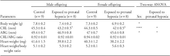
Morphological characteristics of male and female offspring from normoxic and hypoxic pregnancies are provided in Table 1. At PND-1, there was no statistically significant differences in body weight due to prenatal hypoxia exposure, but male offspring were heavier than female offspring. Both male and female offspring exposed to prenatal hypoxia had a reduced CRL and ABG, indicating reduced intrauterine growth (further referred to as IUGR). Although the percent stillbirths were higher in dams exposed to hypoxia compared with control dams (11.7 v. 1.9%; P<0.0001), litter size was not different among the groups (14 [12–18] v. 15 [11–16]; P=0.14).
Morphological characteristics at postnatal day 1 of male and female offspring born from normoxic dams (control) or dams exposed to hypoxia

ANOVA, analysis of variance; CRL, crown to rump length; ABG, abdominal girth.
Data presented as mean±s.e.m.
*P⩽0.05, ****P<0.0001, † P⩽0.05, ‡ P<0.01 compared with its own control after a Tukey’s post-hoc test.
Fn-14 protein expression, plasma levels of TWEAK, and proliferation and binucleation of cardiomyocytes at PND-1
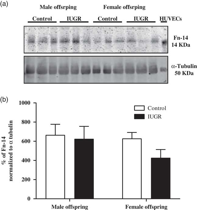
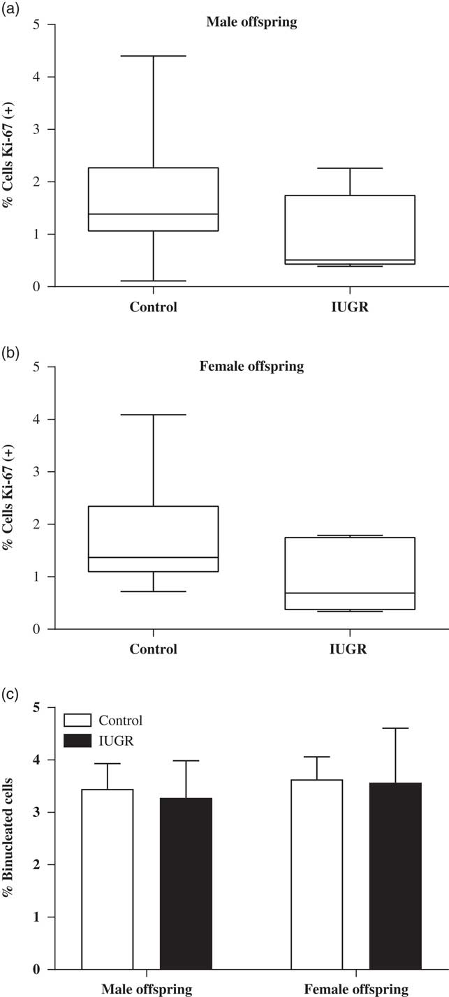
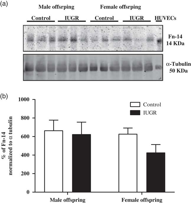
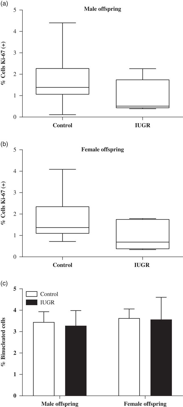
No differences among the groups were found regarding cardiomyocyte Fn-14 protein expression in either male or female (Fig. 1a and 1b) offspring at PND-1. Plasma levels of s-TWEAK were not different in male (25.5±1.4 ng/ml control v. 26±1.7 ng/ml IUGR; P=0.9) or female offspring (22.5±3.6 ng/ml control v. 22.2±0.3 ng/ml IUGR; P=0.9). There were no differences among the groups regarding the proliferation or binucleation of cardiomyocytes at PND-1 (Fig. 2).
Postnatal day 1 cardiac tissue fibroblast growth factor-inducible molecule 14 (Fn-14) protein expression in control and intrauterine growth restriction (IUGR), male and female offspring. All data are presented as a ratio of Fn-14 to α-tubulin expression. (a) Representative image of a Western blot membrane probed for Fn-14 and α-tubulin in male and female, control and IUGR offspring. (b) Summary data of Fn-14 protein expression in male and female control (open bars, n=6) and IUGR (closed bars, n=6) offspring are presented. Human umbilical vein endothelial cell (HUVECs) lysates were used as a positive control. Data are presented as mean±s.e.m. and analyzed by a two-way analysis of variance.
Postnatal day 1 cardiomyocyte proliferation and binucleation in control and intrauterine growth restriction (IUGR), male and female offspring. Cardiomyocyte proliferation was determined by the percentage of Ki-67 positive nuclei in (a) male control (n=8) or IUGR offspring (n=6); and (b) female control (n=9) or IUGR offspring (n=6). Non-normally distributed data are presented as median and 95% confidence interval and analyzed by a Mann–Whitney test. (c) The ratio between the total number of binucleated to mononucleated cardiomyocytes was then determined, summary data in male and female control (open bars, n=8) and IUGR (closed bars, n=5) offspring are presented. Data are presented as mean±s.e.m. and analyzed by a two-way analysis of variance.
Cardiomyocyte stimulation with recombinant TWEAK
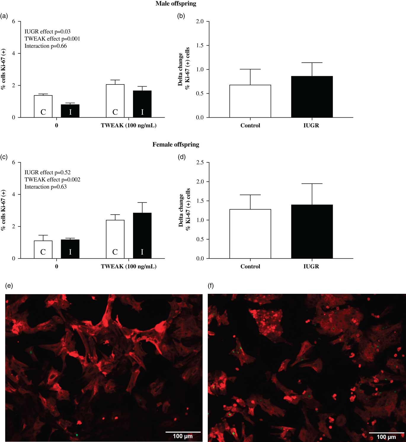
In male offspring, exposed to prenatal hypoxia, cardiomyocyte proliferation was decreased compared with controls after being in culture for 72 h. The addition of r-TWEAK increased cardiomyocyte proliferation to the same extent in both control and IUGR male offspring (Fig. 3a and 3b). The addition of r-TWEAK in the presence of the Fn-14 receptor blocker decreased cardiomyocyte proliferation in both control male offspring [% Ki-67 positive cardiomyocytes; 2.1±0.3 (r-TWEAK) v. 0.6±0.2 (r-TWEAK+Fn-14 receptor blocker); P=0.005], and IUGR male offspring [% Ki-67 positive cardiomyocytes; 1.7±0.3 (r-TWEAK) v. 0.4±0.1 (r-TWEAK+Fn-14 receptor blocker); P=0.0003] (data not shown). The Fn-14 receptor blocker alone had no effect on cardiomyocyte proliferation in control and IUGR male offspring (data not shown).
Proliferation of postnatal day 1 cardiomyocytes exposed to recombinant tumor necrosis factor-related weak inducer of apoptosis (TWEAK) in control and intrauterine growth restriction (IUGR), male and female offspring. (a) Cardiomyocyte proliferation in response to exogenous r-TWEAK was determined by the percentage of Ki-67 positive nuclei in male control (n=5) or IUGR offspring (n=6). (b) Change in proliferation (Ki-67 positive nuclei) in control and IUGR male offspring following the addition of exogenous r-TWEAK. (c) Cardiomyocyte proliferation in response to exogenous r-TWEAK was determined by the percentage of Ki-67 positive nuclei in female control (n=5) or IUGR offspring (n=4). (d) Change in proliferation (Ki-67 positive nuclei) in control and IUGR female offspring following the addition of exogenous r-TWEAK. A two-way analysis of variance was performed to determine the effect of exposure to prenatal hypoxia and/or the addition of recombinant TWEAK on cardiomyocyte proliferation. C, control offspring, I, IUGR offspring. Data are summarized and presented as mean±s.e.m. and analyzed by t-test. Representative immunofluorescence images of proliferating cardiomyocytes in the absence (e) and presence of 100 ng/ml of r-TWEAK (f) (red channel: anti-heavy chain cardiac myosin; green channel: anti-Ki-67).
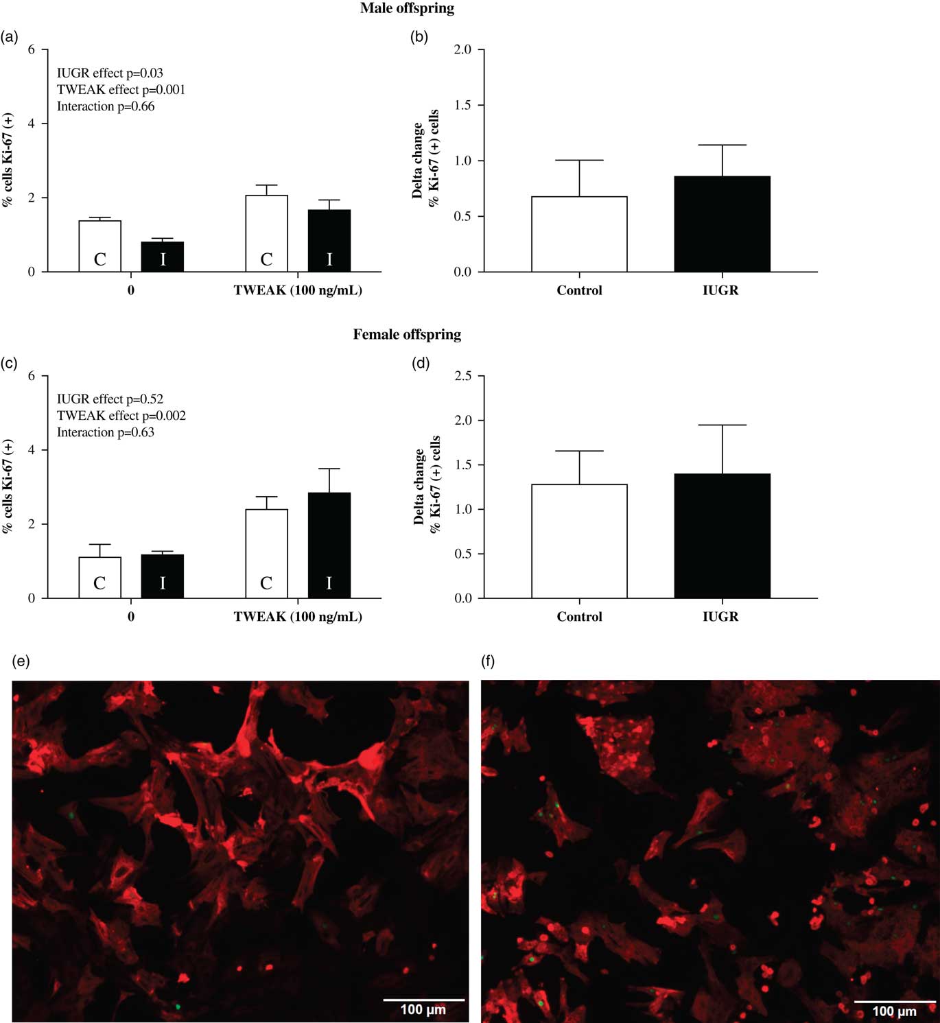
In contrast, in female offspring there were no differences in cardiomyocyte proliferation between control and IUGR offspring; and the addition of r-TWEAK increased proliferation in both control and IUGR offspring (Fig. 3c and 3d). The addition of r-TWEAK in the presence of the Fn-14 receptor blocker decreased cardiomyocyte proliferation in both control female offspring [% Ki-67 (+) cardiomyocytes; 2.4±0.3 (r-TWEAK) v. 0.9±0.2 (r-TWEAK+Fn-14 receptor blocker); P=0.02], and IUGR female offspring [% Ki-67 (+) cardiomyocytes; 2.8±0.7 (r-TWEAK) v. 0.4±0.1 (r-TWEAK+Fn-14 receptor blocker); P=0.009] (data not shown). The Fn-14 receptor blocker alone had no effect on cardiomyocyte proliferation in control and IUGR female offspring (data not shown).
Serum levels of TWEAK in 4-month-old offspring
A series of serum concentrations (1:100; 1:10; 1:2 and neat serum) were tested to determine the serum concentration of s-TWEAK in the 4-month-old offspring experimental groups. Despite this, the values of s-TWEAK were lower than the kit’s sensitivity range (10 pg/ml) and, therefore, remained undetectable.
Fn-14 protein expression in 4-month-old offspring
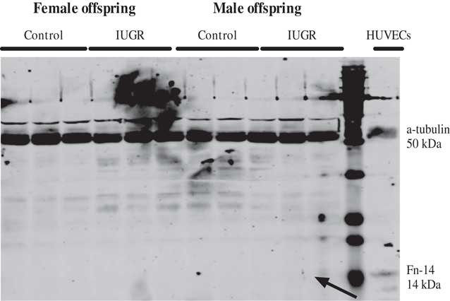
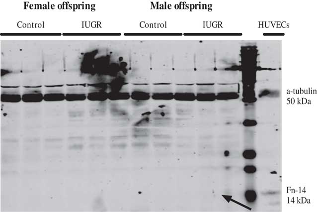
Protein expression of the Fn-14 receptor was not detectable in cardiac tissues from control and IUGR, male and female 4-month-old offspring (Fig. 4).
Cardiac tissue fibroblast growth factor-inducible molecule 14 (Fn-14) protein expression in 4-month-old control and intrauterine growth restriction (IUGR), male and female offspring. Representative image of a Western blot membrane probed for Fn-14 and α-tubulin in male and female, control and IUGR offspring (n=6/group). Human umbilical vein endothelial cell (HUVECs) lysates were used as a positive control. Data are summarized and presented as mean±s.e.m. and analyzed by t-test. Arrow indicates band weight for Fn-14.
Discussion
To the best of our knowledge, this is the first time that the role of the TWEAK/Fn-14 pathway in the susceptibility of hypoxia-induced IUGR offspring to CVD has been assessed. TWEAK is a multifunctional cytokine that has recently gained attention because of its involvement in cardiomyopathy,Reference Mustonen, Sakkinen and Tokola 26 , Reference Chorianopoulos, Heger and Lutz 27 , Reference Novoyatleva, Janssen, Wietelmann, Schermuly and Engel 30 , Reference Jain, Jakubowski and Cui 31 and neonatal cardiomyocyte proliferation.Reference Novoyatleva, Diehl and van Amerongen 23 We found that prenatal hypoxia was not associated with differences in Fn-14 protein expression or cardiomyocyte proliferation at PND-1 in either male or female offspring. However, after being in culture for 72 h, cardiomyocytes displayed reduced proliferation when isolated from IUGR male compared with their controls. The addition of r-TWEAK increased proliferation in both groups to the same extent. In contrast, in female offspring, cardiomyocyte proliferation in culture was not reduced in IUGR offspring and the addition of r-TWEAK increased proliferation to the same extent in both control and IUGR offspring. Cardiomyocyte proliferation among all groups was inhibited by the Fn-14 receptor blocking antibody.
We have previously demonstrated the impact of prenatal hypoxia on reducing fetal weight, CRL and ABG.Reference Rueda-Clausen, Morton, Lopaschuk and Davidge 9 , Reference Reyes, Morton, Kirschenman, DeLorey and Davidge 29 , Reference Rueda-Clausen, Dolinsky and Morton 32 – Reference Shah, Reyes and Morton 34 In those studies, all the measurements were collected either prenatally or within the first 12 h of delivery; whereas the current study measurements were taken 24-h following birth. In this latter subset of animals, hypoxia during pregnancy was associated with a decreased CRL and ABG, but not body weight, in male and female offspring. Thus, milk consumption during this initial 24-h period may have affected the body weight of offspring by the time the measurements were collected.
Heart development is an active process which occurs both prenatally and during early postnatal life.Reference Clubb and Bishop 14 , Reference Black, Siebel, Gezmish, Moritz and Wlodek 35 The fact that there were no differences in the percent binucleation of cardiomyocytes at PND-1 implies that there is still opportunity for growth and differentiation in these cells. Importantly, although we did not find any differences in cardiomyocyte proliferation in our hypoxia-induced IUGR offspring at PND-1; cardiomyocytes from male IUGR offspring proliferated less than controls when in culture for 72 h. These findings suggest that cardiomyocyte proliferation capacity could be compromised, as shown previously by Tong et al. Reference Tong, Xue, Li and Zhang 12 at GD 21 and PND-7 in hypoxia-induced IUGR offspring. Further, a large variation in the proliferation of cardiomyocytes was observed in the control group for both male and female offspring, which reduced the power of our study at PND-1. However, cardiomyocytes demonstrated consistently less proliferation in IUGR offspring compared with controls; suggesting a potential susceptibility to impaired cardiac development. It is important to note, however, that although it has been demonstrated that in rats, hypoxia-induced IUGR offspring have a decrease in cardiomyocyte proliferation.Reference Bae, Xiao, Li, Casiano and Zhang 15 The same stimuli but in other species (e.g. sheep) has shown that there is a decrease in cardiomyocyte number in late gestation with no change in proliferation and a delay in maturation.Reference Louey, Jonker, Giraud and Thornburg 22 , Reference Botting, McMillen, Forbes, Nyengaard and Morrison 36 – Reference Morrison, Botting and Dyer 38 These differences could be due to the fact that in sheep most cardiomyocytes are terminally differentiated soon after birth, whereas in rats, differentiation continues until PND-14.Reference Burrell, Boyn and Kumarasamy 39
We hypothesized that the differences in cardiomyocyte proliferation could be attributed to a decrease in cardiac protein expression of Fn-14. As it has been shown that both TWEAK and its receptor are highly upregulated after a myocardial infarction,Reference Mustonen, Sakkinen and Tokola 26 , Reference Chorianopoulos, Heger and Lutz 27 we hypothesized that these differences would be expressed in adult life at the time of increased susceptibility to CVD observed in our prenatal hypoxia-induced IUGR model. In addition, it has been demonstrated that genetic manipulation of TWEAK, its downstream targets or the induction of its receptor all result in dilated cardiomyopathy, cardiomyocyte hypertrophy and extracellular matrix remodeling in adult rodents.Reference Novoyatleva, Janssen, Wietelmann, Schermuly and Engel 30 , Reference Jain, Jakubowski and Cui 31 Furthermore, mice treated with adenovirus expressing soluble murine TWEAK had a decreased expression of peroxisome proliferator-activated receptor γ coactivator 1-α (PGC1-α), the major regulator of mitochondrial biogenesis.Reference Shi, Jiang and Qiu 40 Decreased PGC1-α expression has been associated with decreased mitochondrial biogenesis and cardiac I/R injury.Reference Sun, Zhao and Yu 41 However, in the current study cardiac protein expression of Fn-14 at PND-1 was not different among the groups. Moreover, in 4-month-old offspring, Fn-14 cardiac protein receptor was not expressed and s-TWEAK serum concentrations were undetectable; thus, it can be surmised that other mechanisms may be involved in the development of cardiac dysfunction observed in the adult IUGR offspring.
There are some study limitations that should be acknowledged. Our findings regarding the role of the TWEAK/Fn-14 pathway in cardiomyocyte proliferation need to be interpreted carefully. We used Ki-67 as a marker of proliferation which is a nuclear protein expressed in all phases of the cell cycle, excluding resting cells in G0. Reference Gerdes, Lemke and Baisch 42 Thus, it could be argued that using this marker, the proliferation effect could be either under or overestimated, when compared with other available markers that target the cardiomyocytes in their S-phase (DNA synthesis), such as bromodeoxyuridine,Reference Gratzner 43 or proliferating cell nuclear antigen (PCNA).Reference van Dierendonck, Wijsman, Keijzer, van de Velde and Cornelisse 44 We used Ki-67 as a proliferation marker, however, based on the fact that cells can enter a quiescent state even after DNA synthesis completion,Reference Drewinko, Yang, Barlogie and Trujillo 45 thus, a proliferation marker is actually indicative of the proliferation potential of the cell rather than a predictor of proliferation.Reference Scholzen and Gerdes 46 In addition, it has been established that PCNA does not work well in very slow proliferative cells in S-phase.Reference van Dierendonck, Wijsman, Keijzer, van de Velde and Cornelisse 44 In addition, alterations in fetal hemodynamics following IUGR are associated with cardiac remodeling.Reference Cohen, Wong, Horne and Yiallourou 47 The fetal circulation is designed to deliver the majority of oxygenated blood to the myocardium and the brain. This is achieved by the presence of intra- and extra-cardiac shunts, which makes the fetal circulation parallel.Reference Murphy 48 During hypoxic conditions, the fetal circulation adapts to increased blood flow resistance in the maternal compartment.Reference Baschat 49 An increase in the right ventricle afterload with a decrease in the left ventricle afterload guarantees redistribution of cardiac output.Reference Baschat 49 The fact that we could not find differences among the groups could be attributed to the use of both left and right ventricles from each pup in our experiments.
In conclusion, activation of the TWEAK/Fn-14 pathway leads to increased proliferation but does not appear to contribute to the reduced cardiomyocyte proliferative capacity observed in prenatal hypoxia-exposed male offspring. Interestingly, the cardiomyocyte proliferative capacity was compromised in male, but not female, IUGR offspring. Previous studies from our laboratoryReference Rueda-Clausen, Morton, Lopaschuk and Davidge 9 , Reference Rueda-Clausen, Morton and Davidge 33 have shown that male IUGR offspring appear to have a greater cardiac dysfunction phenotype compared with females and it could be speculated that this reduced proliferative capacity may have a role to play in the susceptibility of male offspring to CVD. Finally, other mechanisms involved in cardiomyocyte proliferation are likely to have an important role in the long-term consequences of being born growth restricted, and, therefore, further studies are needed.
Acknowledgments
None.
Financial Support
This work was supported by the Canadian Institutes of Health Research (CIHR, Grant numbers: 118160540; 0020054). This research has been funded by the generous support of the Stollery Children’s Hospital Foundation and by generous supporters of the Lois Hole Hospital for Women through the Women and Children’s Health Research Institute (WCHRI). S.T.D. is a Canada Research Chair (CRC) in Maternal and Perinatal Cardiovascular Health. L.M.R. is an Alberta Innovates [Formally Alberta Innovates Health Solutions (AIHS)] – supported graduate student, Grant number: 0014195. A.S. is a Molly Towell Perinatal Research Foundation – supported postdoctoral fellow.
Conflicts of Interest
None.
Ethical Standards
The authors assert that all procedures contributing to this work comply with the ethical standards of the relevant national guides on the care and use of laboratory animals (Canadian Council on Animal Care guidelines) and has been approved by the institutional committee (University of Alberta Health Sciences Animal Policy and Welfare Committee; AUP#00000242).