Introduction
Although Le Fort I osteotomy is one of the most commonly performed surgical procedures for the correction of dental and facial deformities in orthognathic surgery,Reference Chepla, Totonchi, Hsu and Gosain1–Reference Panula, Finne and Oikarinen2 post-operative complications from vascular injuries are rare.Reference Panula, Finne and Oikarinen2 Arterial pseudoaneurysm resulting from blunt or, more commonly, sharp trauma to the vessel wall has been reported,Reference Chepla, Totonchi, Hsu and Gosain1–Reference Zachariades, Rallis, Papademetriou, Papakosta, Spanomichos and Souelem3 but pseudoaneurysm of the sphenopalatine artery following Le Fort I osteotomy is extremely rare. We report a case of acute, massive, life-threatening epistaxis due to a pseudoaneurysm of the sphenopalatine artery as a post-operative complication of orthognathic surgery.
Case report
A 31-year-old man underwent Le Fort I osteotomy with bilateral sagittal split ramus osteotomy of maxillar advancement and mandibular setback for maxillar and mandibular deformities in a local dental clinic. Two weeks later, he presented with a massive epistaxis of both sides and oral bleeding.
Upon physical examination, he was pale, shivering and in a confused state. Nasal packing was performed using Merocel® packs. Complete blood count results demonstrated haemoglobin levels of 7.6 g/dl, haematocrit of 18.8 per cent and platelet count of 475 × 103/μl. Computed tomography (CT) with contrast showed a nodular, vascular, enhancing lesion, measuring 1.2 cm at the posterior aspect of the right maxillary sinus fed by the right internal maxillary artery, which was suggestive of a pseudoaneurysm or other vascular malformation (Figure 1).
Pre-operative axial (a) and coronal (b) computed tomography images, which show a nodular vascular enhancing lesion (arrow) measuring 1.2 cm at the posterior aspect of the right maxillary sinus, suggestive of pseudoaneurysm or other vascular malformation.

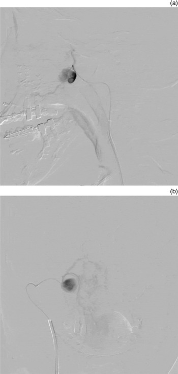
The patient was sent to interventional radiology for angiography and embolisation. Angiography revealed a large 2-cm pseudoaneurysm of the right sphenopalatine artery without collateral circulation between the internal and external carotid branches (Figure 2). Superselective embolisation with lipiodol and N-butyl-2-cyanoacrylate was performed.
Sagittal (a) and coronal (b) superselective angiographic images, which show a central collection of contrast, indicating a 2-cm pseudoaneurysm of the right sphenopalatine artery.

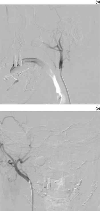
The post-embolisation angiogram showed complete resolution of the pseudoaneurysm (Figure 3). A total of two units of packed red cells and three units of fresh frozen plasma were required to stabilise the patient. He remained asymptomatic during the six-month follow up, with no further episodes of bleeding.
Sagittal (a) and coronal (b) post-embolisation angiographic images showing total obliteration of the pseudoaneurysm, which was achieved by infusion of N-butyl-2-cyanoacrylate into the affected artery and pseudoaneurysm.

Discussion
The Le Fort I osteotomy involves separating the maxilla and the palate from the skull above the roots of the upper teeth.Reference Chepla, Totonchi, Hsu and Gosain1 The operation is frequently carried out with mandibular sagittal split osteotomy for the correction of dentofacial deformity. Osteotome positioning and manipulation are commonly used in procedures in order to mobilise and reposition the mandible and maxilla. The proximity of the osteotome to the internal maxillary artery and branches could increase the potential for injury at this junction.Reference Chepla, Totonchi, Hsu and Gosain1 However, pseudoaneurysm formation from the sphenopalatine artery is extremely rare because its diminutive calibre and deep location in the mid-face have proven protective from blunt and superficially penetrating injuries.Reference Zachariades, Rallis, Papademetriou, Papakosta, Spanomichos and Souelem3
Arterial pseudoaneurysm can occur as a result of blunt or, more commonly, sharp trauma to the vessel wall.Reference Bradley, Elahi and Kawamoto4 A pseudoaneurysm is an abnormal dilatation of an artery resulting from incomplete disruption of the arterial wall, which leads to an expanding lesion between the damaged artery and the surrounding tissues. After disruption of the arterial wall, haemorrhage occurs in the partially transected vessel into the adjacent tissues until the pressure of the formed haematoma counterbalances the arterial pressure, which compresses the defect and stabilises the bleeding.Reference Silva, O'Ryan, Beckley, Young and Poor5 The rarity of a pseudoaneurysm in the external carotid artery is due to the small size of the branches of the external carotid artery, which makes complete cross-cutting more likely than partial laceration. Moreover, larger calibre branches are protected by thicker tissue.Reference Manafi, Ghenaati, Dezham and Arshad6 Most cases of pseudoaneurysm of the external carotid artery have been reported to appear in the superficial temporal artery and facial artery. These branches are more vulnerable because of their large vessel diameter, long course and relatively superficial position.Reference Zachariades, Rallis, Papademetriou, Papakosta, Spanomichos and Souelem3, Reference Pappa, Richardson and Niven7 Our case showed acute massive life-threatening epistaxis due to a pseudoaneurysm of the sphenopalatine artery following orthognathic surgery, which was successfully treated by endovascular embolisation.
The diagnosis of a pseudoaneurysm is often clinical, and this is then confirmed by radiological studies. Although the clinical findings of a pulsatile mass with pain and an audible thrill are highly diagnostic, these common findings are not found in a pseudoaneurysm of the sphenopalatine artery because of the deep location of the artery. The only presentation may be mild to severe oropharyngeal bleeding and epistaxis, as in our case. Computed tomography is recommended as a diagnostic tool because of its capacity to reveal anatomical details of vascular injuries with high definition. However, CT may not allow direct visualisation of a pseudoaneurysm of the sphenopalatine artery, especially if it is small or obscured by a haematoma or oronasal bleeding. Angiography (a radiological technique) can reveal the pathways feeding an injury and enable localisation of the exact site of the bleeding, thereby allowing precise treatment. Therefore, angiography is the diagnostic examination of choice when investigating the major cause of bleeding following a Le Fort I osteotomy.Reference Bynoe, Kerwin, Parker, Nottingham, Bell and Yost8 In the present case, a selective angiogram of the right external carotid artery was performed, which revealed a large pseudoaneurysm irrigated by the sphenopalatine artery.
• Delayed life-threatening epistaxis due to pseudoaneurysm of the sphenopalatine artery is a rare complication following Le Fort I osteotomy
• However, otolaryngologists should be aware of this potential complication and treatment options
• This case report describes an episode of massive epistaxis that occurred two weeks after a Le Fort I osteotomy
• Angiography revealed a pseudoaneurysm of the sphenopalatine artery, successfully treated by endovascular embolisation with N-butyl-2-cyanoacrylate
• High clinical suspicion for pseudoaneurysm should be maintained in patients with delayed post-operative bleeding following orthognathic surgery
Most aneurysms of the face may be treated with vascular ligation or direct repair. However, surgical approaches to a pseudoaneurysm of the sphenopalatine artery can be difficult due to deep location, especially in massive life-threatening epistaxis. Angiographic embolisation has been shown to be safe and effective for the treatment of head and neck haemorrhage.Reference Bynoe, Kerwin, Parker, Nottingham, Bell and Yost8 The use of newer techniques and materials in angioembolisation has further improved success rates and reduced the morbidity associated with surgical exploration. Of the numerous embolising materials, N-butyl-2-cyanoacrylate has the advantages of good penetration and free dispersion in comparison with other materials of embolisation, and provides rapid, permanent induction of thrombosis and occlusion of the pseudoaneurym.Reference Zhang, Xie, You, Mao, Wang and He9 In the present case, superselective endovascular embolisation with N-butyl-2-cyanoacrylate was performed successfully, and the patient remained asymptomatic after more than six months follow up.