I. INTRODUCTION
In recent years, magnetic iron oxide nanoparticles have received considerable attention for their superparamagnetic properties and low toxicity. Hydrophilic superparamagnetic iron oxide nanoparticles (HSPIONs) have attracted especial attention on account of their potential superiority for applications, such as bioseparations,Reference Son, Reichel, He, Schuchman and Lee1–Reference Berensmeier5 drug delivery,Reference Xu, Sun, Li, Wang and Gu6–Reference Luo, Xu, Luo, Wang, Wang, Guo, Lin, Zhao and Wang9 cell imaging,Reference Govindaiah, Park, Jung, Lee, Ryu, Kim and Cheong10–Reference Liu, Sun, Deng, Zou, Li, Guo, Xiong, Gao, Li and Zhao12 and magnetic resonance imaging (MRI).Reference Oh and Park13, Reference Xuan, Wang, Lai, Sham, Wang, Lee, Yu, Cheng and Leung14
To date, HSPIONs could be formed by many technologies, such as coprecipitation,Reference Shi, Neoh, Kang, Shuter, Wang, Poh and Wang15–Reference Gupta and Gupta17 microemulsion method,Reference da Costa, De Grave, de Bakker and Vandenberghe18 sol–gel method,Reference Dave and Gao19 thermal decomposition,Reference Teng and Yang20, Reference Ge, Hu, Biasini, Beyermann and Yin21 and solvothermal method.Reference Wang, Bao, Wang, Zhang and Li22–Reference Deng, Li, Peng, Wang, Chen and Li25 The typical strategy involves iron oxide nanoparticles formationReference Ge, Hu, Zhang and Yin26 and consequent post-synthesis surface modification or polymer encapsulationReference Shi, Neoh, Kang, Shuter, Wang, Poh and Wang15, Reference Yuan, Venkatasubramanian, Hein and Misra27–Reference Li, Huang, Jiang, Ding and Yang29 to make the particles stable in aqueous phase and biocompatible.Reference Wang, Bao, Wang, Zhang and Li22, Reference Taniguchi, Nakagawa, Watanabe, Matsushita and Yoshimura24, Reference Mikhaylova, Kim, Berry, Zagorodni, Toprak, Curtis and Muhammed30, Reference Tartaj, Gonzalez-Carreno, Rebolledo, Bomati-Miguel and Serna31 Requirement of expensive and toxic initial materials, rigorous reaction conditions, low yield, time-consuming, and cost inefficiency might limit the scaling-up and applications of HSPIONs.
Very recently, Li’s group made a breakthrough in this field by using ethylene glycol (EG) as both solvent and reducing agent for solvothermal synthesis of HSPION.Reference Deng, Li, Peng, Wang, Chen and Li25 Morphology and crystallization of iron oxide nanomaterials could be easily tuned by this method.Reference Yourdkhani and Caruntu32–Reference Cheng, Tang and Sheng36 More importantly, the method provided potentials for combining synthesis and surface functionalization in one step. For instance, amine modification was realized and used for detection and purification of biomolecules.Reference Fang, Chen and Zheng2, Reference Chun, Seo, Jung and Lee3, Reference Sun, Wei, Zhang, Ning, Tang, Zhai and Yang37–Reference Zhou, Lu, Guo, Chen, Yang and Wang39 Potential applications include MRI,Reference Govindaiah, Park, Jung, Lee, Ryu, Kim and Cheong10–Reference Xuan, Wang, Lai, Sham, Wang, Lee, Yu, Cheng and Leung14 drug carrier,Reference Xu, Sun, Li, Wang and Gu6–Reference Luo, Xu, Luo, Wang, Wang, Guo, Lin, Zhao and Wang9 sensor,Reference Song, Yang, Liu and Zhao40–Reference Chen, Zhao, Yang, He, Chu, Zhang and Wu42 and composite materials.Reference Zhou, Zhu, Yang and Li43
Herein, we further developed the method by introducing various tuning reagents and systematically investigated the tailoring on surface potential and functional groups. A series of characterizations were implemented to gain deep view of our products. DNA isolation experiments were used to demonstrate the potential of the materials for bioseparation.
II. EXPERIMENTAL SECTION
A. Chemicals
The chemicals outlined below were of analytical purity, purchased from Beijing Chemical Reagent Factory (Beijing, China), and were used as-received without further purification unless otherwise noted.
B. Synthesis of surface-modified magnetic nanoparticles
In a typical synthesis for various organic group modified magnetic nanoparticles, 3.7 mmol ferric chloride (FeCl3·6H2O, AR, ∼1.00 g) was first added into 20 mL EG (AR) under ultrasonication at room temperature to form a clear solution; next, 2.42 mmol anhydrous sodium acetate (NaAc, AR, ∼2.00 g) was poured into the mixture with magnetic stirring to form a uniform suspension, then 5 mL ethanediamine (EDA, AR)/1.00 g polyethyleneimine (PEI, AR, MW = 50,000, 50% solution, Aladdin)/1.50 g polyethylene glycol (PEG, AR, MW = 1000)/1.00 g sodium gluconate (“SG” in abbreviation, AR grade, Aladdin. Note: Dissolving SG in EG needs ultrasonication for >45 min.)/0.15 g polyacrylic acid (PAA, 60% solution, AR, MW = 2000, Aladdin) (Products were named respectively IO-EDA, IO-PEI, IO-PEG, IO-SG, and IO-PAA) were carefully added in the suspension. Finally, the solution was transferred into a 40 mL Teflon-sealed autoclave and reaction was maintained at 180–200 °C for 24 h.
For the synthesis of polyamidoamine (PAMAM)-modified magnetic nanoparticle, PAMAM was first synthesized followed Tomalia’s method,Reference Tomalia, Baker, Dewald, Hall, Kallos, Martin, Roeck, Ryder and Smith44 Michael addition reaction was used for the synthesis of the half generation.Reference Tomalia, Baker, Dewald, Hall, Kallos, Martin, Roeck, Ryder and Smith45 In this addition reaction, a slight excess of methylacrylate (∼10 mol%) was used for the alkylation step. Excess reagents and solvents were removed under vacuum (<1 mmHg) at a temperature of below 40 °C. 0.4 g FeCl2·6H2O was added into the vessel, and the coprecipitation method combined with solvothermal method was carried out with the reaction temperature at 180 °C (note: 200 °C would lead to decomposition of PAMAM). The black or puce products were isolated by outer magnet and rinsed by three cycles of centrifugation/washing/redispersion in water or alcohol, followed by oven-dried at 60 °C for more than 4 h.
C. Characterizations
1. Transmission electron microscopy measurement
The samples were first suspended in ethanol and then placed as the droplet onto a holey carbon-coated copper transmission electron microscopy (TEM) grid. Specimens were allowed to air-dry and imaged at 200 kV using a Hitachi H-800 (Tokyo, Japan).
2. Scanning electron microscope measurement
The samples were first suspended in ethanol and then placed onto a piece of silica wafer, which was treated with methanol, acetone, and isopropanol ordinally before used. Specimens were air-dried and imaged using a Zeiss SUPRA 55 (Oberkochen, Germany).
3. X-ray diffraction measurement
Powder x-ray diffraction (XRD) data were taken on a D/max-Ultima III (Rigaku, Tokyo, Japan) using Cu Kα radiation (40 kV, 30 mA, λ = 1.5418 Å). The samples, as unoriented powders, were step-scanned in steps of 0.02° (2θ) in the range 3–70° using a count time of 4 s per step.
4. Zeta potential measurements
Solid samples were ultrasonicated in deionized water for more than 10 min to form a transparent solution and measured by a Zetasizer instrument (ZS90 Malvern instruments, Worcestershire, UK) to define their surface charge properties.
5. Magnetic property measurements
The magnetization of the magnetic nanohybrid (Beijing, China) was tested on a JSM-13 vibrating sample magnetometer at 298 K and ± 15 kOe applied magnetic field.
6. Fourier transform infrared spectra analysis
The surface composition of the nanoparticles was studied by Fourier transform infrared (FTIR) spectroscopy with a Varian 3100 FTIR (Palo Alto, CA) in the 400–4000 cm−1 spectral range and a resolution of 2 cm−1. The hydrophilic magnetic nanoparticles were mixed with KBr powder, then grounded and compacted into thin disk-shaped pellets. To avoid water molecule contamination, near-infrared lamp was used to dry the disk-shaped pellets.
7. Thermal analysis
The thermal analysis was conducted on a TA instruments (Thermo plus TG8120, Rigaku, Tokyo, Japan) in Ar gas with a heating rate of 10 °C/min from room temperature to 800 °C.
D. Test the capability of extracting nucleic acids
Surface-charged magnetic nanoparticles may take advantage of their surface potentials and plentiful functional groups for extracting biomolecules in various conditions. We chose nearly purified DNA (filtered from cell fracture in advance by Plasmid Maxi Kit from OMEGA, A260 nm/A280 nm = 1.7223) as the model target to separate. In a typical procedure, six kinds of magnetic particles were weighted separately (0.02 g each time after dryness) and washed with PBS buffer twice under assistance of ultrasonication and magnets. The supernatant was decanted. Model mixture was prepared by transforming plasmid into Escherichia coli and then cultivating and proliferation. Then 16 μL of 4.66 g/L model mixture (A260 nm/A280 nm = 1.7223)Reference Zhang, Cui and Wan46 containing DNA [ultraviolet–visible (UV–vis) absorption peak at 260 nm] and protein (UV–vis absorption peak at 280 nm) and 1.0 mL binding buffer consisting of 20% PEG-8000 and 2 M NaCl were added into the microtube, the mixture was stirred for 15 min and kept still for 10 min. The supernatant was decanted after magnetic nanoparticles (MNPs) being drawn to the wall by magnet, the particles were washed twice using binding buffer and 70% ethanol, and the magnetic particles in the tube were dried at low temperature to remove excess ethanol. Finally, the DNA adsorbed on the magnetic particles was eluted by a common elution buffer: TE buffer (10 mM Tris-HCl, 1 mM ethylenediaminetetraacetic acid, and pH = 8.0). DNA quantification was realized by UV absorbance at 260 nm on a UV–vis Bio-Spectrophotometer (Varian CARY-100) and repeated for three times. The purity was determined on the basis of UV absorption ratio at 260–280 nm (A260/A280).
III. RESULTS AND DISCUSSION
Magnetite nanoparticles were formed by high-temperature hydrolysis of Fe3+ upon addition of NaAc in the presence of modificatory reagents. Under the reductive atmosphere provided by EG at high temperature, hydrolysis product, Fe(OH)3, partially transforms to Fe(OH)2 and finally multivalent magnetic iron oxide.Reference Deng, Li, Peng, Wang, Chen and Li25 Here, typical positively charged organic molecules (EDA, PEI, and PAMAM) and typical negatively charged organic molecules (SG and PAA) were chosen to adjust the surface charge and type of functional groups.
A. XRD characterizations
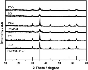
The structure and phase purity of crystalline was determined by powder XRD, as shown in Fig. 1. The sharp, strong peaks confirmed the products were well crystallized. It can be seen that IO-EDA, IO-PEI, IO-PAMAM, IO-PEG, IO-SG, and IO-PAA presented typical reflections of face center cubic iron oxide (JCPDS no. 65-3107), which were indexed into the (220), (311), (400), (422), (511), and (440) peaks. Calculations with the Debye–Scherrer formula for the strongest peak (311) gave grain sizes of IO-EDA, IO-PEI, IO-PAMAM, IO-PEG, IO-SG, and IO-PAA for hydrophilic magnetic nanoparticles with sizes of 20.73, 19.01, 8.73, 21.77, 9.22, and 6.81 nm, respectively. The result implies that the primary nanocrystals do not grow significantly with increasing size of magnetic nanoparticles while the primary crystalline could be also tuned by those modifactory agents.
X-ray diffraction patterns, marked by corresponding modified agents.

B. Size and morphology characterization
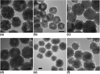
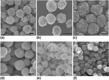
The size and shape of products were examined by TEM and scanning electron microscope (SEM). Figure 2 showed the typical TEM images of the as-prepared magnetite nanoparticles. The average diameter of the samples are between 100 and 200 nm. Former reports of magnetic nanoparticles synthesis showed that the size could be tuned by adjusting reaction temperature and timeReference Liu, Sun, Deng, Zou, Li, Guo, Xiong, Gao, Li and Zhao12, Reference Wang, Bao, Wang, Zhang and Li22: usually longer time and higher temperature would make bigger ones. Here, we introduced various tuning reagents to synthesize magnetic nanoparticles and investigated the effect of modifactory agents. Results indicated that the amount and type of modificatory reagents indeed affected the size of hydrothermal products, this may be due to the instinct of modifactory agents, crystals will certainly behave different under various conditions. SEM images of Fig. 3 coincided well with that of TEM images, which showed that surface morphology of six types of particles were coarse instead of smooth sphere, and they all had secondary structure consisting primary crystal in tens of nanometers, making them superparamagnetic. The properties of coarse surface and superparamagnetic may benefit bimolecular conjugation and bioseparation.
Transmission electron microscopic images of (A) IO-EDA; (B) IO-PEI; (C) IO-PAMAM; (D) IO-PEG; (E) IO-SG; (F) IO-PAA. scale bar=100 nm.

Scanning electron microscopic images of (A) IO-EDA; (B) IO-PEI; (C) IO-PAMAM; (D) IO-PEG; (E) IO-SG; (F) IO-PAA. scale bar=100 nm.

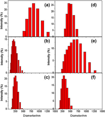
Dynamic light scattering (DLS) analysis (Fig. 4) gave the diameter around 200–300 nm for polymer (e.g., PEI, PEG, and PAA) wrapped IO nanoparticles, which fitted well with their TEM images. However, small molecules wrapped nanoparticles (IO-EDA and IO-SG) were about 1000 nm in diameter according to DLS results, which were attributed to aggregation in certain degree due to the relative weak repulsion induced by much thinner hydrated shell in comparison with magnetic attraction.
Dynamic light scattering measurement of six kinds of magnetic nanoparticles. Respective Z-average diameter (nm): (A) IO-EDA, 1391.2; (B) IO-PEI, 190.9; (C) IO-PAMAM, 239.2; (D) IO-PEG, 468.8; (E) IO-SG, 816.9; (F) IO-PAA, 300.1.

C. Surface properties characterization
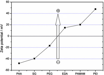
Various organic molecules were introduced to increase the solubility of IO beads and tune the surface charge according to our requirements. We categorized these molecules into three kinds: basic (EDA, PEI, and PAMAM), neutral (PEG), and acidic (SG and PAA). The PEG had no active groups except of terminal hydroxyl groups. Since they are not readily binding to Fe2+ and Fe3+, so the loading amount was limited [∼3% showed by thermogravimetric analysis (TGA)], which surely cannot change much of surface charges. Basic molecules modification on IO beads will easily convert –NH2 group into –NH3+ form, which endows positive charge on surface. On the contrary, acidic coating leads to negative charges by deprotonation. Surface properties were explored by zeta potential measurement and FTIR characterization. Zeta value graph (Fig. 5) showed that zeta values of IO-EDA, IO-PEI, IO-PAMAM, IO-PEG, IO-SG, and IO-PAA are 16.8, 47.6, 20.6, −14.8, −39.6, and −47.8 mV, respectively. It indicated that a wide range of zeta value of MNPs could be tuned by our one-pot solvothermal method. Surface functional groups determine the zeta value of magnetic nanoparticles: amine or amino-terminated PEI, EDA, and PAMAM render positive charges, while carbonate-terminated SG and PAA render negative charge, neutral surface functionalization using PEG inherit the weak negative charge state of crude magnetic nanoparticles. The surface functional groups could help to form stable suspension. We noticed that the small molecular EDA-modified magnetic nanoparticles precipitate in a few hours due to relatively thin hydrated shell and magnetic attraction.Reference Wang, Bao, Wang, Zhang and Li22 But, long-chain macromolecule (e.g., PEG, PEI) modification extends the suspension stability by thickening the hydrated shell and/or strengthening the repulsive electrostatic force.
Zeta potential analysis: surface charge properties could be tuned by different modifying agents.

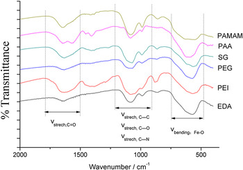
The IR spectroscope (Fig. 6) was used to reveal the existence of modificatory reagent on the six kinds of nanocrystalline surface. Three series of bands appears in common. Strong bands around 590 cm−1 are indexed to the Fe–O stretching modes of the magnetite for all samples.Reference Roca, Marco, Morales and Serna47 A series of bands around 1000–1200 cm−1 corresponds to the stretching vibration of C–C and/or C–O bonds. Another common vibration locates around 1500–1750 cm−1. For IO-PAA and IO-SG, it reveals the presence of carboxyl group (–C=O, at 1650 cm−1), which reveals the loading of PAA and SG onto the MNPs. For other samples without carboxyl group (e.g., PEI), existence of vibrations at 1640 cm−1 is attributed to scissor vibration of –CH2–. Weak –C–O–C– ether vibration band around 1400 cm−1 (antisymmetric stretch) indicates the partial esterification between SG molecules (Fig. 4). Band of 1150 cm−1 clearly shows the presence of C–N bond in the samples of IO-EDA, IO-PEI, and IO-PAMAM, implying the successful loading of organic layer.
Fourier transform infrared spectra of (A) IO-EDA; (B) IO-PEI; (C) IO-PAMAM; (D) IO-PEG; (E) IO-SG; (F) IO-PAA.

D. Magnetic property
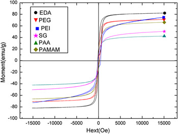
Better magnetic properties can benefit applications in applications like bioseparation or drug delivery. The hysteresis curves (Fig. 7) exhibited that the saturation magnetization of hydrophilic magnetic nanoparticles was between 42 and 82 emu/g, and the field dependence of the magnetization at 300 K showed low remanence (σ r) and a very small coercivity (jHc < 60 Oe), which indicated that six particles are all superparamagnetic. Values of some HSPIONs were comparable with those of Fe3O4 nanoparticles having macro- and nanosizes obtained by other aqueous approaches or with that of the bulk material (92 emu/g for magnetite),Reference Wan, Huang, Yan and Liu48 which is strong enough for bioseparation applications. Saturation magnetization of six products varied possibly due to the size dependence of magnetic nanoparticles and also amount difference of organic molecules on the surface.Reference Taniguchi, Nakagawa, Watanabe, Matsushita and Yoshimura24, Reference Mikhaylova, Kim, Berry, Zagorodni, Toprak, Curtis and Muhammed30 Some studies suggested that the existence of surfactant or polymers on the surface of Fe3O4 nanoparticles quenched surface moments and reduced magnetic moment in such nanoparticles.Reference Wan, Huang, Yan and Liu48
Magnetization curves of six hydrophilic magnetic nanoparticles.

E. TGA analysis
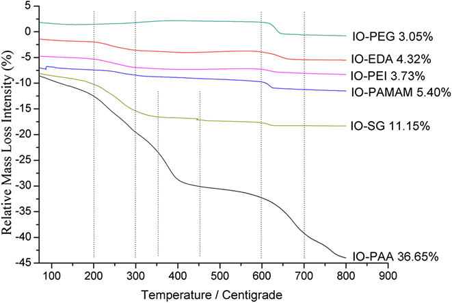
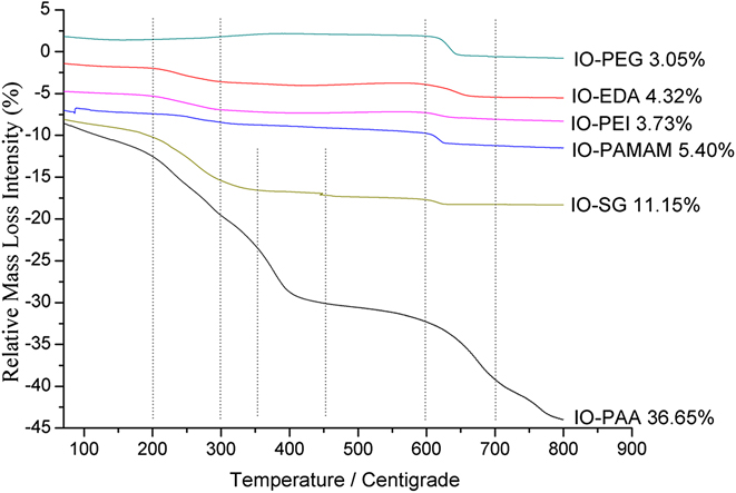
The TGA analysis (Fig. 8) was used to compare the organic molecule amounts on the magnetic iron oxide nanoparticles by showing different decomposition behavior in 100–700 °C. First, weight loss is observed between the temperatures of 50–110 °C which is due to the evaporation of physically adsorbed water on the surface of HSPIONs. Other two typical stages: one is 200–300 °C and the other is 600–700 °C. The former is attributed to the decomposition of modifactory reagents to NH3 or CO2 (formed by initially partial decomposition).Reference Wang, Zhou, Ma, Li and Gu28, Reference Mikhaylova, Kim, Berry, Zagorodni, Toprak, Curtis and Muhammed30 The latter was probably caused by carbothermic reduction to form α-Fe or γ-Fe.Reference Hoch, Mack, Hydutsky, Hershman, Skluzacek and Mallouk49 Special weight loss of IO-SG and IO-PAA was spied at the range of 350 and 450 °C, it may be caused by loss of CO or CO2 molecule formed by carbonization.Reference Zhi, Wang, Lu, Ma and Luo50
Thermal deposition behavior under heating under inert gas (Ar). Relative mass loss intensities have been marked behind respective curves
F. Extracting DNA from model mixture
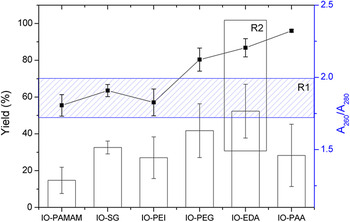
The functionalized magnetic particles are widely used in bioseparations through specificReference Wang, Bao, Wang, Zhang and Li22, Reference Mikhaylova, Kim, Berry, Zagorodni, Toprak, Curtis and Muhammed30, Reference Wang, Shi, Van Antwerp, Cao, Swanson, Bi and Baker51 and nonspecific molecular binding.Reference Narain, Gonzales, Hoffman, Stayton and Krishnan52, Reference McBain, Yiu, El Haj and Dobson53 Here, we investigated the effects of surface modification for nonspecific DNA extraction (data shown in Fig. 9). Protocols were adopted as Taylor et al.Reference Taylor, Hurst, Davies, Sachsinger and Bruce54 described. A model mixture of DNA and protein was used to test the DNA-extracting capability of modified MNPs. The recovery yields and purities of DNA isolated by magnetic particles of various functional groups were determined by yield and ratio of A260 to A280. Commonly, A260/A280 ratio at 1.7–2.0 is considered of high purity.
Recovery yields and purities of DNA samples extracted with different magnetic particles: R1 region stands for the best A260/A280 ratio of pure DNA; R2 region shows that IO-EDA has the best yield and the suitable ratio of A260/A280 among those hydrophilic superparamagnetic iron oxide nanoparticles.

As can be seen from Fig. 8, in terms of both recovery yield (higher than 80%) and purity (A260/A280 = 1.7–2.0, as the shadow zone marked), IO-EDA was unambiguously the best candidate (marked with R2 in a rectangle). The second best is IO-PEG. All the others lack of enough recovery yield or selectivity, or both. This indicated that the type and amount of surface functional groups both had significant effects on the recovery yield and purity of isolated DNA.
It had been expected that the positively charged amino- and imine-containing macromolecules functionalized magnetic nanoparticles (IO-PEI and IO-PAMAM) could have reasonable recovery yield by electronic static attraction because DNA is negatively charged. However, the very low yield and selectivity suggested that the charge did not play a dominant role. Possibly, only aminos work for selective adsorption, but these two mostly contain imine groups. The relative large steric hindrance of imine in macromolecules makes it hard to form hydrogen bonds.
Surprisingly, negatively charged IO-SG and IO-PAA had better performance, suggesting the carboxyl groups have some specific interaction with DNA, possibly by hydrogen bonds. PAA even showed the highest recovery yield, which should relate to its far higher organic molecules mass loading ratio than other nanoparticles, as revealed by TGA (Fig. 8). But, the relatively low selectivity suggested their wide but nonspecific interactions with DNA and protein.
The surface of IO-EDA magnetic particles contain mostly –NH2 groups, which made the particles positively charged. TGA data showed the relative high loading of modifactory molecules onto the surface of MNPs (4.3%, higher than PEI and PEG). They could interact with DNA in a more specific way, associated with electrostatic pull,Reference Yoza, Matsumoto and Matsunaga55 which lead to much better recovery yield and purity. IO-PEG also got reasonable yield and the ratio of A260 to A280, though the mass loading is quite low. This is due to the capability of PEG to transfer DNA from B-type to A-type.Reference Taylor, Hurst, Davies, Sachsinger and Bruce54 Such results confirmed the former reports that ways of DNA binding onto modified magnetic particles are through electrostatic pullReference Yoza, Matsumoto and Matsunaga55 or absorption onto the modified iron oxide nanoparticles in high salt solution.Reference Taylor, Hurst, Davies, Sachsinger and Bruce54
IV. CONCLUSION
In summary, a facile one-pot solvothermal method by introducing various tuning reagents was developed to prepare surface-modified magnetite nanoparticles with tunable magnetism, sizes, suspension stability, and surface charge. These hydrophilic magnetite nanoparticles are evidenced excellent candidates for bioseparation by selectively extracting DNA molecules from a model of cell fractures. Results indicated that PEG and amino-containing organic modules can benefit from the selective extraction of DNA molecules, which should be inspirative for further designing such magnetic nanoparticles for bioapplications.
ACKNOWLEDGMENTS
This work was supported by National Natural Science Foundation of China, the Foundation for Authors of National Excellent Doctoral Dissertations of People’s Republic of China, the Program for New Century Excellent Talents in Universities, the 973 Program (2011CBA00503, 2011CB932403), the National Transgenic Major Program (2008ZX08012-001), and the Fundamental Research Funds for the Central Universities.