Introduction
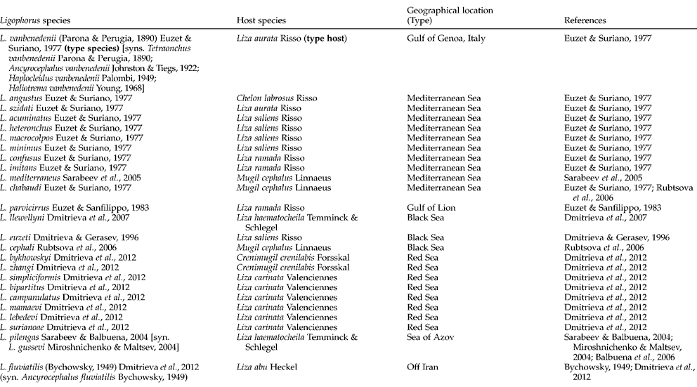
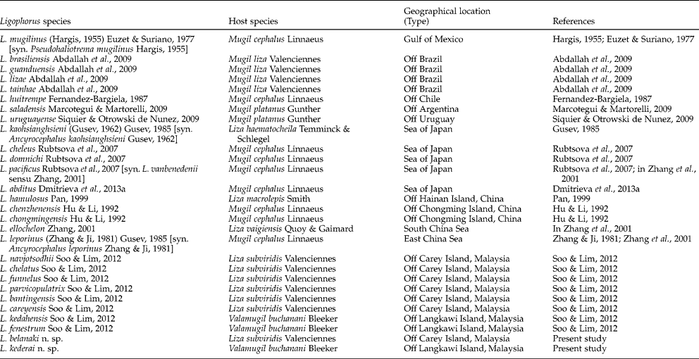
To date 50 species of Ligophorus Euzet & Suriano, Reference Euzet and Suriano1977 have been described from mugilids from off the Mediterranean region, the Red Sea and off Iran (table 1) and the Gulf of Mexico to off the South American coast (Argentina, Brazil, Chile and Uruguay) and off China, Japan and Malaysia (table 2) (see Soo & Lim, Reference Soo and Lim2012). Six Ligophorus species have been described from Liza subviridis (L. navjotsodhii, L. chelatus, L. funnelus, L. parvicopulatrix, L. bantingensis and L. careyensis) and two species from Valamugil buchanani (L. kedahensis and L. fenestrum) caught off Peninsular Malaysia (see Soo & Lim, Reference Soo and Lim2012) (table 2). In a recent survey (2012–2013), we discovered two new Ligophorus species from Liza subviridis off Carey Island and from Valamugil buchanani off Langkawi Island, which were not found in our earlier survey due to their low abundance and low prevalence. The descriptions of the two new species are given herein.
A list of Ligophorus species off European regions, Red Sea and off Iran.

A list of known and new species of Ligophorus off South American and Asia-Pacific regions.
In our recent publication on Ligophorus, principal component analysis (PCA) and numerical taxonomy (NT) analyses were done to provide a more objective and comprehensive comparison of new species with known species and between congeneric species (see Soo & Lim, Reference Soo and Lim2012). PCA and NT analyses are also used here to facilitate the comparison of the present two new species with known Ligophorus species. PCA is used to affirm the status of the two new Ligophorus species based on morphometric data of these two new species and the eight known species from Peninsular Malaysia (see Soo & Lim, Reference Soo and Lim2012). NT analyses are done to determine how the two new Ligophorus species are related to each other and to the 50 known Ligophorus species, based on categorized metric and morphological characters (see Soo & Lim, Reference Soo and Lim2012). The NT analyses also assist in delimiting the number of species for comparison.
Materials and methods
Collection of fish hosts and monogeneans
The mullet hosts, L. subviridis (52 specimens) and V. buchanani (26 specimens) were collected in the coastal waters off Carey Island (2°51′N, 101°22′E) and off Langkawi Island (6°21′N, 99°46′E) on the west coast of Peninsular Malaysia between the years 2010 and 2013. Monogeneans were collected from freshly killed or frozen fish and processed for morphological investigation. Briefly, the monogeneans were removed from the gills, pipetted on to clean glass slides, covered with cover slips and cleared in modified ammonium–picrate–glycerin (Lim, Reference Lim1991). The specimens were carefully flattened to properly expose the hard and soft anatomical structures for morphological study. These ammonium–picrate–glycerin specimens were later washed and dehydrated through a graded ethanol series and mounted in Canada Balsam without staining (Lim, Reference Lim1991). Some specimens were fixed in AFA (acetic acid–formalin–alcohol) on glass slides, stained in Gomori's triple stain and mounted in Canada Balsam after dehydration in increasing ethanol series (Lim, Reference Lim2006; Lim & Gibson, Reference Lim and Gibson2010). The stained and unstained specimens were studied under bright-field and phase-contrast microscopes. Images of the hard and soft anatomical structures of the Ligophorus species were captured using a Leica digital camera and image analysis software (QWin Plus). The hard and soft parts were illustrated using a digitizing tablet (WACOM) and Adobe Illustrator software. Type specimens of the two new species were deposited at the Museum of Natural History, London (BMNH) and Zoological Museum University of Malaya, Kuala Lumpur (MZUM).
Morphometrics
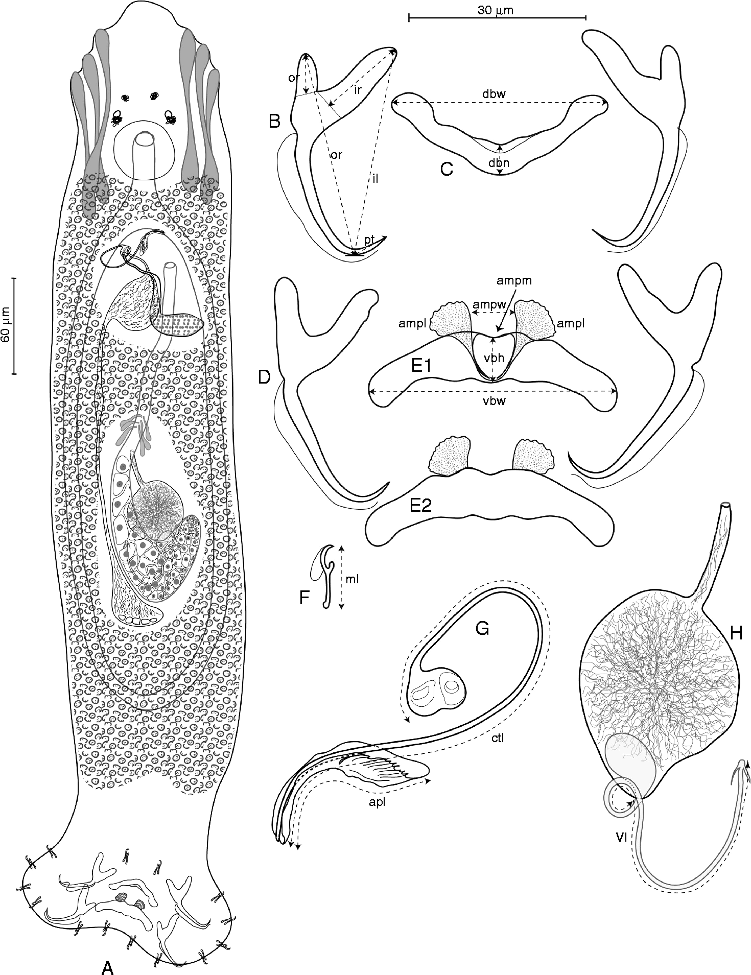
The diagnostic sclerotized hard parts of the haptor (anchors, bars and marginal hooks), vaginal tube and the male copulatory organ (copulatory tube and accessory piece) of 55 specimens belonging to the two new Ligophorus species, which had been properly flattened (stained and unstained), were measured in micrometres (μm) using the measuring option in the Leica QWin software, according to the parameters in fig. 1B, C, E1, F, G and H. It should be noted that the inverted V-shape ventral bars of Ligophorus species possess a highly diverse antero-median protuberance (AMP) which consists of a median piece and two lateral membranous or non-membranous pieces (see Soo & Lim, Reference Soo and Lim2012). In this study, the distance between the two lateral pieces (ampw) was measured (see fig. 1E1). A total of 19 parameters of these diagnostic hard parts were measured: 10 parameters from dorsal and ventral anchors (inner root, outer root, inner length, outer length and point), one parameter from the marginal hook (hook length), three parameters from the ventral bar (length, width and distance between lateral pieces of the AMP), two parameters from the dorsal bar (length and width), two parameters from the male copulatory organ (length of copulatory tube and length of accessory piece) and one parameter from the female organ (length of vaginal tube) (fig. 1). The ‘draw line’ option in Leica QWin software was used to trace and measure the curves and coils of the male copulatory organ and the vaginal tube (see fig. 1G, H). The mean values and the minimum–maximum range (within parentheses) of these measurements were used in the descriptions of the new species (see figs 1, 2, 7, 8; table 3). The morphometric data were further analysed using PCA (see below).
Entire worm of Ligophorus belanaki n. sp. (dorsal view) (A) and sclerotized hard parts of dorsal anchors (B), dorsal bar (C), ventral anchors (D), ventral bar (ventral view) (E1), ventral bar (dorsal view) (E2), marginal hook (F), male copulatory organ (G) and vaginal tube and seminal receptacle (H). Parameters measured: ir, inner root; or, outer root; il, inner length; ol, outer length; pt, point; dbh, dorsal bar height; dbw, dorsal bar width; vbh, ventral bar height; vbw, ventral bar width; ampw, distance between lateral pieces of AMP (antero-median protuberance); ampl, lateral piece of AMP; ampm, median piece of AMP; ml, marginal hook length; ctl, male copulatory tube length; apl, male accessory piece length; vl, vaginal tube length. Illustrated using a digitizing tablet (WACOM) and Adobe Illustrator software.
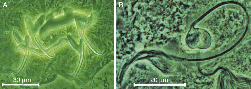
Photomicrographs of sclerotized hard parts of Ligophorus belanaki n. sp., captured using Leica digital camera and an image analysis software (QWin Plus): (A) anchors and bars; (B) male copulatory organ.
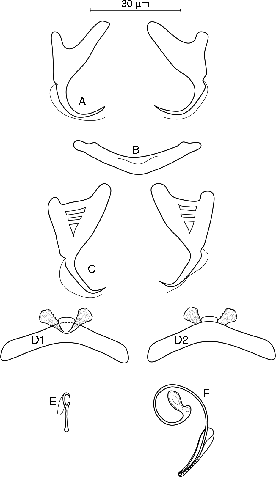
Sclerotized hard parts of Ligophorus kederai n. sp.: (A) dorsal anchors; (B), dorsal bar; (C) ventral anchors; (D1), ventral bar (ventral view); (D2), ventral bar (dorsal view); (E) marginal hook; and (F) male copulatory organ. Illustrated using a digitizing tablet (WACOM) and Adobe Illustrator software.
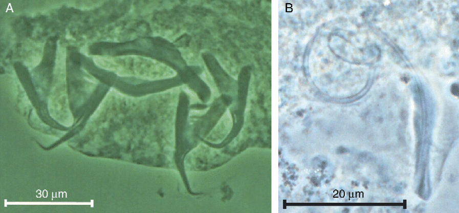
Photomicrographs of sclerotized hard parts of Ligophorus kederai n. sp., captured using Leica digital camera and an image analysis software (QWin Plus): (A) anchors and bars (B) male copulatory organ.
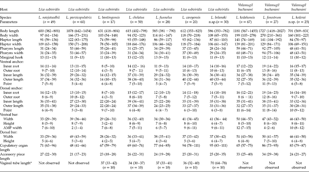
Morphometrics (μm) of eight known and two new Ligophorus species obtained from Liza subviridis and Valamugil buchanani; n=number of specimens measured, and range in size given in brackets.
* Not used in the morphometric analysis due to difficulty in obtaining good specimens.
Principal component analysis (PCA) (Pearson, Reference Pearson1901)
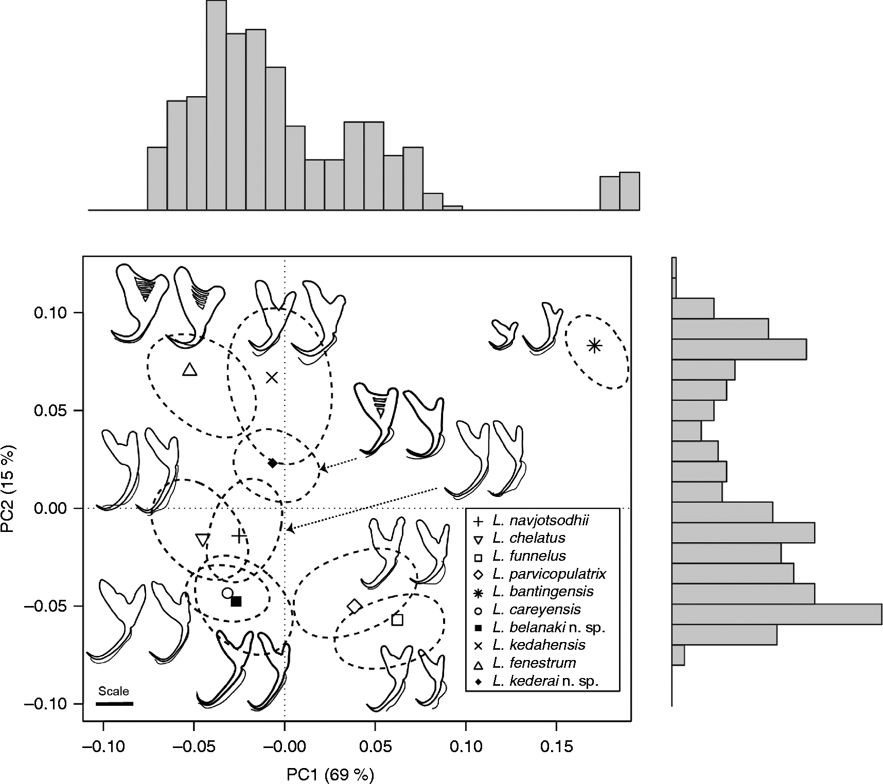
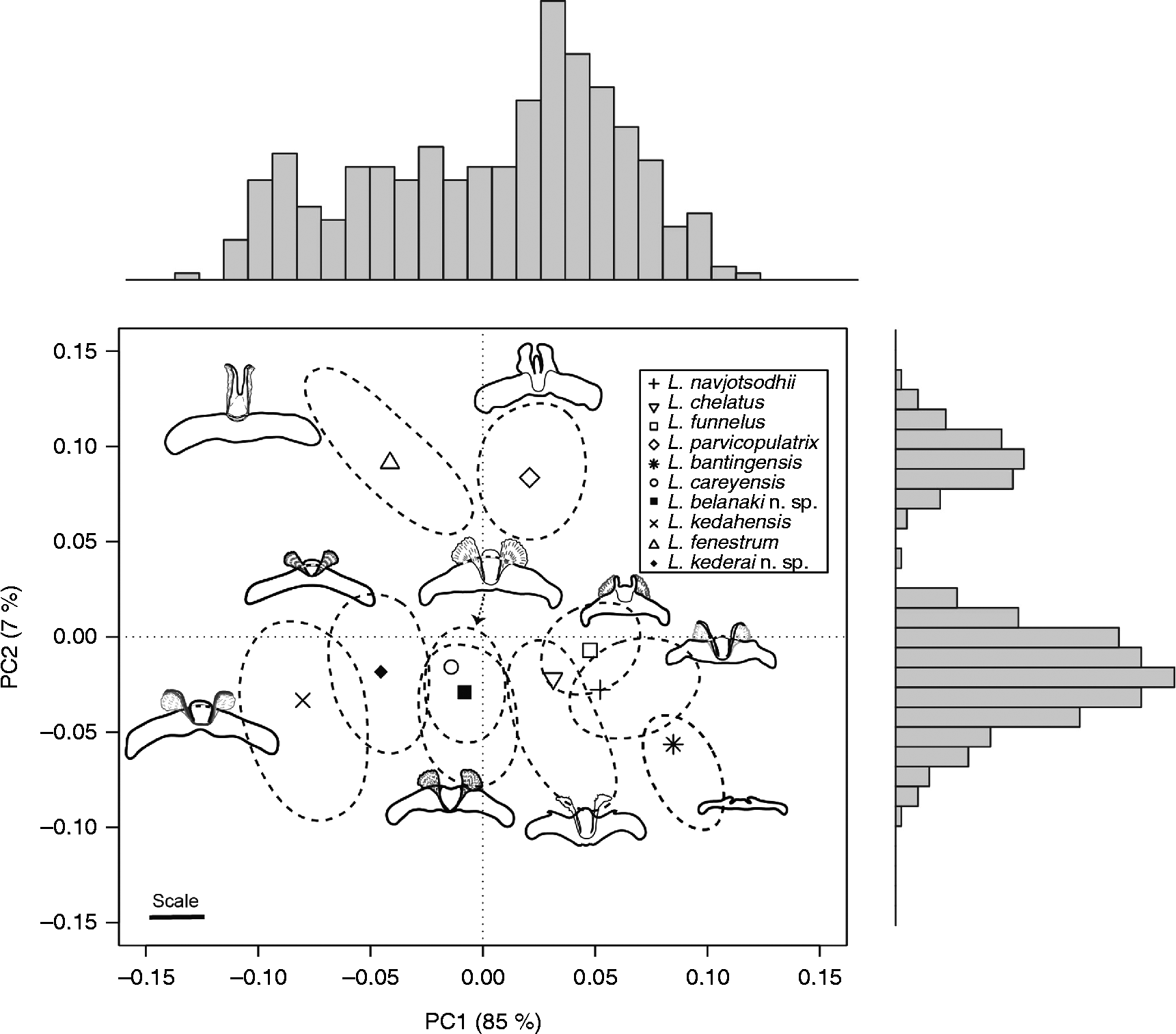
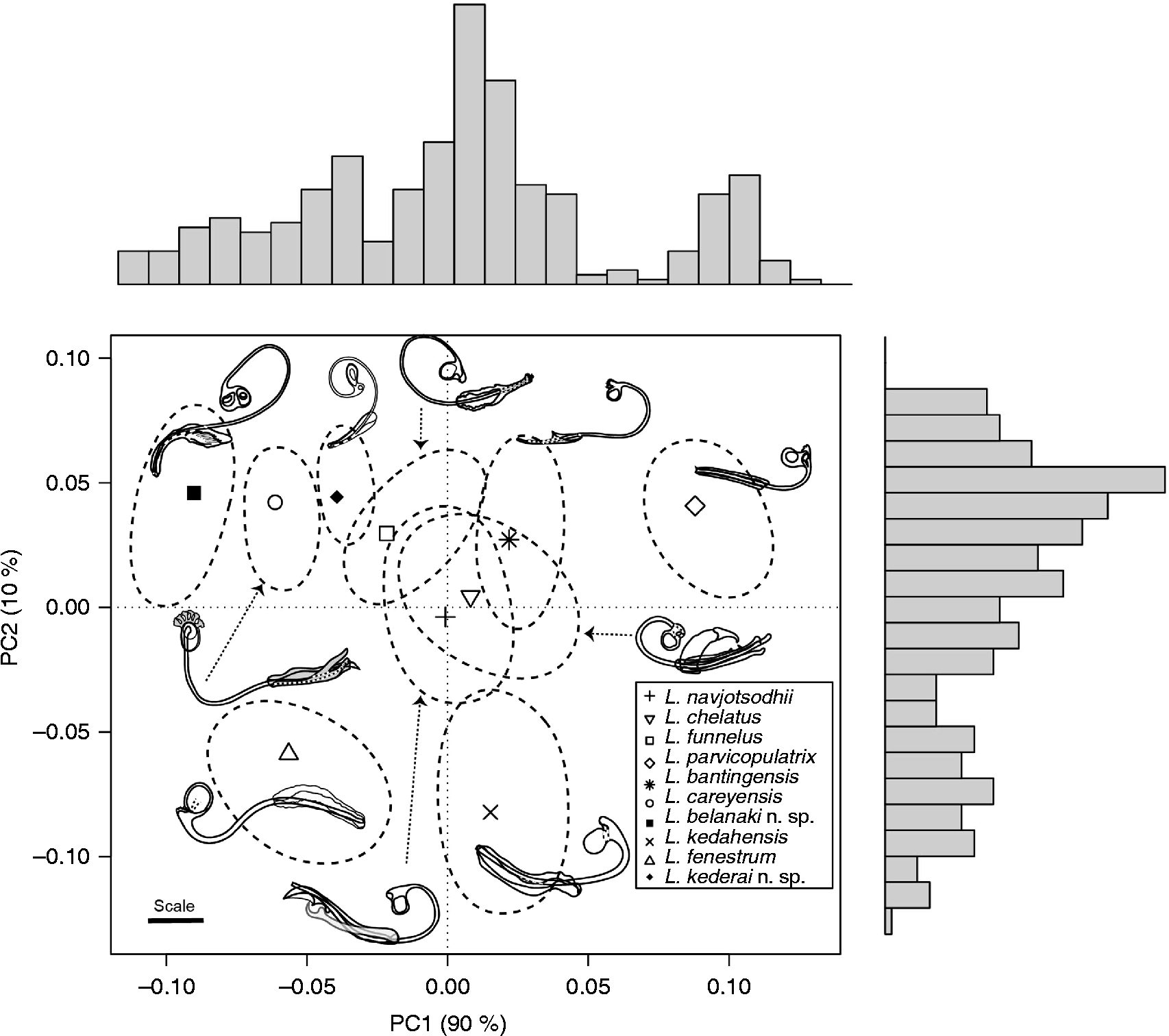
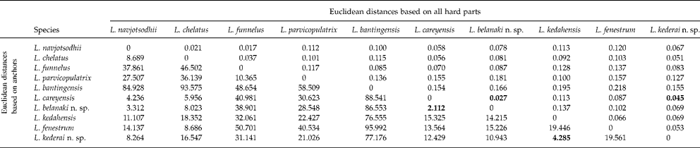
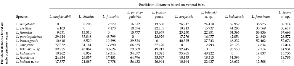
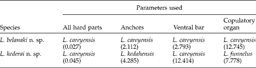
Morphometric data of the 55 specimens of the two new species and the 318 specimens of the eight Ligophorus species described by Soo & Lim (Reference Soo and Lim2012) were analysed using PCA in R (version 2.15.1; R Core Development Team, 2008). The morphometric data of all the hard parts measured excluding the length of vaginal tube (18 parameters), of only the anchors (10 parameters), ventral bars (three parameters) and copulatory organ (two parameters) were analysed separately. The length of the vaginal tube was excluded from these analyses because the vaginal tube was only observed in five out of the ten Ligophorus species and the number of measurements taken was low due to poor visibility of the fine vaginal tube (table 3). The results of the four PCAs were presented in the form of scatterplots to view the important distinguishing characters as a two-dimensional PCA plot (see figs 3–6). The scattering of all the 373 individuals was indicated only in the PCA scatterplot resulting from all the 18 parameters (see fig. 3). In the scatterplots of the anchors, bars and copulatory organs, only the centroids (mean scores) for each of the ten clusters were given. This was done so that the respective diagnostic features could be included into the figures to aid in comparison (see figs 4–6). Horizontal and vertical barplots were given for the two principal component axes for all the scatterplots (see figs 3–6). The Euclidean distance between the centroids of each cluster in the four PCA scatterplots were determined in R (version 2.15.1; R Core Development Team, 2008) and tabulated in tables 4 and 5. The shortest Euclidean distance for the two new species and their nearest neighbouring species were summarized in table 6.
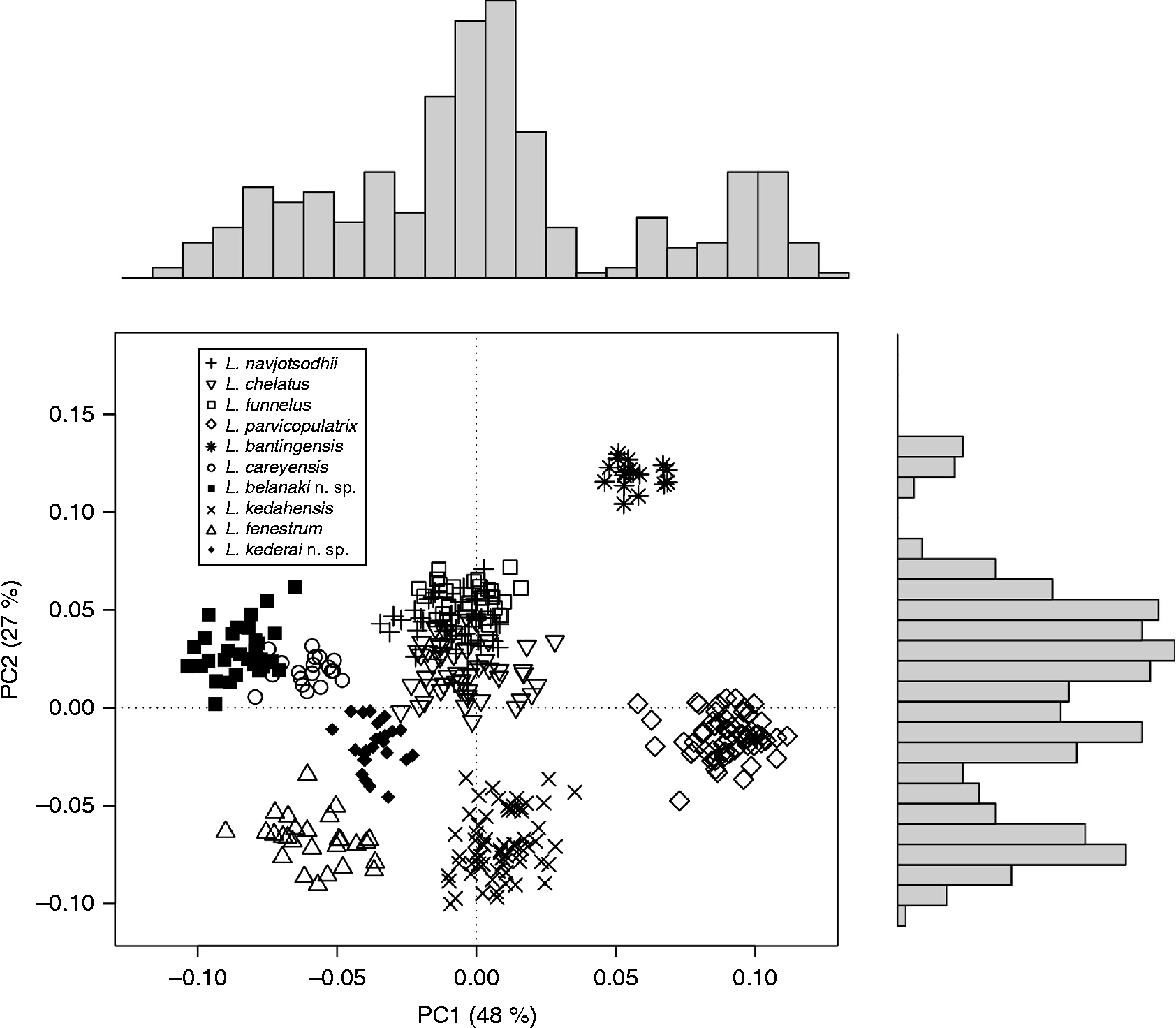
Principal component analysis (PCA) scatterplot of 373 Ligophorus specimens based on all hard parts (marginal hook, anchors, bars and copulatory organ). Vertical and horizontal bar plots indicate one-dimensional summary of the principal component axes, PC1 and PC2 (PC1, index of total variation of overall size of hard parts; PC2, index that contrasts the copulatory tube (length); ventral anchor (outer length, inner root and outer root); dorsal anchor (inner length and outer root); and ventral and dorsal bar (width)).
Principal component analysis (PCA) scatterplot of the two new and eight known Ligophorus species based on the anchors, showing only the centroids for each Ligophorus species. Vertical and horizontal bar plots indicate one-dimensional summary of the principal component axes, PC1 and PC2 (PC1, index of total variation of overall size of anchors; PC2, index that contrasts the ventral anchor (inner length and outer length) and dorsal anchor (inner length, outer length and point)). Scale bar = 20 μm.
Principal component analysis (PCA) scatterplot of the two new and eight known Ligophorus species based on the ventral bar, showing only the centroids for each Ligophorus species. Vertical and horizontal bar plots indicate one-dimensional summary of the principal component axes, PC1 and PC2 (PC1, index of total variation of overall size of ventral bars; PC2, index that contrasts the ventral bar width and AMP width). Scale bar = 20 μm.
Principal component analysis (PCA) scatterplot of the two new and eight known Ligophorus species based on the copulatory organ, showing only the centroids for each Ligophorus species. Vertical and horizontal bar plots indicate one-dimensional summary of the principal component axes, PC1 and PC2 (PC1, index of total variation of overall size of copulatory organ; PC2, index that contrasts the copulatory tube length). Scale bar = 20 μm.
Data matrix of Euclidean distances between PCA centroids of Ligophorus species from Peninsular Malaysia containing (a) the Euclidean distances between PCA centroids of Ligophorus species based on all hard parts (see fig. 3) and (b) the Euclidean distances between PCA centroids of Ligophorus species based on anchors (see fig. 4); the shortest Euclidean distances are in bold.
Data matrix of Euclidean distances between PCA centroids of Ligophorus species from Peninsular Malaysia containing (a) the Euclidean distances between PCA centroids of Ligophorus species based on ventral bars (see fig. 5) and (b) the Euclidean distances between PCA centroids of Ligophorus species based on male copulatory organ (see fig. 6); the shortest Euclidean distances are in bold.
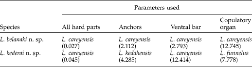
The clustering of the two new Ligophorus species with known Ligophorus species from Peninsular Malaysia based on PCA (principal component analysis); numbers in brackets are the shortest Euclidean distances between the new species and its closest neighbouring species based on tables 4 and 5.
Numerical taxonomy (NT) analysis (Sneath & Sokal, Reference Sneath and Sokal1973)
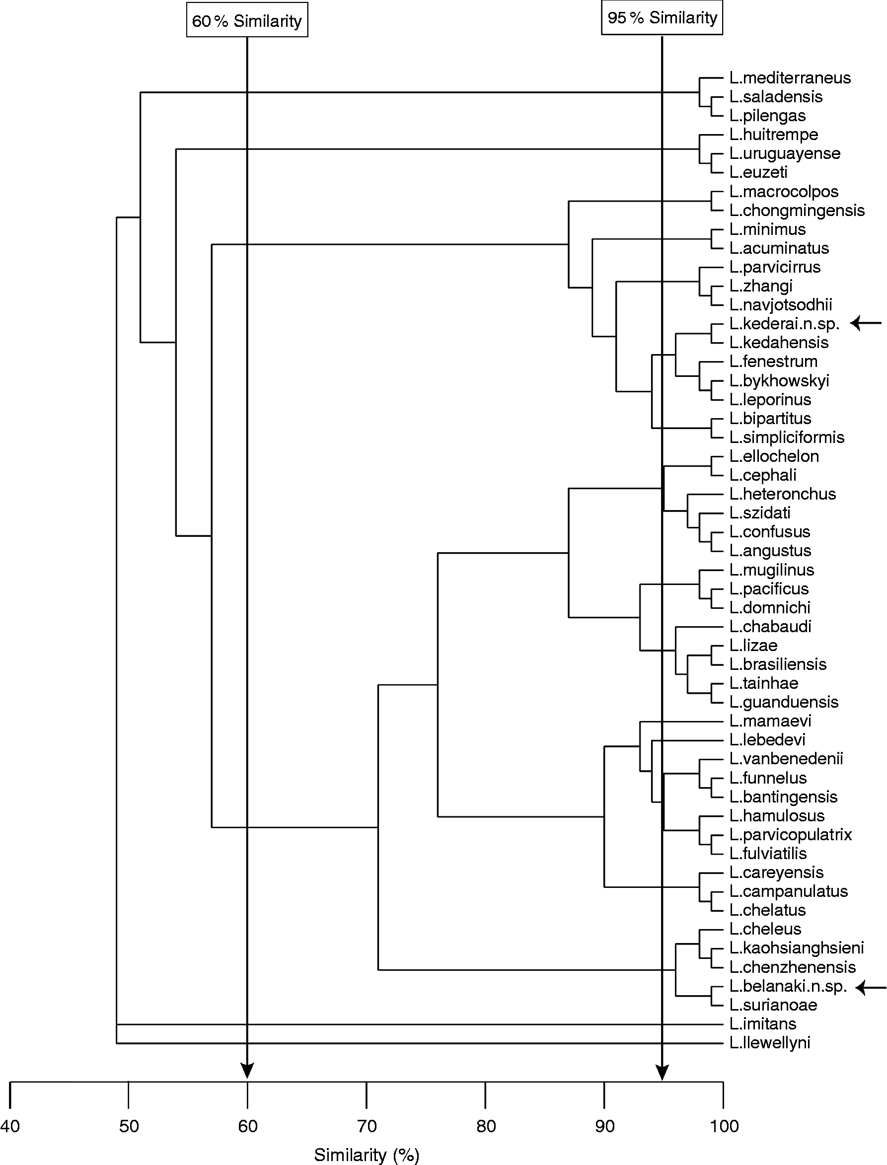
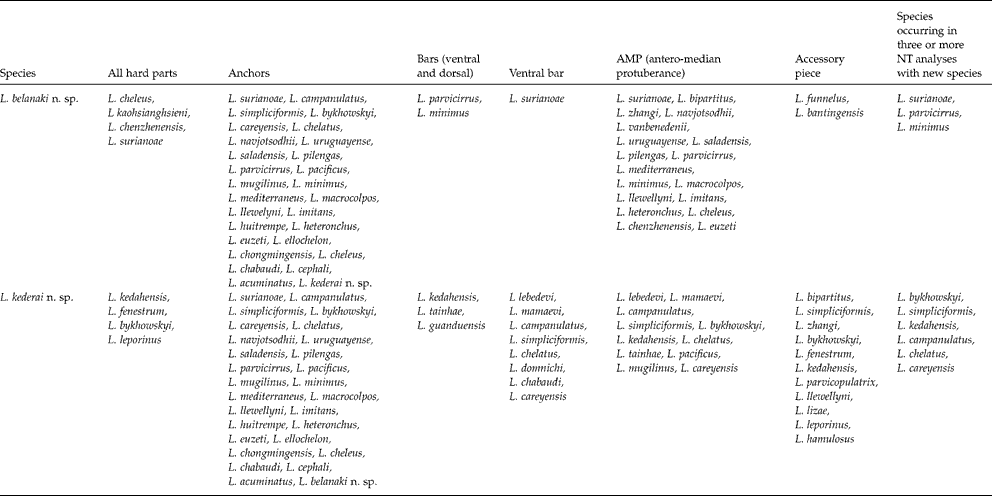
The procedures to collect and analyse morphological and metric data from Ligophorus species and to summarize the results in the form of dendrograms were given in Soo & Lim (Reference Soo and Lim2012). Briefly, a total of 60 character states representing the hard parts (of the haptor and copulatory organs) used in differential diagnoses have been identified, categorized and coded by Soo & Lim (Reference Soo and Lim2012). The two new species and previous 50 species were characterized based on the 60 character states (table available upon request). Next, a data matrix based on Jaccard's Index of Similarity was generated and a neighbour-joining method was used to summarize and cluster the 52 species in the form of dendrograms. The calculation of Jaccard's Index of Similarity and clustering were done in R (version 2.15.1; R Core Development Team, 2008). In this study, six dendrograms have been constructed based on the characters states of all the five diagnostic features (anchors, bars, ventral bars only, AMP and copulatory organs) and on each of the five diagnostic features separately. Although six dendrograms were generated, only one dendrogram based on all the hard parts is presented in this paper (see fig. 9) for brevity. The results from this and the other five dendrograms were summarized in table 7.
Dendrogram of 52 Ligophorus species based on characteristics of all hard parts (anchors, bars and copulatory organ). The arrows on the far right indicate the positions of the new species (L. belanaki n. sp. and L. kederai n. sp.).
The clustering of the two new Ligophorus species with 50 known Ligophorus species based on numerical taxonomy (NT) analyses at 95% similarity level.
Results
The results from the PCA (see figs 3–6) and from the six dendrograms generated from the NT analyses (see fig. 9) are summarized and tabulated (see tables 6 and 7). Since the results of the morphometric analyses are to facilitate differential diagnoses of the two new species, it is appropriate to discuss them prior to the descriptions of the two species to avoid excessive repetitions. In the differential diagnoses of the two new species, comparisons are only made with species which have the shortest Euclidean distance with the two new species in the PCA (see table 6) and with species that shared three or more similar characters with the new species in the NT analyses (see table 7). By doing this, we are able to limit our comparison to a manageable number of morphometrically and morphologically related species.
Clustering of Ligophorus species using PCA
The 373 specimens of Ligophorus (eight previously described and the two new species) from off Peninsular Malaysia are separated into ten clusters which correspond to the eight known and two new Ligophorus species in all the four PCA scatterplots (see figs 3–6). Based on the 18 parameters, the 30 specimens of L. belanaki n. sp. and the 25 specimens of L. kederai n. sp. are grouped closest to L. careyensis (see fig. 3; table 4). In this PCA scatterplot, the first principal component axis (PC1, x-axis), which accounts for 48% of the total variations, is an index of the overall size of all the hard parts and separates the 373 individuals into six groups as shown in the horizontal bar plot (see fig. 3). The second principal component (PC2, y-axis), which explains 27% of the total variation, is an index that contrasts the copulatory tube (length); ventral anchor (outer length, inner root and outer root); dorsal anchor (inner length and outer root); ventral and dorsal bar (width) against the other parameters, separating the Ligophorus specimens into four groups (see fig. 3). In the three PCA scatterplots based on the anchors, ventral bar and copulatory organ, L. belanaki n. sp. is clustered closest to L. careyensis (see figs 4–6; tables 4 and 5). Ligophorus kederai n. sp. is clustered closest to L. kedahensis in the PCA scatterplot based on the anchors (see fig. 4; table 4), to L. careyensis in the PCA scatterplot of the ventral bar (see fig. 5; table 5) and to L. funnelus in the PCA scatterplot of the copulatory organ (see fig. 6; table 5). The information from the four PCA scatterplots is summarised in table 6 and will be used in the differential diagnosis of the two new Ligophorus species (see Descriptions and figs 1, 2, 7, 8).
Clustering of Ligophorus species using NT analysis
In the dendrograms generated in the NT analyses, L. belanaki n. sp. and L. kederai n. sp. are separated into different groups based on the morphology of their hard parts. Based on the dendrogram resulting from the use of all characters, the 50 known and two new Ligophorus species are grouped into four main clusters at 60% similarity level and into 16 groups at the 95% similarity level (see fig. 9; table 7). When only the anchors are used, 10 groups are generated, 17 groups when the two bars are used, 11 groups when only the ventral bar (inclusive of shape and size of AMP) is used, 9 groups when the shape of AMP is used and12 groups when the accessory piece is used (table 7). Although L. belanaki n. sp. and L. kederai n. sp. are similar to four and four known Ligophorus species, respectively, at the 95% level of similarity using all hard parts, they are different in the detailed structures of the bars and male copulatory organ (see Descriptions, fig. 9 and table 7).
Descriptions
Ligophorus belanaki n. sp
Type host. Liza subviridis Valenciennes, 1836.
Type locality. Off Carey Island, Banting, Malaysia (2°51′N, 101°22′E).
Type specimens. Holotype NHMUK 2013.8.16.1 and three paratypes NHMUK 2013.8.16.2–2013.8.16.4 in the Natural History Museum, London; 28 paratypes in the University of Malaya collection MZUM(P)2013.327(P) − 2013.354(P).
Materials studied. Thirty-two specimens studied and 30 specimens measured.
Etymology. This species is named after ‘belanak’, the Malaysian name of the fish host, Liza subviridis.
Description. Figures 1A–H, 2A, B. Body elongate, 586 (353–782) × 108 (65–153), 3 pairs of head organs, 2 pairs pigmented eye spots, posterior with lenses and bigger than anterior pair. Mouth subterminal, ventral; pharynx ovoid, 28 (24–34) × 29 (24–35); intestine bifurcates posterior to pharynx, rejoins posterior to testis and anterior to peduncle forming cyclocoel. Haptor well demarcated; size 87 (61–110) × 106 (61–147); 14 larval type marginal hooks, similar, length 11 (9–13); two pairs of anchors; 2 dorsal anchors, inner length 35 (31–38), outer length 33 (31–36), inner root 14 (10–18), outer root 7 (5–11), point 6 (3–8); 2 ventral anchors, inner length 36 (30–41), outer length 40 (33–44), inner root 14 (10–16), outer root 10 (6–13), point 7 (5–9); 2 connecting bars: V-shaped dorsal bar, 37 (30–42) × 6 (4–7); ventral bar inverted V-shape, 41 (36–44) × 6 (4–7), AMP consists of two membranous lateral pieces and a flattened median piece, distance between lateral pieces, 9 (6–11). Testis single, elongate, ovoid, comparatively small, postero-dorsal to ovary, spermatozoa observed in anterior region; vas deferens leaves anterior region of testis, to sinistral-ventral side, ascends intercaecally, distending, forming seminal vesicle, narrows as vas efferens to enter into smaller lobe of bilobed initial part of copulatory tube. Single elongate gourd-shaped prostatic reservoir with prostatic duct leaving reservoir to enter bigger lobe of bilobed initial of copulatory tube. Copulatory organ consists of copulatory tube, length 95 (83–111) with bilobed initial part and simple funnel-like groove accessory piece, 25 (20–35). Ovary J-shaped, elongate with recurved posterior region; oviduct arises from anterior part of ovary, continues anteriorly as ootype surrounded by Mehlis' gland; proceeds anteriorly as uterus to open near male copulatory organ. Sclerotized vaginal opening, median, at level of recurved portion of ovary; vaginal tube thin, long, sclerotized, 70 (64–78) (n= 10 stained specimens) leading to sperm-filled ovoid seminal receptacle at midbody, dorsal to ovary, duct from seminal receptacle enters oviduct. Vitellarium in lateral fields approximately co-extensive with intestinal caeca, confluent just posterior to intestinal bifurcation, just anterior to ovary and just posterior to cyclocoel.
Differential diagnosis
Ligophorus belanaki n. sp. is similar to L. careyensis in having ventral and dorsal anchors with long inner lengths and short points, narrow V-shape dorsal bars with shoulders and curved ventral bars (table 3). However, they are different in the length of the vaginal tube; 70 (64–78) μm in L. belanaki n. sp. and 36 (32–40) μm in L. careyensis (table 3) and in the morphology of the copulatory organ and median piece of AMP: ornamented bilobed initial part with two opposing accessory pieces and a raised median piece in the AMP of L. careyensis (Soo & Lim, Reference Soo and Lim2012) compared to a non-ornamented bilobed initial part with funnel-shaped groove accessory piece and flat median piece in the AMP of L. belanaki n. sp. (figs 1E1, G, 2A, B). Ligophorus belanaki n. sp. is clustered closest to L. careyensis in the PCA scatterplots for all 18 parameters (fig. 3, table 4) and in the PCA scatterplot for the anchors and ventral bar (figs 4 and 5, tables 4 and 5), thus confirming our observation that the two species have similar anchors and bars (table 6). In the PCA scatterplot for the copulatory organ, L. belanaki n. sp. is also grouped closest to L. careyensis (fig. 6, table 5) based mainly on the copulatory tube length (table 3). Although L. belanaki n. sp., L. funnelus and L. bantingensis possess funnel-shaped accessory pieces, in the PCA scatterplot based on the male copulatory organ, L. belanaki n. sp. is not grouped with L. funnelus and L. bantingensis (fig. 6) because of the length of the copulatory tube, which is 95 (83–111) μm in L. belanaki n. sp., 77 (64–85) μm in L. funnelus and 67 (59–79) μm in L. bantingensis (table 3).
In the NT analyses, L. belanaki n. sp. is grouped with three species, L. surianoae, L. parvicirrus and L. minimus, in having similar anchors, bars and AMP with two lateral membranous pieces and flat median piece (table 7). Ligophorus belanaki n. sp. is different from these species in having a male copulatory organ with funnel-shaped groove accessory piece (fig. 1G), compared to the grooved tubes with expanded distal parts in L. minimus and L. parvicirrus and the grooved tube with expanded proximal part in L. surianoae.
Ligophorus kederai n. sp.
Type host. Valamugil buchanani Bleeker, 1854.
Type locality. Off Langkawi Island, Kedah, Malaysia (6°21′N, 99°46′E).
Type specimens. Holotype NHMUK 2013.8.16.5 and one paratype NHMUK 2013.8.16.6 in the Natural History Museum, London; 23 paratypes in the University of Malaya collection MZUM(P)2013.37(P)–2013.52(P), MZUM(P)2013.55(P)–2013.59(P), MZUM(P)2013.239(P)–2013.240 (P).
Materials studied. Twenty-five specimens studied and 25 specimens measured.
Etymology. This species is named after ‘kedera’, the Malaysian name of the fish host, Valamugil buchanani.
Description. 7A–F, 8A, B. Body elongate, 701 (569–831) × 160 (101–223), 3 pairs of head organs, 2 pairs pigmented eye spots, posterior with lenses and bigger than anterior pair. Mouth subterminal, ventral; pharynx size 48 (41–51) × 48 (43–51). Haptor well demarcated, size 84 (70–97) × 106 (69–151); 14 larval type marginal hooks, similar, length 11 (10–12); two pairs of anchors; 2 dorsal anchors, inner length 33 (32–36), outer length 30 (28–31), inner root 16 (14–18), outer root 9 (7–10), point 10 (9–12); 2 ventral anchors with fenestration, inner length 35 (34–39), outer length 35 (32–36), inner root 16 (15–17), outer root 11 (8–13), point 6 (5–8); 2 connecting bars: dorsal bar broad inverted U, 46 (41–50) × 6 (4–8); ventral bar inverted V, 46 (43–50) × 8 (6–11), AMP consists of two membranous lateral pieces and a raised median piece, distance between lateral pieces, 10 (8–12). Testis single, comparatively small, spermatozoa observed in anterior region; vas deferens leaves anterior region of testis on sinistral side, ascends intercaecally, distending forming seminal vesicle, narrows as vas efferens to enter into smaller lobe of bilobed initial part of copulatory tube. Single prostatic reservoir with prostatic duct entering bigger lobe of bilobed initial part of copulatory tube. Male copulatory organ consists of copulatory tube, length 83 (79–87) with bilobed initial part and a simple grooved boat-like accessory piece, 24 (21–27). J-shaped ovary elongate with recurved posterior region; oviduct arises from anterior region of ovary, continues anteriorly as ootype surrounded by Mehlis' gland; proceeds anteriorly as uterus to open near male copulatory organ. Vaginal opening and tube not observed. Sperm-filled ovoid seminal receptacle at midbody near ovary. Vitellarium in lateral fields approximately co-extensive with intestinal caeca, confluent just posterior to intestinal bifurcation, just anterior to ovary and just posterior to cyclocoel.
Differential diagnosis
Ligophorus kederai n. sp. has a male copulatory organ with simple grooved boat-like accessory piece (figs 7F and 8B), similar to the copulatory organ of L. kedahensis and L. fenestrum, which are from the same host, V. buchanani (see Soo & Lim, Reference Soo and Lim2012). This new species is different in having only the ventral anchors fenestrated (figs 7C and 8A), compared to L. fenestrum where both the ventral and dorsal anchors are fenestrated and L. kedahensis with non-fenestrated anchors (see Soo & Lim, Reference Soo and Lim2012). The shapes of the anchors in the two species are also different: L. kederai n. sp. has anchors with narrower base and longer points (fig. 7C) compared to broader base and shorter points in L. fenestrum (see Soo & Lim, Reference Soo and Lim2012). The 25 specimens of L. kederai n. sp. are grouped closest to L. careyensis in the PCA scatterplot of all 18 parameters (fig. 3, table 4) and in the PCA scatterplot of the ventral bars (fig. 5, table 5). It differs from L. careyensis mainly in having fenestrated ventral anchors, slightly broader ventral bar (46 (43–50) μm) and male copulatory organ with simple boat-like accessory piece with non-ornamented initial part (fig. 7F, table 3) as compared to non-fenestrated ventral anchors, slightly narrower ventral bar (41 (34–45) μm) and male copulatory organ with two opposing accessory pieces and ornamented initial part in L. careyensis (Soo & Lim, Reference Soo and Lim2012). In the PCA scatterplot of the anchors, L. kederai n. sp. is grouped closest to L. kedahensis (fig. 4, table 4), indicating that both these species have anchors with similar morphometries, but morphologically, L. kederai n. sp. is different in having fenestrated ventral anchors (fig. 7C). In the PCA scatterplot of the copulatory organ, L. kederai n. sp. is grouped closest to L. funnelus (fig. 6, table 5) based on the copulatory tube length. Ligophorus kederai n. sp. is different from L. funnelus in having a simple boat-like accessory piece compared to funnel-shape accessory piece in L. funnelus.
In the NT analyses, L. kederai n. sp. is grouped with six species, L. bykhowskyi, L. simpliciformis, L. kedahensis, L. campanulatus, L. chelatus and L. careyensis, in having similar AMP (with two lateral membranous pieces and a raised median piece) and anchors with short points ( ≤ 10 μm) and long inner lengths ( ≥ 30 μm) (table 6). Ligophorus kederai n. sp. differs from these six species in having fenestrated ventral anchors (figs 7C and 8A, table 7).
Discussion
The type species of Ligophorus, L. vanbenedenii (Parona & Perugia, Reference Parona and Perugia1890) Euzet & Suriano, Reference Euzet and Suriano1977 had been assigned and reassigned to different ancyrocephalid genera (Tetraonchus, Ancyrocephalus, Haplocleidus and Haliotrema) prior to its assignment as the type species of Ligophorus (tables 1 and 2) (see Soo & Lim, Reference Soo and Lim2012). Ligophorus vanbenedenii has several characteristics (single prostatic reservoir, vas deferens not overlapping the right intestinal branch and vagina opening at the medio-ventral part of the body) which prompted its reassignment into the genus Ligophorus by Euzet & Suriano (Reference Euzet and Suriano1977). Taxonomically, Ligophorus has been assigned to Ancyrocephalidae Bychowsky, 1937 by the majority of the researchers on Ligophorus (14 studies) and also to Ancyrocephalinae Bychowsky, 1937 in Dactylogyridae Bychowsky, 1933 by others (four studies). The 14 studies which ascribed Ligophorus to Ancyrocephalidae are Euzet & Suriano (Reference Euzet and Suriano1977), Euzet & Sanfilippo (Reference Euzet and Sanfilippo1983), Dmitrieva & Gerasev (1996), Mariniello et al. (Reference Mariniello, Ortis, D'Amelio and Petrarca2004), Sarabeev & Balbuena (Reference Sarabeev and Balbuena2004), Sarabeev et al. (Reference Sarabeev, Balbuena and Euzet2005), Balbuena et al. (Reference Balbuena, Rubtsova and Sarabeev2006), Dmitrieva et al. (Reference Dmitrieva, Gerasev and Pron'kina2007), Dmitrieva et al. (Reference Dmitrieva, Gerasev, Merella and Pugachev2009), Marcotegui & Martorelli (Reference Marcotegui and Martorelli2009), Siquier & Owtrowski de Nunez (Reference Siquier and Otrowski de Nunez2009), Dmitrieva et al. (Reference Dmitrieva, Gerasev, Gibson, Pronkina and Galli2012), Soo & Lim (Reference Soo and Lim2012) and Dmitrieva et al. (Reference Dmitrieva, Gerasev and Gibson2013a). The other four studies which ascribed Ligophorus to Ancyrocephalinae in Dactylogyridae are Rubtsova et al. (Reference Rubtsova, Balbuena, Sarabeev, Blasco-Costa and Euzet2006), Rubtsova et al. (Reference Rubtsova, Balbuena and Sarabeev2007), Abdallah et al. (Reference Abdallah, de Azevedo and Luque2009) and Blasco-Costa et al. (Reference Blasco-Costa, Miguez-Lozano, Sarabeev and Balbuena2012). The validity of Ancyrocephalidae and Ancyrocephalinae in Dactylogyridae is a longstanding issue and has been elaborated by Lim et al. (Reference Lim, Timofeeva and Gibson2001) and summarized herein. Briefly, to resolve the heterogeneity of the Ancyrocephalidae and the paraphyletic nature of the Ancyrocephalidae, Kritsky & Boeger (Reference Kritsky and Boeger1989) proposed two options for revising the Ancyrocephalidae and favoured the one considering the Ancyrocephalidae as a junior synonym of the Dactylogyridae. Lim (Reference Lim1998), however, disagreed with this move, while recognizing the paraphyletic nature of Ancyrocephalidae, and proposed that the Ancyrocephalidae be left intact within the Dactylogyridea until further studies have been carried out. In this paper we will be retaining Ligophorus in Ancyrocephalidae until more intensive molecular and morphological studies of the different ancyrocephalid genera are done.
The two new species, L. belanaki n. sp. and L. kederai n. sp., bring the total number of Ligophorus species described off Peninsular Malaysia to ten species, and to 52 species globally. Ligophorus belanaki n. sp. and L. kederai n. sp. were collected from Liza subviridis and Valamugil buchanani, respectively. To date L. subviridis harbours seven species (six known species and L. belanaki n. sp.) and V. buchanani harbours three species (two known species and L. kederai n. sp.) (see Soo & Lim, Reference Soo and Lim2012).
As already noted, the ventral bar of Ligophorus is basically an inverted V-shape bar with an antero-median protuberance (AMP) consisting of a median piece and two lateral membranous or non-membranous ear-like processes arising from the median piece (see Soo & Lim, Reference Soo and Lim2012) and has been shown to be an important diagnostic feature for Ligophorus species (fig. 5, tables 4 and 5). The AMP of L. belanaki n. sp. and L. kederai n. sp. consists of two lateral membranous pieces but they differ in the morphology of the median piece, which is flat in L. belanaki n. sp. (figs 1E, 1E and 5) and raised in L. kederai n. sp. (figs 7D, D2 and 5).
To date, amongst the Ligophorus species, fenestrated anchors only occur in two described and one unnamed species: L. fenestrum (dorsal and ventral anchors) (Soo & Lim, Reference Soo and Lim2012) and L. kederai n. sp. (ventral anchors) (figs 7C and 8A) found on V. buchanani off Langkawi Island, and Ligophorus sp. 14 (ventral anchors) from Liza affinis off North Vietnam (South China Sea) (Dmitrieva et al., Reference Dmitrieva, Gerasev, Kolpakov, Nguen and Ha2013b). The fenestrations in all three species are different: in L. fenestrum and L. kederai n. sp. fenestrations are made up of three or more slit-like apertures, compared to the single aperture in Ligophorus sp. 14. Fenestration in anchors is not limited to these Ligophorus species or to any particular group of monogeneans and can be found in species from different genera, such as in most Thaparocleidus species from pangassids (single fenestrum in ventral anchors) (see Pariselle et al., Reference Pariselle, Lim and Lambert2001a, Reference Pariselle, Lim and Lambertb, Reference Pariselle, Lim and Lambert2002, Reference Pariselle, Lim and Lambert2003, Reference Pariselle, Lim and Lambert2004, Reference Pariselle, Lim and Lambert2005a, Reference Pariselle, Lim and Lambertb, Reference Pariselle, Lim and Lambert2006) and in Haliotrema banana Lim & Justine, Reference Lim and Justine2007 (single fenestrum in dorsal and ventral anchors) (see Lim & Justine, Reference Lim and Justine2007). The occurrences of fenestrated anchors in a few species of unrelated genera seem to suggest that fenestration in anchors is of specific importance rather than generic importance in monogeneans. Despite the presence of fenestrated anchors, L. fenestrum and L. kederai n. sp. are valid Ligophorus species possessing characteristics of Ligophorus, such as a vas deferens which does not overlap the intestinal caecum, male copulatory organ with bilobed initial part, J- to U-shaped ovary, seminal receptacle within the curve of the J- to U-shaped ovary and ventral bar with AMP. Hence there is no justification to erect a new genus to accommodate the Ligophorus species with fenestrated anchors. Although Ligophorus species are specific to mugilids, currently we have too few Ligophorus species with fenestrated anchors to conclude whether they are restricted to specific mugilid host species or not. The fact that L. fenestrum and L. kederai n. sp. are from V. buchanani might suggest close relationships of the two species caused by speciation within the host species. However, it should be noted that V. buchanani is also host to L. kedahensis which has non-fenestrated anchors (Soo & Lim, Reference Soo and Lim2012). The relationship of L. fenestrum, L. kederai n. sp. and Ligophorus sp. 14 is difficult to explain since the host of the latter species is L. affinis. The ecological advantage or disadvantage of fenestrations in anchors is difficult to speculate. Fenestration is probably caused by a ‘reduction’ in sclerotized materials (‘scleroporosis’) in a specific area, forming holes or windows or fenestrum or apertures. However, without any knowledge of the developmental history of these Ligophorus species, in particular that of the anchors, it is difficult even to speculate how these holes are formed and their function(s), if any.
The copulatory organ of L. belanaki n. sp. and L. kederai n. sp. consists of a copulatory tube with bilobed initial part and accessory piece (figs 1G, 8B). The bilobed initial part is a common feature in all the 52 Ligophorus species. The bilobed initial part of the copulatory tube of L. belanaki n. sp. and L. kederai n. sp. lacks ornamentation, bringing the number of Ligophorus species without ornamentation to 28 species, and 24 species with ornamentation. Of the ten Ligophorus species from Malaysia, only L. careyensis possesses ornamentation (see Soo & Lim, Reference Soo and Lim2012) (fig. 6). In the majority of Ligophorus species, the accessory piece is not connected to the initial part of the male copulatory organ by any structure. To date only six Ligophorus species – L. angustus, L. cephali, L. confusus, L. ellochelon, L. heteronchus and L. szidati – were described and illustrated as having a male copulatory organ with a muscular sheath surrounding the copulatory tube, attaching at its distal end to the accessory piece and at its proximal end to the initial part of the male copulatory organ. It should be noted that such a muscular sheath is lacking in the other 46 Ligophorus species, including these two new species and all the eight known Ligophorus species on Malaysian mugilids.
Although not illustrated, the soft anatomical structures of the male and female reproductive parts of L. kederai n. sp. are similar to those of L. belanaki n. sp. and other known Ligophorus species (see Soo & Lim, Reference Soo and Lim2012), in having the testis situated postero-dorsal to the ovary, vas deferens traversing intercaecally along the sinistral-ventral side, J-shaped ovary, vaginal opening (if observed) located at midbody near the J-ovary and a seminal receptacle located at the recurved part of the ovary. The generic characteristics of Ligophorus were emended in Soo & Lim (Reference Soo and Lim2012) to include the fact that the ovary is J- to U-shaped and that the vaginal system is not obvious in all members of the genus if not sclerotized.
Ligophorus belanaki n. sp. is observed to have a sclerotized vaginal tube (fig. 1H) which brings the number of Ligophorus species with sclerotized vaginal tubes to 39. A sclerotized vaginal tube was not observed in the present 25 specimens of L. kederai n. sp. and, to date, there are 12 other species without obvious vaginal tubes (L. acuminatus, L. fluviatilis, L. leporinus, L. minimus, L. parvicirrus, L. navjotsodhii, L. kedahensis, L. fenestrum, L. bykhowskyi, L. simpliciformis, L. zhangi, L. bipartitus). Ligophorus parvicopulatrix from L. subviridis has no obvious vaginal tube but it has a large sclerotized vaginal opening (see Soo & Lim, Reference Soo and Lim2012). The vaginal system of Ligophorus (when observed) consists of a sclerotized vaginal pore, undulating to a coiled vaginal tube and obvious seminal receptacle. The vaginal system is not easily observed in non-stained specimens.
Previous and present studies have shown that the use of analytical methods in taxonomic studies is becoming increasingly common (Rubtsova et al., Reference Rubtsova, Balbuena, Sarabeev, Blasco-Costa and Euzet2006, Reference Rubtsova, Balbuena and Sarabeev2007; Dmitrieva et al., Reference Dmitrieva, Gerasev and Pron'kina2007, Reference Dmitrieva, Gerasev, Gibson, Pronkina and Galli2012; Tan et al., Reference Tan, Khang and Lim2010; Soo & Lim, Reference Soo and Lim2012). Multivariate analyses (PCA and NT) can be used to facilitate comparative diagnoses because of rising numbers of species being described, especially between species which are morphologically and morphometrically similar (see Soo & Lim, Reference Soo and Lim2012) and also to distinguish between inter- and intra-specific differences (Tan et al., Reference Tan, Khang and Lim2010). PCA could separate species and detect morphovariants, but one has to bear in mind that PCA requires data from large numbers of specimens (see Tan et al., Reference Tan, Khang and Lim2010; Soo & Lim, Reference Soo and Lim2012) for well-defined clusters to be generated. Hence metrical analyses (such as PCA and NT) are only complementary and supplementary in assisting in the clustering of species, thereby reducing the number of species for comparison to a manageable level. Such analytical tools are not obligatory tools in taxonomy, since it is highly unrealistic to require taxonomists to measure hundreds of specimens for purely taxonomic investigations. It should be noted that although metric analyses will provide a more objective comparison of new and known species, the morphometric data used do not take shape information into consideration. This is elaborated in fig. 6 where Ligophorus species with funnel-shape accessory piece (L. belanaki n. sp., L. bantingensis and L. funnelus) are clustered separately, while L. belanaki n. sp. and L. careyensis are clustered closer, based mainly on the copulatory tube length, although their accessory pieces and initial parts are morphologically different: funnel-shaped groove accessory piece and unornamented initial part in L. belanaki n. sp. (fig. 1G) and two opposing accessory pieces with ornamented initial part in L. careyensis (see Soo & Lim, Reference Soo and Lim2012). The same is observed for the anchors and ventral bars (figs 4 and 5). Since morphometric data do not include morphological shapes, which are important taxonomic characteristics, geometric morphometry (Adams et al., Reference Adams, Rohlf and Slice2004; Vignon & Sasal, Reference Vignon and Sasal2010) could be a potentially relevant analytical tool in differential diagnosis, and this is currently being explored.
Acknowledgements
The authors would like to thank Dr Jean-Lou Justine and Dr David I. Gibson for procurement of literature; Dr Evgenija V. Dmitrieva for sending us photomicrographs for our use; Dr Khang Tsung Fei for advice in using the R software for analyses; to Mr Liew Kim Seng for assisting in collecting and preparing stained and unstained specimens and to Yap Fook Choy for assisting in field work. This paper forms part of the PhD thesis of the first author.
Financial support
The field trips were funded by the Klang Islands Expedition Grant (OS27-B27518 OCAR TNC (P&I) 2009) and the Biodiversity Database Flagship Project grant (FL001/2011 – Biodiversity Information Management in Malaysia: Current Status and Way Forward) to the corresponding author.
Conflict of interest
None.