This manuscript highlights the imaging sessions held at the 7th World Congress of Pediatric Cardiology & Cardiac Surgery in Barcelona, Spain, 16–21 July, 2017. We had specialists from all around the world come talk about the versatility and diagnostic power of the current and upcoming imaging tools in paediatric cardiology and cardiac surgery.
Imaging was one of many tracks at the conference, including catheterisation, general cardiology, electrophysiology, intensive care/anaesthesia, heart failure/pulmonary arterial hypertension, surgery, and adult CHD. Imaging Track presentations were broken down into 4 days, focusing on specific topics each day. Clinical and research work as well as interesting cases were presented by international specialists from Australia, Austria, Belgium, Brazil, Canada, Denmark, France, Hong Kong, India, Israel, Italy, Japan, the Netherlands, New Zealand, Nigeria, Norway, Poland, Spain, South Africa, Sweden, Switzerland, Taiwan, Turkey, United Kingdom, United Arab Emirates, and United States of America.
Day 1 overview
Day 1 covered “Basics and Frontiers of Advanced Imaging”. The morning session specifically included basics of advanced imaging by two-dimensional and three-dimensional echocardiography, MRI, CT, and strain imaging to assess cardiac morphology, dimensions, areas, volumes, and systolic and diastolic function. Dr Girish Shirali from Children’s Hospital Mercy, United States of America gave a great review on three-dimensional imaging by echocardiography. A session led by Dr Steven Colan from Boston Children’s Hospital, United States of America, reviewed the reliability and reproducibility of imaging by two-dimensional echocardiography. In the “Ventricular Volume and Variability study” that followed 169 children with dilated cardiomyopathy, left ventricular mass, volumes, and ejection fraction by 5/6 area-length method based on three-beat average demonstrated the highest reproducibility.Reference Margossian, Chen and Sleeper 1 As expected, serial acquisitions by a single imager with measurements performed by a single observer resulted in the best reproducibility over time.Reference Margossian, Chen and Sleeper 1
Dr Luc Mertens from the Hospital for Sick Children, Canada gave great examples on the difficulties of applying adult guidelines in assessment of diastolic function to children due to age and heart rate dependency. Dr Bart Bijnens from University of Leuven, Spain and Dr Yui-Fai Cheung from the University of Hong Kong, Hong Kong, discussed strain and its current clinical use with spectacular slides to demonstrate that strain enables functional assessment of right ventricular function, ventricular–ventricular interactions, mechanics of functional single ventricles, and atrial function in CHD that permits early detection of impaired myocardial function. We then moved on to MRI and covered its applications to assess anatomy, ventricular function, and tissue characterisation, led by Dr Andrew Powell from Boston Children’s Hospital, United States of America.
Another interesting presentation by Dr Emanuella Valsangiocoma from Children’s Hospital Zurich, Zurich, Switzerland, concentrated on “Real-time 3-Dimensional Echocardiographic Quantification of Left Ventricular Volumes”, drawing conclusions that measurement of ventricular volumes and ejection fraction by three-dimensional echocardiography is highly reproducible; however, that it consistently underestimates ventricular volumes and ejection fraction compared with cardiac MRI.Reference Mor-Avi, Jenkins and Kuhl 2 She noted that a major gap is lack of normal data from infancy to adulthood, and that current data on morphologically normal hearts need validation for different CHD conditions. Lastly, she concluded that the semi-automated summation disc method best performs for ventricles of abnormal morphology. As discussed by Dr Anthony Hlavecek and Dr Israel Valverde from the Medical University of South Carolina, United States of America and Hospital Universitario Virgen del Rocío, Spain/United Kingdom, respectively, an important advance in the field of imaging is that the radiation dose of CT has diminished significantly over the last decade bringing CT back into the front fields of cardiac imaging in CHD, when appropriate.
In the afternoon sessions, there were key lectures discussing frontiers of advanced imaging including three-dimensional printing and holograms and cardiac computer modelling. Dr John Simpson from King’s College Hospital, London touched on the fascinating development on three-dimensional echocardiography in CHD and the consensus guidelines. Now, patient-specific modelling and modelling of valves can be performed by three-dimensional echocardiography. Then Dr Elchanan Bruckheimer from Schneider Children’s Medical Center of Israel captivated the audience with the tool of interactive live holography which allows specialists to visualise the heart in all views, slice, measure, and see real-time volumes during interventions. Dr Ajit Yoganathan from Georgia Institute of Technology College of Engineering, United States of America gave a brilliant talk on cardiac computer modelling, and how it can allow for more precise surgical planning, preoperatively exploring blood flow characteristics and different surgical scenarios. He then demonstrated his work he has pioneered in collaboration with Emory University School of Medicine, United States of America in planning of the Fontan Pathway/total cavopulmonary connection. We also got inspired by the upcoming tools of vortex imaging and blood speckle imaging, which took us to a mini-futuristic voyage into the next decade.
Day 2 overview
Day 2 was the “Potpourri Day”, covering topics including “Imaging in the Catheterisation Laboratory and Operating Room”, “Fetal Echocardiography”, “Telemedicine”, and “Bridging Session of Imaging and Rheumatic Fever and Kawasaki Disease”.
We had great presentations reviewing how echocardiography can guide surgical interventions and catheterisation procedures by Dr Nancy Ayres from Texas Children’s Hospital, United States of America and Dr Simone Pedra from the Hospital for the Heart, São Paulo, Brazil. Dr Gerald Marx from Boston Children’s Hospital, United States of America gave a spectacular review on epicardial imaging. Transoesophageal echocardiography can be contraindicated in some patients because of small patient size, cranio-facial abnormalities, oesophageal abnormalities, difficulty in monitoring blood pressure, or because of operations involving pulmonary vein repair.Reference Dragulescu, Golding and Van Arsdell 3 Interesting, epicardial imaging in congenital heart surgery has been resurging in some centres, with special interest by the surgeons to improve their imaging skills, which has improved intraoperative detection of residual lesions in certain situations. To ensure we covered the “little hearts” in our potpourri day we had a great discussion on what is new and upcoming in fetal medicine by Dr Sally Ann Cur from the Netherlands.
In the rapidly expanding field of telemedicine, Dr Craig Sable from Children’s National Health System, Washington, United States of America gave several great examples of implementations of telemedicine in paediatric cardiology that improved outcomes and reduced cost. For example, Webb et al conducted a multicentre study tracking patients without critical heart disease followed by telemedicine versus patients who received routine clinical care. In the multicentre study, the percentage of patients transported for the telemedicine group was half of that of the control patients (5 versus 10%). Total length of stay was 1 day for the telemedicine population versus 2.6 days for the control patients.Reference Webb, Waugh and Grigsby 4
Another interesting and upcoming topic of telemedicine is parental involvement. At Lucile Packard Children’s Hospital at Stanford, we have tested the reliability of parental acquisition of echo images on children with heart transplant patients using hand-held devices at home. Preliminary results show that parents can be successfully trained to take images on their own children that can be reliably interpreted by trained echocardiography specialists. All parents reported that they are “comfortable” imaging their children. This method of tele-care could allow patients who live far away from a specialised centre to receive long-term care with professionals via live-video conferencing.
We ended the day bridging Rheumatology and Imaging and had a great presentation on lumen imaging of coronary arteries by MRI by Dr Gerald Greil from the University of Texas, Southwestern, United States of America.
Day 3 overview
Day 3 included discussions on “Imaging and Anatomy”, “Imaging in Heart Failure and Transplant”, “The never-ending debates on Ventricular Septal Defect and Double Outlet Right Ventricle”, “Peri-procedural Imaging”, and “Right Ventricular Imaging”.
We kicked the day off with great discussions on reaching consensus in terms of nomenclature of ventricular septal defects, led by Dr Amy Juraszek from the University of Florida College of Medicine, United States of America and Dr Meryl Cohen from Children’s Hospital of Philadelphia, United States of America. Next, it was a hot debate on double outlet right ventricle by Dr Robert Anderson from the University College London, United Kingdom and Dr Leo Lopez from the Nicklaus Children’s Hospital, United States, Finally, there was a great presentation of vascular slings and embryology by Dr Juberan Pushparajan from the Evelina Children’s Hospital, United Kingdom and Dr Vera Aiello from Sao Paulo University School of Medicine, Brazil.
We then moved on to talk about new guidelines that have been published in the Journal of American Society of Echocardiography on multimodality imaging of patients with transposition of the great arteries, highlighting potential long-term complications by Dr Xavier Iriart from University Hospital of Bordeux, France.Reference Cohen, Eidem and Cetta 5
This topic was followed by a spectacular talk on unbalanced atrioventricular canal defects by Dr Meryl Cohen from Children’s Hospital of Philadelphia, United States. Dr Cohen reviewed various of her and others’ work on echocardiographic variables investigated to define unbalance and potentially predict suitability for single versus two ventricle repairs. Dr Leo Lopez from Nicklaus Children’s Hospital, United States of America, presented the importance of diagnosing systemic venous anomalies. Systemic venous anomalies are often associated with other CHD, and could be the aetiology of unexplained cyanosis. It can also help with venous access, venous cannulation during surgery, and surgical timing for staged palliation of single ventricle patients. Most importantly, Dr Lopez noted that for trainees it is prudent to know about all the potential systemic venous anomalies to be able to diagnose them throughout their career, an important topic that came up during the Imaging Sessions frequently.
I had the opportunity to give a talk on “Functionally” Single Ventricles, and what to cover for each condition by echocardiography to guide the clinical and surgical team in the best possible way. Dr Rebecca Beroukhim from Boston Children’s Hospital, United States of America, presented the use of MRI in cardiac tumours with pristine images. This was followed by a great review of aortapathies and imaging by Dr Julie DeBacker from Ghent University, Belgium. Dr Norman Silverman from Stanford University, United States of America “took the hit” and gave a terrific talk on laterality disorders or “heterotaxy”.
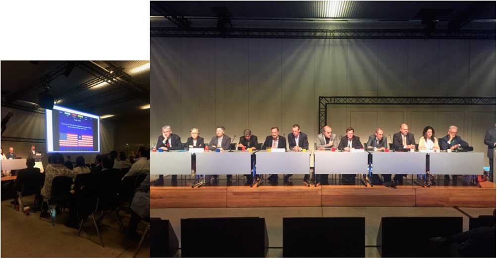
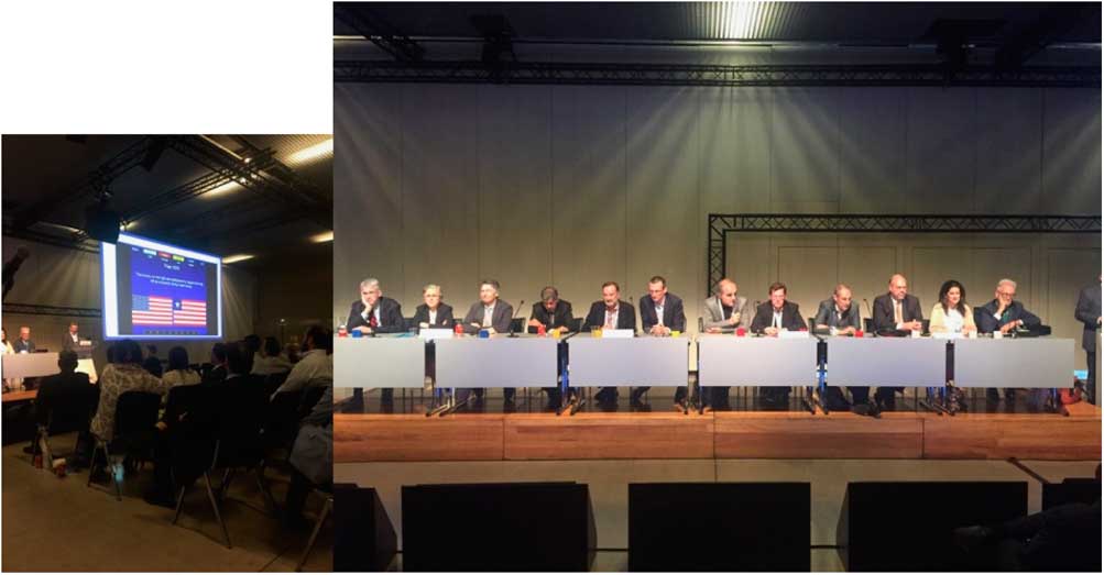
It was very entertaining to watch Jeopardy (Fig 1) run by Dr Michael Brook from University of San Francisco, United States of America. The contestants were all very “brave men” since Meryl Cohen from Children’s Hospital of Philadelphia, United States of America “killed it”.
Moments from Jeopardy.
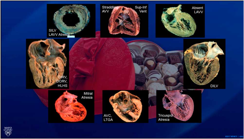
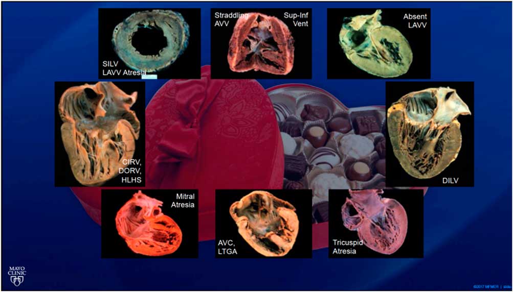
We adjourned this day with great discussions on right ventricular imaging led by Dr Aurelio Secinaro from Università degli Studi di Torino, Italy, Dr Tal Geva and Dr Yasir Qureshi from the Boston Children’s Hospital and Mayo Clinic, United States of America, Dr Mark Friedberg from University of Toronto, Canada, and Dr Jan Marek from the Great Ormond Street Hospital, United Kingdom. The take home message was that the right ventricle is not an “obscure” entity anymore and that it should be assessed using a multimodality approach that includes two-dimensional and three-dimensional imaging by echocardiography, in addition to CT and MRI. Some of the discussions focussed on the multimodality imaging guidelines by the American College of College of Cardiology on repaired tetralogy of Fallot patients,Reference Chen, Dusenbery, Valente, Powell and Geva 6 right ventricular imaging in single ventricle patients and Ebstein/Uhl anomaly patients, as well as assessment of right ventricular performance in pressure loaded right ventricles. The quote and picture of the day (Fig 2), or probably the Imaging Sessions overall, was that “Single ventricles are just like a box of chocolates. You never know what you will get!” by Dr Quereshi.
Box of Chocolates (from Dr Quereshi’s talk).
Day 4 overview
Day 4 honed in on “Bridging Imaging and Pathology” and on “Fetal Sessions”. The imaging and pathology sessions included great reviews of congenital anomalies tricuspid valves and coronary arteries by Dr Benjamin Eidem, Dr Robert Anderson, Dr Lucille Houyel, and Dr Vitor Guerra from the Mayo Clinic, United States of America, the University College London, United Kingdom, Hôpital Marie Lannelongue, France, and Tulane University, United States of America, respectively.
The fetal sessions started off with a great review of the current guidelines and the “finally established” consensus on standards for performance of fetal echocardiography by Dr Mary Donofrio from Children’s National Health System, Washington, United States of America. Consensus says that certain aspects such as assessment of cardiac position, situs, two-dimensional assessment of all cardiac chambers, color Doppler interrogation of all valves/vessels/and septae, pulse Doppler interrogation of all valves and ductus venosus, assessment of cardiac rate and rhythm, qualitative function, and video clips with sweeps of cardiac structures are mandatory. Following these guidelines will allow for standardisation of imaging across subspecialties’ for better detection of CHD and improvements in in-utero and postnatal care for fetuses with CHD. However, there remains limitations such as limited utility of current referral indications, and limited experience with in utero therapy and specialised delivery room management.Reference Rychik, Ayres and Cuneo 7
Dr Gerald Tulzer from Children’s Heart Center Linz, Kepler University Hospital, Austria touched on defining boundaries for a fetal interventional programme in Europe, and how to establish standards and centralise care, an important consideration for all surgical and fetal centres in the world. Dr Christopher Lindblade from the Phoenix Children’s Hospital, United States of America and Dr Orhan Uzun from the Cardiff’s University Hospital of Wales, United Kingdom gave great talks on assessment of heterotaxy and arrhythmia in fetal life. Dr Queralt Ferer from University Hospital Vall d’Hebron, Spain reviewed how the newer modalities are being used more frequently in assessment of fetal cardiac function. Finally Dr Gurur Biliciler-Denktas from the University of Texas, United States of America, concluded the fetal sessions with interesting fetal cases.
A “hot topic” was the placenta, as discussed by Jack Rychik from Children’s Hospital of Philadelphia, United States of America. The connection between the abnormal placenta in CHD is relatively unknown but could uncover significant answers. Major unanswered questions are on optimal methods for characterisation of the placenta in utero: placental size, placental function, and placental blood flow: What is the relationship between placental characteristics and clinical outcomes? Can we modify elements of placental anatomy, function, or blood flow to improve human development prior to birth?
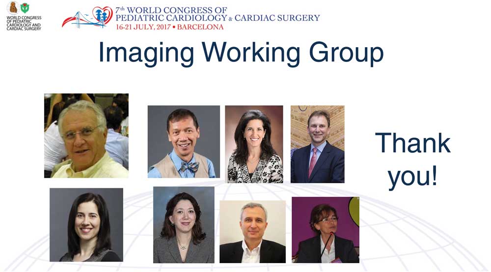
The 7th World Congress in Barcelona, Spain was an immense success and a great privilege to be part of (Imaging Working Group Fig 3). The 4-day conference celebrated the field of Pediatric Cardiology and Cardiovascular Surgery, acknowledging the work of international specialists, captivating the audience and opening the dialogue for improving care worldwide.
Imaging Working Group.
Acknowledgements
The author would like to thank the Organizing Committee of the 7th World Congress of Pediatric Cardiology and Cardiac Surgery for this wonderful opportunity to be part of this very important academic mission.
Financial Support
This research received no specific grant from any funding agency, commercial or not-for-profit sectors.
Conflict of Interest
None.