Introduction
In order to study the biodiversity of free-living nematodes in the Beikedidao Bay of Bohai Sea, China, sediment samples were collected from an intertidal sandy beach in May 2019. Abundance of free-living nematodes is 4214 ind 10 cm−2 in these habitats. The dominant species are Bathylaimus australis Cobb, Reference Cobb1894, Nudora gourbaultae Vincx, Reference Vincx1989 and Chromadorita longispiculata Gagarin, Reference Gagarin2012. Among the species from the habitats, an unrecorded species was identified as Deontolaimus holovachovi sp. nov. Meanwhile, an unrecorded species from the East China Sea is described as Campylaimus zhoui sp. nov.
The genus Deontolaimus de Man (Reference de Man1880) was established with the type species Deontolaimus papillatus from marine or brackish sediment. A total of 19 valid species have now been listed in Nemys (Bezerra et al., Reference Bezerra, Decraemer, Eisendle-Flöckner, Hodda, Holovachov, Leduc, Miljutin, Mokievsky, Peña Santiago, Sharma, Smol, Tchesunov, Venekey, Zhao and Vanreusel2021). The genus Camacolaimus de Man (Reference de Man1889) was considered a junior synonym of Deontolaimus by Holovachov & Boström (Reference Holovachov and Boström2015) based on re-examination of type material of Camacolaimus tardus de Man (Reference de Man1889) and C. barbatus Warwick (Reference Warwick1970) which all possess alveolar supplements. The species originally belonging to the genus Camacolaimus were transferred to the genus Deontolaimus. Holovachov & Boström (Reference Holovachov and Boström2015) reviewed Deontolaimus, emended the genus diagnosis and described three additional new species. Meanwhile, they provided an identification key to 17 valid species. According to a recent article of Holovachov (Reference Holovachov2020), four species (Camacolaimus bellus, C. bulbimangani, C. glandulosus and C. iaculator) described by Bussau (Reference Bussau1993) are now recognized valid species which are transferred to the genus Deontolaimus. Therefore, there are now 23 valid species in the genus Deontolaimus around the world.
The genus Campylaimus Cobb (Reference Cobb1920) is a broadly distributed but relatively uncommon genus of marine and brackish nematodes with 23 nominal species (Bezerra et al., Reference Bezerra, Decraemer, Eisendle-Flöckner, Hodda, Holovachov, Leduc, Miljutin, Mokievsky, Peña Santiago, Sharma, Smol, Tchesunov, Venekey, Zhao and Vanreusel2021). It was first established by Cobb with the type species C. inaequalis Cobb (Reference Cobb1920). Subsequently, the genus was reviewed by Tchesunov (Reference Tchesunov1978), Huang & Zhang (Reference Huang and Zhang2006), Villares et al. (Reference Villares, Martelli, Lo Russo and Pastor2013) and Fadeeva et al. (Reference Fadeeva, Mordukhovich and Zograf2016). Recently, Holovachov (Reference Holovachov2019) provided an identification key to species of the genus. In Chinese sea areas, only one species in the genus, Campylaimus gerlachi Timm (Reference Timm1961) from the Yellow Sea has been recorded by Huang & Zhang (Reference Huang and Zhang2006).
Materials and methods
Intertidal sediment samples were collected in multiple locations of a sandy beach along the coast of the Beikedidao Bay of the Bohai Sea between 38°13′–38°14′N 117°56′–117°57′E using a sawn-off syringe with a 2.6 cm inner diameter in May 2019. The samples were taken to a depth of 8 cm and divided into three sections (i.e. 0–2, 2–5 and 5–8 cm), then fixed with 10% formalin in seawater for long-term preservation. The sublittoral sediment samples from the East China Sea were obtained using a 0.1 m2 improved Gray–O'Hara box at Station DH 5-3 during the Open Research Cruise of National Natural Science Foundation of China by the RV ‘Dongfanghong 2’ in October 2012. In the laboratory, sorting and mounting of nematodes were performed as previously described (Sun et al., Reference Sun, Huang and Huang2018; Qiao et al., Reference Qiao, Jia and Huang2020). The descriptions were made from glycerin mounts using a differential interference contrast microscope (Leica DM 2500). Line drawings were made with the aid of a camera lucida. All measurements were obtained using Leica LAS X version 3.3.3, and all curved structures were measured along the curved median line. Type specimens have been deposited in the Marine Biological Museum of Chinese Academy of Sciences, Qingdao.
Abbreviations are as follows: a, the ratio of body length to maximum body diameter; b, ratio of body length to pharynx length; c, ratio of body length to tail length; c′, ratio of tail length to cloacal or anus body diameter; V%, position of vulva from anterior end expressed as percentage of total body length.
Results
Systematics
Order PLECTIDA Gadea (Reference Gadea1973)
Family CAMACOLAIMIDAE Micoletzky (Reference Micoletzky1924)
Genus Deontolaimus de Man (Reference de Man1880)
Deontolaimus holovachovi sp. nov.
(Figures 1–3)
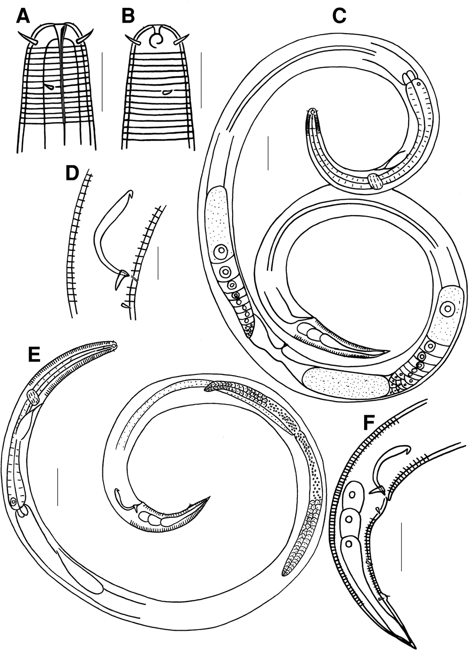
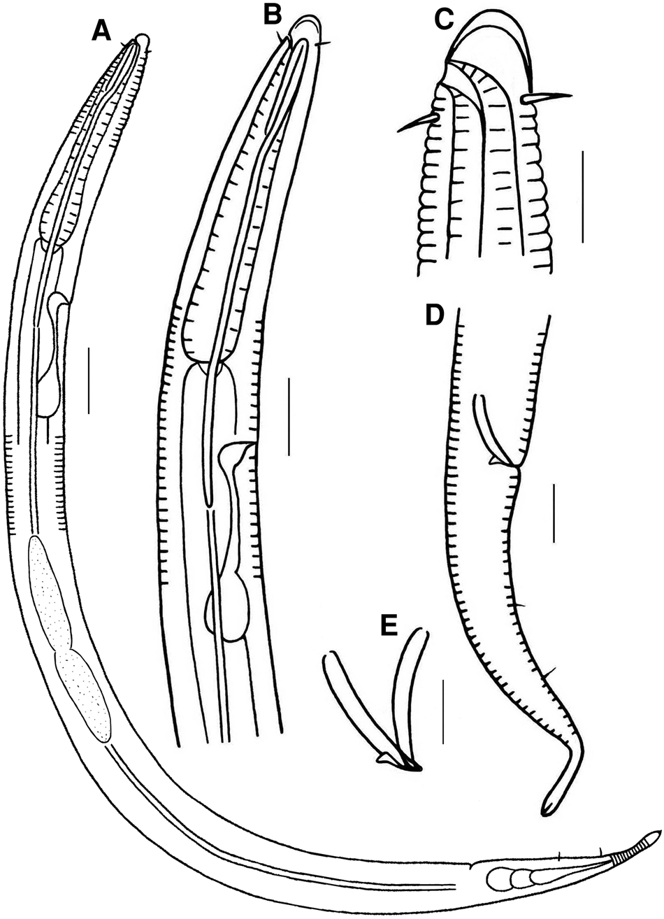
Deontolaimus holovachovi sp. nov. (A) anterior end of holotype; (B) anterior end of female; (C) view of entire female; (D) cloacal region of holotype; (E) view of entire holotype; (F) tail end of male. Scale bars: A, B, D = 10 μm; C, E = 30 μm; F = 20 μm.

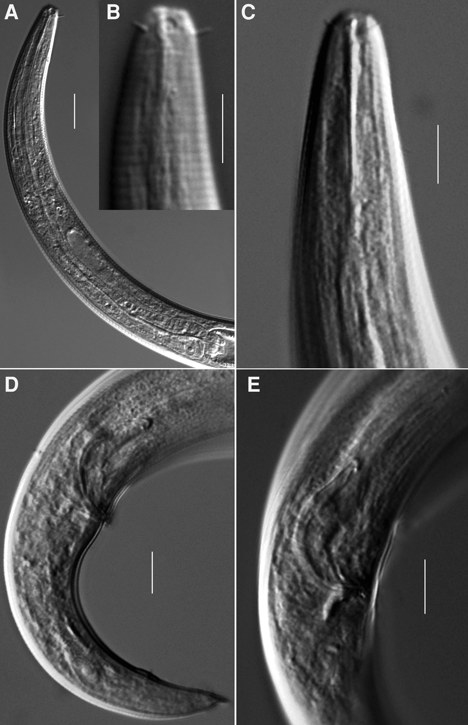
Deontolaimus holovachovi sp. nov. (A) pharyngeal region of holotype; (B) anterior end of female; (C) anterior end of female; (D) male tail; (E) cloacal region of male. Scale bars: A = 20 μm, B–E = 10 μm.

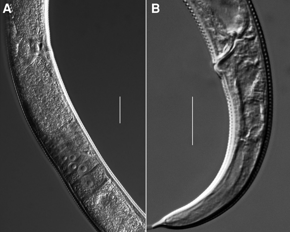
Deontolaimus holovachovi sp. nov. (A) vulva region of female; (B) female tail. Scale bars: A, B = 20 μm.

Type material
Holotype male was collected from Stations BHY4 (middle of the intertidal zone), on the slide BHY4-17. Paratypes, ♂2 from station BHY4, on slide BHY4-15; ♂3, ♂4, ♂5 from station BHY5, on slide BHY5-8; ♀1 from station BHY4, on slide BHY4-19; ♀2 from station BHY4, on slide BHY4-18; ♀3, ♀4 and ♀5 from station BHY5, on slide BHY4-8.
Type locality and habitat
Intertidal beach of Beikedidao Bay in the Bohai Sea, China. Station BHY4 (38°13′55″N 117°56′39″ E), fine sand with broken shells. Paratype at Station BHY5 (38°13′53″N 117°56′43″E), silt with broken shells.
Etymology
The species is named in honour of Dr Oleksandr Holovachov, Swedish Museum of Natural History, for his contributions to nematode taxonomy.
Measurements
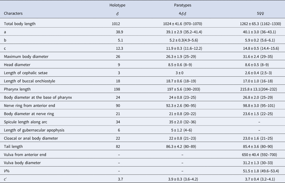
Measurement data are given in Table 1.
Measurements (in μm except a, b, c, c′ and V%) of Deontolaimus holovachovi sp. nov.

Description
Males: Body slender, cylindrical over most of its length, tapering in anterior half of pharyngeal region and posteriorly on tail. Cuticle annulated, 1.5–2 μm thick; annules without ornamentation. Anterior-most annule appearing posterior to cephalic setae base. Lateral alae not observed. Labial region rounded, continuous with body contour. One circle of six small papilliform outer labial sensilla visible on the anterior surface of labial region. Four cephalic sensilla setiform, 3 μm long; their bases located at the level with posterior portion of amphideal fovea, about 4 μm from the anterior body end. A pair of lateral stout cervical setae (2 μm long) located at the middle of onchiostyle, 9–13 μm from the anterior body end, and another pair of cervical setae situated at the mid-length between nerve ring and anterior body end, 46–52 μm from the anterior body end. Amphidial fovea unispiral, 3 μm wide, located just in front of cephalic setae bases. Ocelli absent. Alveolar supplements in pharyngeal region not observed under the light microscope. Nerve ring surrounding pharynx just anterior to its middle. Secretory-excretory system consisting of a large renette cell located on left-hand side of body along anterior part of intestine, an excretory ampulla just posterior to nerve ring and a short connecting duct, opening at level with nerve ring. Buccal cavity funnel-shaped, with a long dorsal onchiostyle, 18–19 μm long. Pharynx cylindrical, expanding posteriorly, not forming a bulb. Cardia large, oblong in shape, 10 μm long and 8 μm wide, embedded in intestine.
Reproductive system diorchic, both testes opposed, outstretched. Spicules paired, curved in the middle with the proximal half swollen and the distal half narrow, proximal end in a sharp right-angled ventral bend. Gubernaculum 3 μm long, plate-like with paired dorso-caudal apophyses. Precloacal supplement absent. Three pairs of postcloacal subventral stout setae present: the first pair located just posterior to cloacal opening, the second pair located at about mid-length of tail, and the third pair situated at one-third from tail terminus. A midventral papilla with a short seta at its top present at one-third from tail terminus. Tail elongated conical, ventrally curved. Three caudal glands present. Spinneret sclerotized, conical with acute tip, 3–4 μm long.
Females: Similar to males in most respects except body slightly larger; tail without postcloacal sensilla, lateral alae more distinct, 3 μm wide, extending from anterior part of pharynx to the middle of tail. Reproductive system didelphic with two opposed, reflexed ovaries. Anterior ovary located on right side of intestine, posterior ovary located on left side of intestine. Oviduct tubular. Spermathecae indistinct. Uterus broad. Egg oblong. Vagina straight, not thickened, about 0.25 times vulval body diameter long. Vulva located in the mid-body length.
Differential diagnosis and discussion
Deontolaimus holovachovi sp. nov. is characterized by the combination of short cephalic setae (2.5–3 μm long); unispiral amphidial fovea; excretory pore located at the level with nerve ring; lateral alae present, two pairs of lateral cervical setae present; arcuate spicules, 32–36 μm long, the proximal half swollen with hooked proximal end, the distal half narrow; gubernaculum plate-like with dorso-caudal apophysis; a midventral caudal papilla with a short seta located at posterior third of tail, lacking obvious pharyngeal alveolar supplements.
The new species is similar to Deontolaimus tardus (de Man, Reference de Man1889) Holovachov & Boström (Reference Holovachov and Boström2015), D. timmi Holovachov & Boström (Reference Holovachov and Boström2015) and D. catalinae Holovachov & Boström (Reference Holovachov and Boström2015) in body size, spicules shape, relatively short cephalic setae and amphideal fovea structure, but differs from D. tardus in having slender body (vs plump in shape), smaller body size (0.97–1.33 mm vs 1.6–2.34 mm), relatively longer cephalic setae (2.5–3 μm vs 1.5–2.2 μm), shorter and different shape spicules (32–36 μm vs 58–59 μm), shorter gubernacular apophysis (4–6 μm vs 8–9 μm) and longer tail (c′ = 3.6–4.2 vs 2.5–2.8). It differs from D. timmi in having relatively longer body (0.97–1.33 mm vs 0.71–0.94 mm), longer cephalic setae (2.5–3 μm vs 1.5–2 μm), gubernaculum with apophysis (vs without apophysis), tail possessing a distal midventral papilla with a short seta. The new species differs from D. catalinae by having longer cephalic setae (2.5–3 μm vs 1.5 μm), excretory pore opening at level with nerve ring (vs opening near the anterior end, i.e. short distance posterior to onchiostyle base), presence of a distal caudal midventral papilla with a short seta (vs absence), spinneret acute and sclerotized (vs short and unsclerotized). Moreover, the present species resembles D. cylindrocaudatus (Chitwood, Reference Chitwood1951) Holovachov & Boström (Reference Holovachov and Boström2015), D. guillei (de Bovee, Reference De Bovee1977) Holovachov & Boström (Reference Holovachov and Boström2015) and D. lorenzeni Holovachov & Boström (Reference Holovachov and Boström2015) in some aspects. However, the new species is distinguished from D. cylindrocaudatus by having shorter cephalic setae (2.5–3 μm vs 5–8 μm) and shorter tail (c′ = 3.6–4.2 vs 6); it differs from D. guillei by smaller body size (0.97–1.33 mm vs 2.72–3.17 mm), shorter spicules (32–36 μm vs 63–65 μm) and relatively longer tail (c′ = 3.6–4.2 vs 2.4–3.2). It differs from D. lorenzeni by shorter cephalic setae (2.5–3 μm vs 5 μm), longer spicules (32–36 μm vs 20–21 μm) and broad conical spinneret not offset from tail (vs spinneret narrow and dorsally curved, sharply offset from tail).
Order ARAEOLAIMIDA De Coninck & Schuurmans Stekhoven (Reference De Coninck and Schuurmans Stekhoven1933)
Family DIPLOPELTIDAE Filipjev (Reference Filipjev1918)
Genus Саmpylaimus Cobb (Reference Cobb1920)
Campylaimus zhoui sp. nov.
(Figures 4–6)
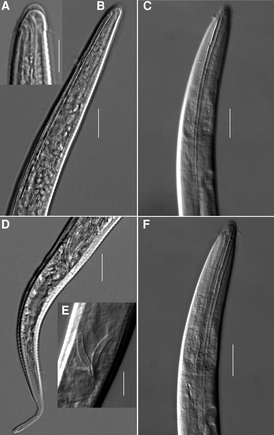
Campylaimus zhoui sp. nov. (A) view of entire male; (B) lateral view of male anterior region; (C) lateral view of male anterior end; (D) lateral view of male posterior region; (E) magnified spicules and gubernaculum. Scale bars: A = 30 μm; B, D = 20 μm; C, E = 10 μm.

Campylaimus zhoui sp. nov. (A) lateral view of male anterior end; (B) sublateral view of male anterior end; (C) lateral view of male anterior end; (D) lateral view of male posterior end; (E) magnified spicules and gubernaculum; (F) lateral view of male anterior end. Scale bars: A, E = 10 μm; B, C, D, F = 20 μm.

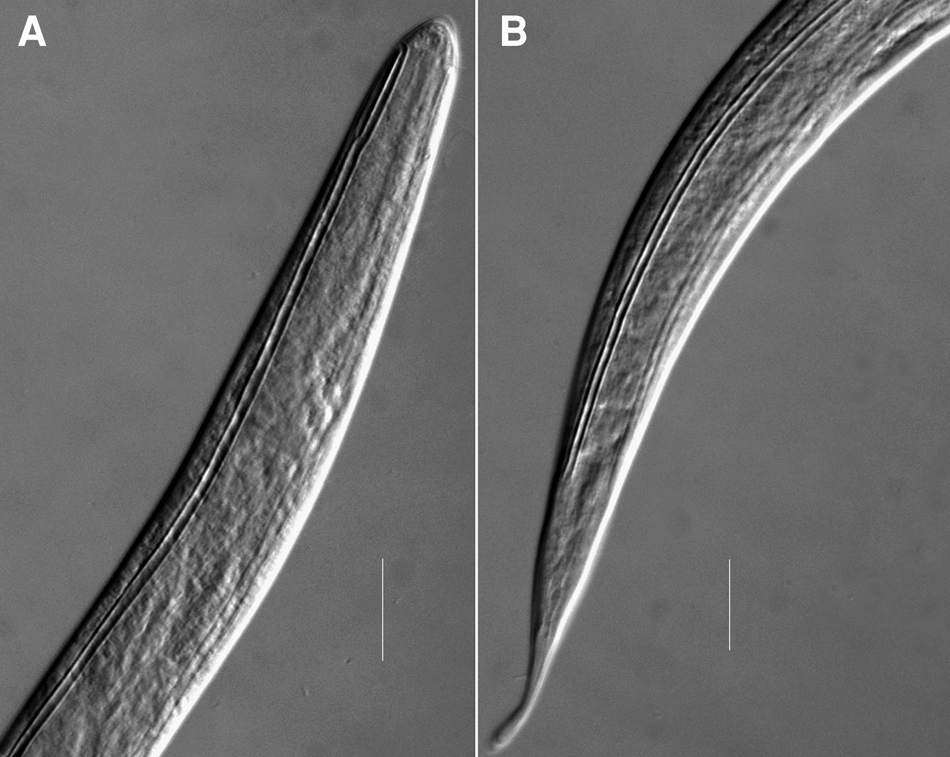
Campylaimus zhoui sp. nov. (A) anterior end of juvenile; (B) tail end of juvenile. Scale bars: A, B = 20 μm.

Type material
Two males and one juvenile were discovered at Station DH 5-3 in the East China Sea. They mounted on slide DH 5-3-4-3 and DH 5-3-4-4 in glycerin, respectively.
Type locality and habitat
Seafloor sediment of sublittoral region in the East China Sea. Station DH5-3: 122°49′34″E 28°2΄26″N, water depth 74 m, clay and sandy sediment.
Etymology
The species is named in honour of Orofessor Zhenbo Zhou, for his kind help in samples collection.
Measurements
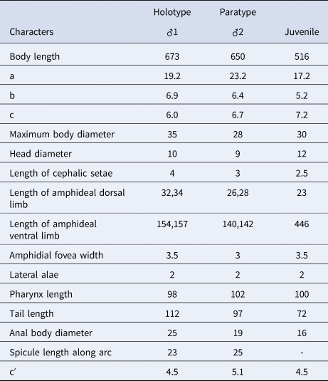
All measurement data are given in Table 2.
Measurements (in μm except a, b, c, c′ and V%) of Campylaimus zhoui sp. nov.

Description
Males: Body spindle-shaped. Cuticle annulated, extending from the oral opening to the tail end. Annules nearly equal in width except at most anterior section and tail section, each annule about 2 μm wide. Anterior end with a cuticular cap. Oral opening small, rhomboid, displaced subterminally on the dorsal side of body. Stoma unarmed. Inner labial and outer labial sensilla invisible. Four short cephalic setae 4 μm long, asymmetrically placed. The position of two laterodorsal cephalic setae slightly far from head end and the position of two lateroventral cephalic setae slightly near head end. Amphideal fovea elongated, loop-shaped with unequal limbs. Dorsal limb extending for a short distance anteriorly, equal to 3–3.3 labial region diameters in length. Ventral limb extending along pharyngeal region to level of anterior part of intestine, 4.7–5.2 times the length of dorsal limb. Anteriormost edge of amphid positioned at the level with or just anterior to oral opening. Anterior portion of amphidial fovea 4 μm wide, ventral limb 2.5–3 μm wide. Space between the limbs absent. Amphideal gland not observed. Lateral ala 2–2.5 μm wide, extending along the entire body from the ventral limb boundary towards the tail end, terminating at junction of conical portion and cylindrical portion of tail. Boundary between ventral amphid limb and lateral alae well-defined. Pharynx cylindrical for most of length, gradually widening to the base with a small conical cardia. Nerve ring indistinct. Secretory-excretory system consisting of a single renette cell and a short duct (30 μm in length), which extending anteriorly to an ampulla and opening posterior to the pharyngo-intestinal junction, about 120 μm from the anterior end.
Reproductive system diorchic, with both opposed, outstretched short testes. Spicules equal in length, symmetrical, slightly curved, proximal end not cephalated, 23–25 μm long. Gubernaculum plate-like with small dorsal apophysis. Precloacal supplements absent. Tail conico-cylindrical with a clavate tip, 4.5–5.1 anal body diameter long. Conical part accounting for four-fifths of tail length. Two pairs of subventral small caudal setae 4 μm long.
Female: not found.
Juvenile: Similar to males in the body shape. Anteriormost edge of amphid positioned at level with oral opening. Dorsal limb of amphid equal to 1.9 labial region diameters in length. Ventral limb of amphid extending along entire body beyond anus, equal to 19.4 times the length of dorsal limb. Boundary between ventral amphid limb and lateral alae not defined. The genital primordium not observed.
Differential diagnosis and discussion
Campylaimus zhoui sp. nov. is characterized by having long ventral limb of the amphid extending along pharyngeal region to level of anterior part of intestine, 4.7–5.2 times the length of dorsal limb (19.4 times the length of dorsal limb in juvenile); excretory pore opening posterior to the pharyngo-intestinal junction; spicules symmetrical, slightly arcuate without proximal capitulum; gubernaculum with small dorsal apophysis; precloacal supplements absent.
The new species is the most similar to C. amphidialis Fadeeva et al. (Reference Fadeeva, Mordukhovich and Zograf2016) and C. longispiculus Holovachov (Reference Holovachov2019) in the size and shape of the amphid with ventral limb being more than three times as long as the dorsal limb. Nevertheless, it differs from C. amphidialis by the position of secretory-excretory pore (opening posterior to the pharyngo-intestinal junction vs apically on anterior end), spicules cylindrical without proximal manubrium (vs conoid with rounded manubrium), gubernaculum with small dorsal apophysis (vs without apophysis), precloacal supplements absent (vs presence of two precloacal supplements). The new species is distinguished from C. longispiculus in having relatively longer ventral limb of the amphid, (4.7–5.2 vs 2.8–3.5 times the dorsal limb in male), shorter spicule without proximal manubrium (23–25 μm vs 28–35 μm long with rounded manubrium), gubernaculum with small dorsal apophysis (vs without apophysis), precloacal supplements absent (vs presence of two precloacal supplements).
Acknowledgements
The authors are thankful to all crew members of RV ‘Dongfanghong 2’ for their kind help in samples collection and Mrs Chunyan Qiao for her kind help with sorting specimens and mounting slides. We are sincerely grateful to two anonymous reviewers for providing valuable criticisms and improving the manuscript.
Financial support
This work was supported by the National Natural Science Foundation of China (No. 41676146).