Brief report
A 17-year-old male presented to the emergency room with persistent chest pain. At 4 days before admission, he complained of sore throat and fever. He was diagnosed with streptococcal pharyngitis using a rapid antigen detection test and was started on a course of amoxicillin. A day before admission, he developed severe substernal chest pain while playing basketball. Chest pain increased with recumbence and deep inspiration and was associated with shortness of breath. He denied having syncope, arthralgia, rashes, or a history of trauma. His biological father had poorly defined CHD requiring surgical repair at 6 years of age, and his paternal grandfather had cardiac disease at 55 years of age. There was no other family history of cardiac disease or of sudden unexplained death. In the emergency room, the initial electrocardiogram showed non-specific ST-segment changes in the inferolateral leads and a mildly elevated troponin level of 1.39 ng/ml.
The patient was transferred to the paediatric ICU for management of presumed myocarditis. He was afebrile with heart rate of 70, blood pressure of 107/65, respiratory rate of 25, and normal oxygenation on minimal supplemental oxygen. His weight was 81.2 kg. Physical examination demonstrated the teenager to be in mild distress. Mild pharyngeal erythema without exudates or cervical lymphadenopathy was noted. The heart sounds were normal, and there was no jugular venous distension or peripheral oedema. There was no sternal tenderness or rash.
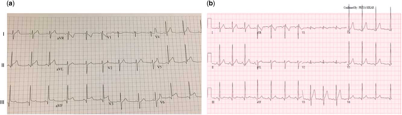
Laboratory data 7 hours after initial presentation were significant for elevated levels of troponin (28.8 ng/ml), creatine kinase-MB (129.7 mcg/L), and C-reactive protein (10 mg/dl). Streptolysin O antibody level was elevated (580 IU/ml; normal <150 IU/ml). Viral studies were negative, including a respiratory viral panel and tests for human herpesvirus-6, adenovirus, human immunodeficiency virus, and parvovirus. Serological tests for Epstein–Barr virus and cytomegalovirus were negative. Echocardiography revealed no structural defects, no regional wall motion abnormalities, and mildly diminished left ventricular systolic function with an ejection fraction of 49%. In light of ongoing severe chest pain, a repeat electrocardiography was performed, demonstrating normal sinus rhythm with ST-segment elevation in II, III, aVF, and V4-6, indicative of inferolateral acute myocardial infarction or dissection (Fig 1). The cardiology department was consulted, and after administration of anti-platelet therapy, the patient immediately underwent coronary angiography, which demonstrated normal coronary arteries. A cardiac MRI result obtained 3 days after presentation demonstrated transmural delayed enhancement of the mid-septum in a circumferential pattern, extending to the apex, consistent with myocarditis. Considering the recent diagnosis of streptococcal pharyngitis, the patient was presumed to have non-rheumatic streptococcal myocarditis. He was treated with a 10-day course of amoxicillin and with intermittent non-steroidal anti-inflammatory medication.
Electrocardiogram demonstrating ST-segment elevation in II, III, aVF, and V4-6 on arrival to the paediatric ICU ( a ) and normalised electrocardiogram with resolution of ST-segment elevation 4 months after discharge ( b ).

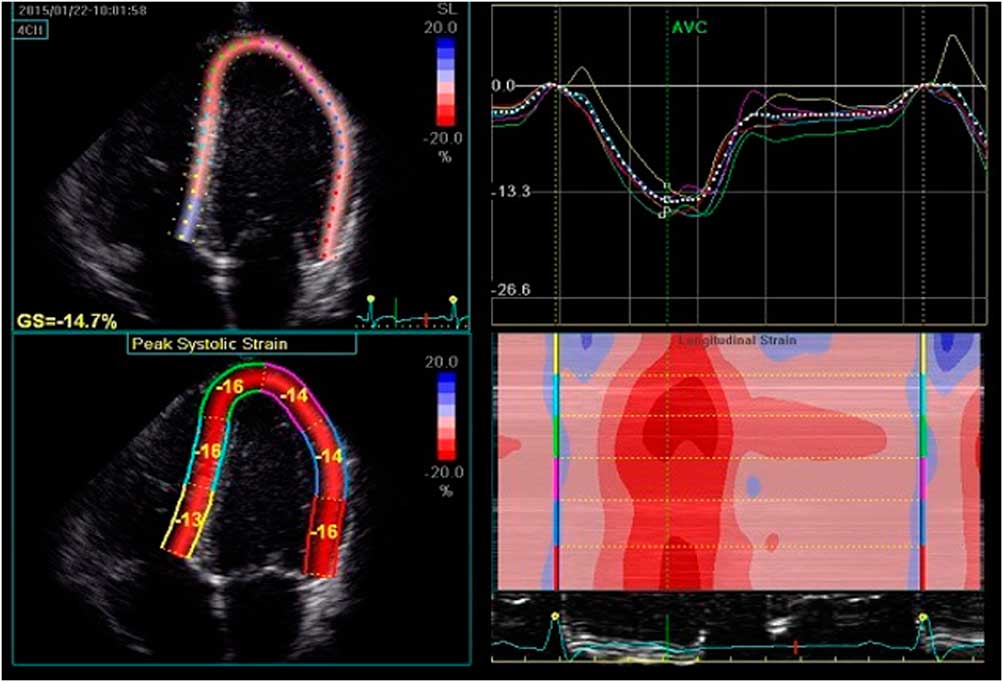
Over the course of 6 days, the patient’s symptoms resolved and troponin and creatine kinase-MB levels normalised. A repeat echocardiogram showed an ejection fraction of 59% and global left ventricular longitudinal peak systolic strain of −14.7% (5th percentile for age −19.9%, Fig 2). The patient was discharged on lisinopril to be taken at a dose of 5 mg daily with instructions to refrain from strenuous physical activity for 6 months. A month later, echocardiography demonstrated improved left ventricular longitudinal peak systolic strain of −16.4%. At 4 months after acute illness, an electrocardiogram showed normal sinus rhythm without ST-segment abnormalities (Fig 1) and echocardiography continued to show normal right and left ventricular function with improving global longitudinal peak systolic strain of −17.3%. Exercise stress echocardiography was performed at that time, which was negative for ischaemia. These data all support the clinical conclusion of myocardial recovery.
Two-dimensional strain imaging derived by speckle tracking. Apical four-chamber view demonstrating global left ventricular longitudinal peak systolic strain of −14.7%, represented with a white dotted line.

Discussion
The association of streptococcal infection and cardiac disease is most commonly described with acute rheumatic fever; however, several published case reports describe concomitant streptococcal pharyngitis and myocarditis, termed as acute non-rheumatic streptococcal myocarditis.Reference Gore and Saphir 1 – Reference Said and Severin 8 The largest case series described 35 cases of fatal myocarditis associated with nasopharyngeal infections in men aged 20–30 years.Reference Gore and Saphir 1 Streptococcal pharyngitis and/or streptococcal bacteraemia was demonstrated in 12 of these patients. Since that publication, there have been at least 30 other cases described in the literature, predominately in young men.
The pathophysiology of acute non-rheumatic streptococcal myocarditis remains poorly understood, and is rarely described in paediatric literature. Given the concurrence of pharyngitis and myocarditis, it seems unlikely to be an immune-mediated phenomenon as is the case with acute rheumatic fever. In their autopsy study, Saphir and Gore described both focal and diffuse mononuclear inflammatory changes within the myocardium.Reference Gore and Saphir 1 They also noted absence of bacteria in the myocardium, suggesting myocarditis was not a direct result of bacterial invasion. Other authors have speculated that myocardial injury may be toxin-mediated, especially given the concurrence of cardiac symptoms with pharyngitis. The observation that most patients develop myocarditis after initiation of antibiotics, as did the patient presented here, further supports this notion as large amounts of toxin may be released during bacteriolysis.Reference Karjalainen 7 , Reference Khavandi, Whitaker, Elkington, Probert and Walker 9
Myocarditis can mimic an acute myocardial infarction with chest pain, electrocardiogram changes, and troponin leak. In a case series of 34 adults with clinical signs and symptoms of acute myocardial infarction but with normal coronary artery anatomy, 32% had histology consistent with myocarditis on biopsy.Reference Dee, Walkman, Southern, Fallon, Hutter and Palacios 10 In adults, acute ST-elevation in a coronary distribution appears to be a common presentation of non-rheumatic streptococcal myocarditis.Reference Aguirre, Jurado and Porres-Aguilar 2 , Reference Upadhyay, Gainor, Stamm, Weinberg, Dec and Ruskin 3 , Reference Mokabberi, Shirani, M, Go and Schiavone 5 , Reference Karjalainen 7 , Reference Khavandi, Whitaker, Elkington, Probert and Walker 9 We are, however, unaware of a previously published report of a child presenting in this manner.
The long-term prognosis for acute non-rheumatic streptococcal myocarditis treated with antibiotics appears to be excellent. Most reports document resolution of symptoms and normalisation of electrocardiographic, echocardiographic, and cardiac MRI findings within weeks to months.Reference Upadhyay, Gainor, Stamm, Weinberg, Dec and Ruskin 3 , Reference Chikly, Durst, Lotan and Chen 6 , Reference Karjalainen 7 , Reference Khavandi, Whitaker, Elkington, Probert and Walker 9 Left ventricular strain assessment by echocardiography is a relatively new, non-invasive technique used in this case to assess sub-clinical ventricular dysfunction. Strain analysis may be a useful tool to provide insight into the time course of myocarditis resolution. To our knowledge, there has only been one report of recurrence of symptomsReference Chikly, Durst, Lotan and Chen 6 and no reports of development of acute rheumatic fever; therefore, routine antibiotic prophylaxis does not appear to be warranted.
In conclusion, coronary catheterisation should be considered in children presenting with acute chest pain and electrocardiogram changes suggestive of ischaemia of the coronary artery. In children with a recent history of pharyngitis, acute chest pain, localised electrocardiogram changes, and angiographically normal coronary arteries, acute non-rheumatic streptococcal myocarditis should be considered, particularly given the ease of treatment and favourable prognosis.
Acknowledgements
The authors extend gratitude to their colleagues in paediatrics, both at our institution and in the community who cared for this patient.
Financial Support
This research received no specific grant from any funding agency, commercial, or not-for-profit sectors.
Conflict of Interest
None.