Case report
A 6-year-old boy was referred for cardiological evaluation at our centre. He was born in Pakistan at term via caesarean section. He was discovered in infancy to have right lung hypoplasia for which he had been followed by several pulmonologists and paediatricians. He had not seen a cardiologist previously. His parents report that he had a history of frequent and longer duration upper-respiratory infections than his peers and sibling. His mother was concerned by perceived tachycardia and a prominent impulse in the precordium. He was otherwise asymptomatic, with the family denying poor growth, low energy, or other issues. The family immigrated to the United States of America and re-established care with a community pulmonologist. On the basis of the patients’s history, he was sent for a CT scan of the chest. This demonstrated cystic changes of the right lower lobe and ground-glass appearance. There was concern for right pulmonary artery and right lung hypoplasia. On the basis of this study, he was referred to a cardiologist and paediatric surgeon at our centre.
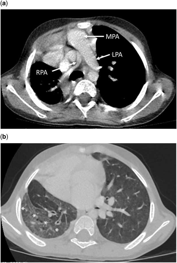
At that visit, the patient was found to be of normal size: 64th percentile for weight and 66th percentile for height for his age and sex. He was acyanotic, and cardiac exam demonstrated normal precordial activity and a 2/6 systolic ejection murmur audible at the left upper-sternal border. A 12-lead electrocardiogram demonstrated sinus rhythm with a rate of 95 beats per minute with occasional premature ventricular complexes. An echocardiogram demonstrated a patent oval foramen and otherwise normal intra-cardiac anatomy, with normal ventricular size and function. His right pulmonary artery was notably much smaller in diameter (3 mm z-score −4.8) than his left, which was dilated (13 mm, z-score of 3.1). Doppler interrogation demonstrated no acceleration across the right ventricular outflow tract or pulmonary artery. Flow in the left pulmonary artery was laminar, but on the right there was apparent reversal of antegrade flow with reflux back into the main pulmonary artery. The left pulmonary veins were seen returning normally to the left atrium, but the right pulmonary veins were not seen. There was insufficient tricuspid valve regurgitation to measure right ventricular systolic pressure with Doppler echocardiography. He was also seen at this time by a member of our Department of Pediatric Surgery with the intention of determining whether any surgical intervention was required. Review of the previously acquired CT angiogram of the chest demonstrated that the right pulmonary artery was hypoplastic, and no right pulmonary veins could be clearly identified entering the left atrium (Fig 1a). There were cystic changes in the right lower lobe with ground-glass appearance compared with the remainder of the lung (Fig 1b). The case was discussed and, based on this discussion, a referral was made for cardiac catheterisation to clarify the diagnosis, determine whether there was significant pulmonary vascular disease, and to assess whether systemic to pulmonary artery collaterals were supplying the right lung. Specifically, there was concern that the collaterals were potentially a source of volume load to the contralateral lung.
CT angiogram of the chest. ( a ) Axial chest image at level of pulmonary bifurcation demonstrating right pulmonary artery hypoplasia. ( b ) Axial chest image demonstrating ground-glass appearance and cystic changes of right lower lobe (*) compared with remainder of the lung. LPA=left pulmonary artery; MPA=main pulmonary artery; RPA=right pulmonary artery.

The catheterisation was performed with the patient receiving general anaesthesia with an initial fraction of inspired oxygen of 21%. Systemic arterial saturation in the descending aorta was 97%. Saturation in the superior caval vein was 77% and there was no step-up through the right heart to the left pulmonary artery. On the other hand, the saturation in the right pulmonary artery was 97% on multiple measurements. Pressure measurements demonstrated normal right-sided pressures with a right ventricular pressure of 28/6 with a trivial gradient to the left pulmonary artery (21/11, mean 16). The interatrial septum was crossed through the existing interatrial communication. The left atrial pressure was the same as both left upper and left lower pulmonary veins. Pulmonary capillary wedge pressures in the left lung were equal and normal (mean of 10 mmHg); however, in an upper and lower branch of the right pulmonary artery, pulmonary capillary wedge pressures were markedly abnormal with a dampened arterial waveform and elevated pressures of 28/17, mean 25, and 39/30, mean 35, respectively.
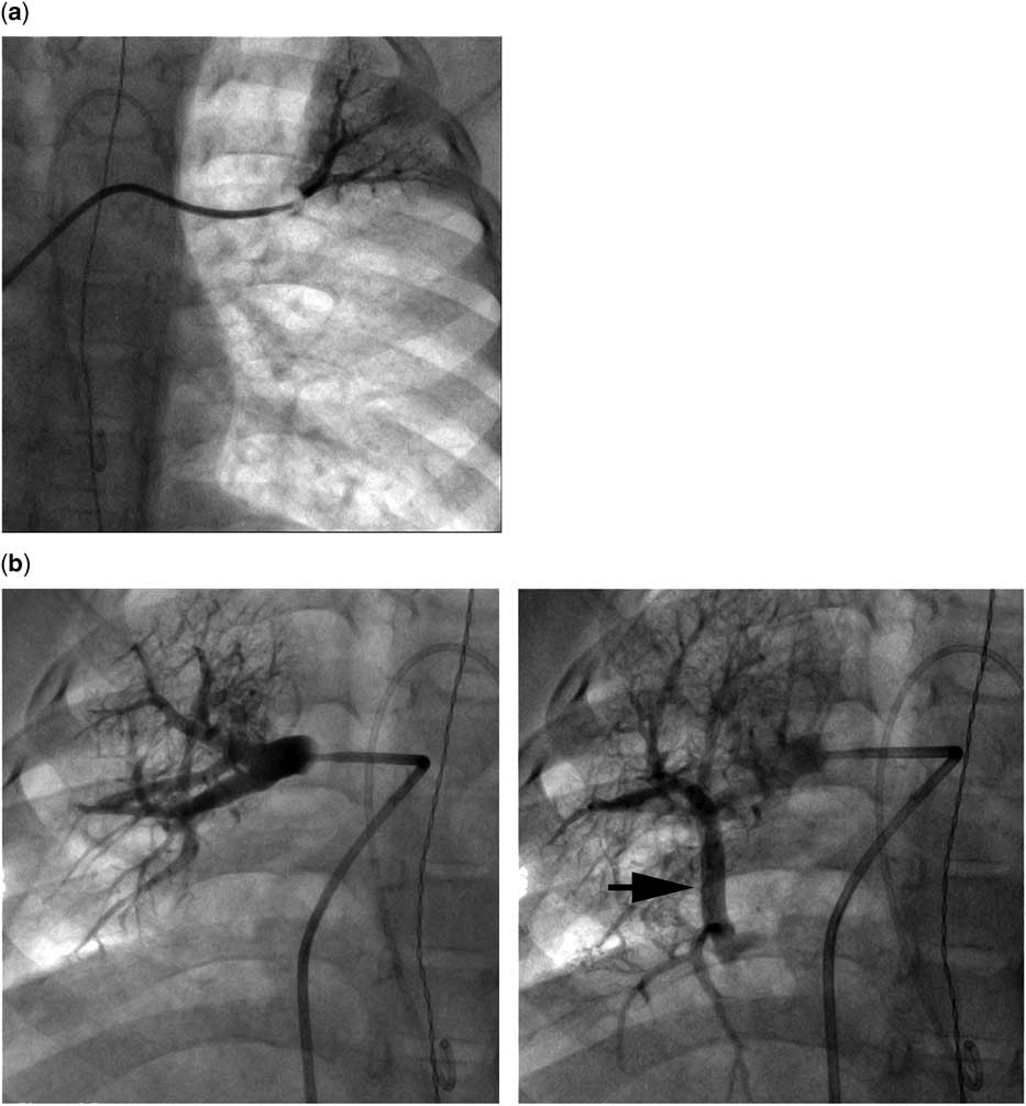
Power-injection angiography was performed in the main pulmonary artery, during which the left pulmonary artery opacified, as did the proximal right pulmonary artery (Supplementary Fig 1a); however, contrast never reached the origin of the lobar branches of the right pulmonary artery and was almost immediately washed out, refluxing back to the main and left pulmonary arteries, consistent with an alternative source of pulmonary blood flow (Supplementary Fig 1b). Levophase demonstrated normal pulmonary venous return from the left lung, but no contrast was seen returning from the right lung. Wedge angiographies were performed in the upper and lower lobes of both lungs. In the left lung (Fig 2a), these demonstrated normal distal pulmonary artery architecture without evidence of pulmonary vascular disease. In the right lung, however (Fig 2b), there were markedly abnormal distal pulmonary arteries, and the venous phase of the injection demonstrated contrast drainage into a large caliber vertical vein. This contrast did not reach the heart, and, eventually, was seen to drain to the azygos vein and the superior caval vein. On the basis of these angiograms the diagnosis of scimitar syndrome with right pulmonary vein atresia was made.
Pulmonary artery wedge angiograms. Selective hand-injection angiograms of layered contrast and saline were performed through antegrade wedge catheters with the balloon inflated. ( a ) In the upper-lobe branch of the left pulmonary artery, wedge angiograms demonstrated normal distal branch pulmonary artery distal vasculature with no evidence of pulmonary vascular disease. ( b ) In the main right pulmonary artery, wedge angiograms demonstrated a small caliber pulmonary artery with abnormal distal vasculature (left). Transit time was relatively rapid with limited blush phase. In the venous phase (right), a large vertical vein (black arrow) drained inferiorly, but did not reach the heart. Instead, contrast was seen reversing direction and draining into the azygos (not pictured).

Power-injection angiograms in the aorta demonstrated four moderate-sized collateral vessels originating from the aorta – three from the thoracic aorta and one larger collateral originating below the diaphragm – and supplying segments of the right lung (Supplementary Fig 2a). These were engaged with a retrograde endhole catheter and selective angiographies were performed (Supplementary Fig 2b and c). These initial angiograms were used to identify the depth at which each collateral vessel uniquely supplied lung tissue (and importantly did not perfuse the systemic circulation, especially the spine). Once a catheter was advanced sufficiently past this point, a series of embolisation coils were deployed in each vessel. In total, 20 0.038″ MREye coils (Boston Scientific, Boston, Massachusetts, United States of America) of various lengths and diameters were used in the four collateral vessels. Post-intervention angiograms demonstrated successful occlusion of the collaterals. There were no complications with the procedure and the patient was discharged the next morning after overnight observation.
The patient was seen 1 month after catheterisation. The patient’s mother and grandparents reported that they felt that there was a significant reduction in both the patient’s heart rate at rest and precordial activity, both of which were confirmed at that examination. At 8 months after the initial intervention the patient continues to be well with no signs of recurrent or progressive disease and no episodes of respiratory illness.
Discussion
Scimitar syndrome consists of partial anomalous pulmonary venous return of the right pulmonary veins to the inferior caval vein or inferior cavoatrial junction along with other anomalies including abnormal lung lobation, lung hypoplasia and simultaneous pulmonary artery hypoplasia, dextrocardia or dextroposition, systemic arterial supply to the right lung, and pulmonary sequestration along with a number of other intracardiac defects, most commonly atrial septal defects. Patients present either in infancy or later in life. The “infant form” is associated with more severe disease and risk for early mortality between 33% and 60%.Reference Dupuis, Charaf, Brevière and Abou 1 – Reference Huddleston, Exil, Canter and Mendeloff 3 Patients presenting in childhood and adulthood with the “adult form” have significantly lower morbidity and mortality.Reference Dupuis, Charaf, Brevière, Abou, Rémy-Jardin and Helmius 4
Previous series have reported stenosis of the anomalous pulmonary vein in infants presenting with scimitar syndrome.Reference Dupuis, Charaf, Brevière and Abou 1 , Reference Gao, Burrows, Benson, Rabinovitch and Freedom 2 Stenosis of the anomalous pulmonary vein, along with pulmonary parenchymal hypoplasia and left to right shunt through systemic to pulmonary artery collaterals, anomalous pulmonary venous return, and other intra-cardiac defects, is implicated with the development of pulmonary arterial hypertension that is the major source of morbidity and mortality in these patients. Stenosis of the contralateral veins is much rarer, and, to our knowledge, only two cases have been reported.Reference Gao, Burrows, Benson, Rabinovitch and Freedom 2 , Reference Argueta-Morales, Meador, Nykanen and DeCampli 5 Although operative correction of scimitar syndrome with reimplantation or baffling of the anomalous vein is frequently complicated by stenosis of the treated vein,Reference Huddleston, Exil, Canter and Mendeloff 3 , Reference Eckhauser, O’Byrne, Weinberg, Ravishankar, Spray and Gaynor 6 – Reference Najm, Williams, Coles, Rebeyka and Freedom 8 to our knowledge, pulmonary vein stenosis or atresia before surgical intervention has not been reported in a patient with scimitar syndrome presenting after a year of age. It is possible that this is due to a difference in the propensity for developing pulmonary vein stenosis between “infant” and “adult” forms of the disease and that this is part of the reason that the former group of patients presents earlier in life and has greater morbidity. It is also possible that the rates of stenosis of the anomalous vein are similar, but that the infant scimitar syndrome patient is more vulnerable to any issue that aggravates or induces pulmonary hypertension, resulting in presentation.
Given the large caliber confluence, we hypothesised that progressive stenosis and atresia developed after birth and likely after infancy. We emphasise that it is not possible to retrospectively determine when the anomalous right pulmonary vein(s) became occluded. In the infantile form of scimitar syndrome, atresia of a pulmonary vein has been associated with clinical decompensation and development of pulmonary vascular disease. In this case, the loss of the right pulmonary veins did not result in haemodynamic embarrassment, specifically the development of pulmonary vascular disease. Despite receiving what is likely the entirety of antegrade pulmonary blood flow the right ventricular pressure is only mildly elevated. In addition, there is no evidence of stenosis of either left pulmonary vein. As noted, however, we can only speculate as to when the right pulmonary vein became occluded.
In the catheterisation laboratory, profound loss of the density of the distal pulmonary arteries was noted, presumably due to both right lung parenchymal hypoplasia and as a response to pulmonary vein occlusion. This suggested that the right lung arterial tree was unlikely to recover even if pulmonary vein flow was restored. In addition, despite some mild symptoms, namely active precordium and relative tachycardia, the patient had no significant limitations to age-appropriate activities and enviable right-sided pressures; moreover, recanalising the relatively long occluded portion of the right pulmonary vein would have been challenging through a transcatheter intervention, and achieving an effective pulmonary vein to left atrial baffle was deemed unfeasible. In light of these factors, the decision was made to not intervene in the catheterisation laboratory. The case was later reviewed with our cardiac surgical team, who also felt that given the favourable current haemodynamics the risk for morbidity of any attempted surgical correction of the right pulmonary vein atresia exceeded the likely benefit.
The presence of retrograde flow in the right pulmonary artery on a transthoracic echocardiogram raised concern for the anatomic abnormality and led to a referral for cardiac catheterisation. Systemic to pulmonary collaterals have been reported in cases of congenital heart anomalies, pulmonary vein atresia or stenosis, lung pathology, and in some postsurgical states such as single ventricle patients between their second-stage surgeries and Fontan completions. Evidence of reversal of flow without another plausible anatomic explanation led to the hypothesis that flow was due to abnormal vessels.
Haemodynamically, the systemic to pulmonary collaterals are a volume load on the right lung. This would ordinarily also represent a volume load on the left heart, but as there is no connection between the right lung to the left atrium, this must represent a left to right shunt, both from venous drainage to the azygos and also backfilling of the main pulmonary artery and left pulmonary artery, and potential volume load for the left heart and the left lung, with the potential to accelerate development of pulmonary vascular disease on this side. The absence of a significant step-up from a sample high in the superior caval vein to the rest of the right heart and the absence of left ventricular dilation on transthoracic echocardiography suggest that the collaterals were not a source of haemodynamically significant shunt at the time of the procedure. In spite of this, the decision was made to occlude the collaterals with coils. In making this decision we acknowledged the possibility that Fick underestimated the magnitude of shunting in this unusual anatomic configuration. We also aimed to reduce the risk of flow through the arterial collaterals becoming a significant volume load on the left heart and left lung. Finally, we also aimed to reduce the long-term risk of haemoptysis. In patients with severe obstruction to pulmonary blood flow, systemic to pulmonary collaterals can become large in caliber and torturous and have the potential to erode into the airspaces and cause potentially life-threatening haemoptysis. Massive haemoptysis of this kind has been reported as the presenting finding in several adults with scimitar syndrome.Reference Reddy, Shah, Thorpe and Gibbs 9 , Reference Yamakawa, Shimizu and Michimoto 10 In these cases, occlusion of these collaterals with coils has demonstrated efficacy in the acute therapy of haemoptysis. It was our hope that preemptive occlusion would reduce the risk for bleeding over the course of the rest of the patient’s life.
After discussion, the following goals for longitudinal follow-up were established: monitoring development of pulmonary vascular disease and pulmonary hypertension; and observing for recurrence of systemic to pulmonary collateral vessels, and/or dilation of the true bronchial arteries that can lead to erosion and hemoptysis,Reference Yamakawa, Shimizu and Michimoto 10 with recurrence of a significant volume load and the potential for development of haemoptysis. The only effective means of evaluating these arterial collaterals would be using angiography. As a result, our plan was to supplement data from serial clinical examinations and echocardiograms with a cardiac catheterisation 3–5 years after the initial intervention. Timing of catheterisation was based on the fact that the patient had not yet demonstrated evidence of haemoptysis or other symptoms, and recurrence of previous signs or symptoms, and/or appearance of a new issue would prompt earlier catheterisation.
This case highlights the potential importance of systemic to pulmonary collaterals in patients with acquired pulmonary vein atresia. Also, the unusual clinical course in this case highlights the variability in the natural history of patients with scimitar syndrome. The patient appeared to have anatomy that would predispose him to the development of severe pulmonary hypertension: pulmonary hypoplasia with a very small caliber right pulmonary artery, stenotic then atretic anomalous pulmonary venous return, and several moderate to large caliber systemic to pulmonary collaterals but, ultimately, without intervention and in the face of pulmonary vein atresia has only very-mild right ventricular hypertension and no evidence of vascular disease to the left lung. Ultimately, though only a single case, this should inform the care of children with scimitar syndrome.
Acknowledgements
The authors acknowledge Dr Anthony Sandler from Children’s National Medical Center’s Department of Surgery for his contributions to the care of this patient and his close reading of this manuscript.
Financial Support
Dr M.L.O’B. and R.R.C. receive research support from the National Heart Lung and Blood Institute/National Institute of Health (K23 HL130420-01, HHSN268201500001C, respectively). These agencies had no role in the planning or execution of the study, nor did they influence the manuscript as presented. The manuscript represents the opinions of the authors alone.
Conflicts of Interest
None.
Ethical Standards
This case report describes the clinically indicated care of the case. No study procedures were performed. All patient identifiers have been removed to protect the confidentiality and privacy of the case and their family.
Supplementary Material
To view supplementary material for this article, please visit https://doi.org/10.1017/S1047951117001822