Introduction
The endonasal approach to the lacrimal sac was first described by CaldwellReference Caldwell1 in 1893, and later in 1911 by West;Reference West2 however, its use remained limited due to difficulties in visualising the endonasal structures during the operation. The introduction of the rigid endoscope provided the catalyst for endoscopic endonasal dacryocystorhinostomy (DCR).
McDonogh and MeiringReference McDonogh and Meiring3 published the first clinical study of endoscopic endonasal DCR in 1989. The first cadaver study demonstrating the feasibility of endoscopic endonasal DCR was published by Rice in 1988.Reference Rice4 Endoscopic laser-assisted lacrimal surgery was first successfully performed by Gonnering et al. Reference Gonnering, Lyon and Fisher5 Woog and colleaguesReference Woog, Metson and Puliafito6 reported an overall long term patency rate of 82 per cent using holmium:YAG laser. Since the introduction of endoscopic endonasal DCR, the procedure has continued to evolve with the introduction of various modifications and sophisticated instruments, such as lasers,Reference Woog, Metson and Puliafito6–Reference Alanon Fernandez, Alanon Fernandez, Martinez Fernandez, Cardenas Lara, Rodriguez Domínguez and Ballesteros Navarro9 powered drilling with a microdebrider,Reference Wormald10 intubation of the nasolacrimal system,Reference Sadiq, Hugkulstone, Jones and Downes8, Reference Mullner11, Reference Apaydin, Fisenk, Karayalcin, Akar and Saka12 dacryoendoscopy,Reference Maier, Schmidt and Schmidt13 optical fibre localisation of the lacrimal sac (via transillumination),Reference Zhou, Zhou, Li and Wang14, Reference Menerath, Guichard and Kydavongs15 flexible miniendoscopy combined with Hyb:erbium-YAG laser,Reference Mullner11 lacrimal endoscopyReference Mullner16 and the endoscopic microdrill.Reference Maier, Schmidt and Schmidt13
Three groups of procedures are currently practised: external DCR, endoscopic DCR with contact laser and surgical endoscopic DCR without lasers. Many factors influence the outcome of these different approaches. External DCR has remained the ‘gold standard’ surgical treatment for nasolacrimal duct obstruction, with a success rate of 90–95 per cent.Reference Hartikainen, Antila, Varpula, Puukka, Seppa and Grenman17 Important factors for achieving this success rate are wide bone removal to expose the entire lacrimal sac, and anastomosing the lacrimal sac mucosa and nasal mucosa. This concept has been used by the authors during endoscopic transnasal DCR.
In the present series of endoscopic transnasal DCR procedures, the authors were able to achieve adequate exposure of the lacrimal sac on its anteromedial wall by removing the bone of the frontal process of the maxilla, anterior and superior to the attachment of the middle turbinate, with added removal of thin lacrimal bone in the posteromedial aspect of the lacrimal sac. This created a large surgical window. The lacrimal sac mucosa was incised to create a large posterior flap, which was placed in apposition with a nasal mucosal flap to create a large rhinostomy and to enable primary intention healing.
Materials and methods
Between January 2003 and December 2006, 226 patients with nasolacrimal duct obstruction were treated consecutively by endoscopic transnasal DCR. There were 47 male patients (21 per cent) and 179 female patients (79 per cent). Patients' ages ranged from eight to 74 years. Patients were usually referred by an ophthalmologist with a history of epiphora and nasolacrimal duct obstruction. Patients with suspected canalicular blockage and functional drainage failure were investigated by dacryocystography and lacrimal scintiscan, respectively. These cases were excluded from the study.
There were 11 cases of acute dacryocystitis, for which surgery was planned after one week's antibiotic treatment. There were 48 cases of chronic dacryocystitis with purulent drainage at the medial canthus and a positive regurgitation test. Seven cases of lacrimal fistula were also treated with endoscopic transnasal DCR.
Pre-operatively, a detailed clinical examination was carried out by an ophthalmologist and an ENT surgeon, including regurgitation testing and lacrimal syringing and probing. Endoscopic evaluation was performed in every case, in order to check for access, deviated nasal septum, turbinate hypertrophy or any other associated pathology.
General anaesthesia was used in five early cases in the series, four uncooperative patients and 11 young patients aged eight to 15 years. In the remaining cases, local anaesthesia was preferred and was adequate for the procedure.
The nose was prepared using cotton strips soaked in 4 per cent xylocaine and adrenaline 1:1000, in a ratio of 4:1, 10–15 minutes prior to surgery. This ensured adequate decongestion, mucosal anaesthesia, easy access and a bloodless field. Two per cent xylocaine with 1:200 000 adrenaline was submucosally injected into the lateral nasal wall, superior and anterior to the attachment of the middle turbinate, and then along the maxillary line. External infiltration was performed just below the medial canthus of the eye, in order to anaesthetise and ensure vasoconstriction at the anterior lacrimal crest and lacrimal fossa. The ocular surface was anaesthetised with two drops of 4 per cent xylocaine.
Surgical technique
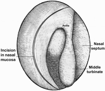
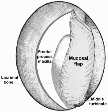
An incision was made into the nasal mucosa on the lateral wall, using a Bald Parker knife with a number 15 blade. The incision was begun 8 mm above the axilla of the middle turbinate and extended 6 mm anterior to the axilla and onto the frontal process of the maxilla. The incision was then extended vertically downward in a ‘C’ shape towards the insertion of the inferior turbinate, and then continued posteriorly up to the insertion of the uncinate process (Figure 1). A mucoperiosteal flap was then elevated backward over the maxillary and lacrimal bone, using a Freer's suction elevator and round knife, and was rolled over posteriorly onto the middle turbinate. The thin lacrimal bone and thick maxillary bone were identified (Figure 2).
Incision to raise the mucosal flap, begun 8 mm above the axilla of the middle turbinate and extended 6 mm anterior to the axilla onto the frontal process of the maxilla.

Elevation of the nasal mucosal flap and exposure of the frontal process of the maxilla and the lacrimal bone. The flap is rolled posteriorly onto the middle turbinate.

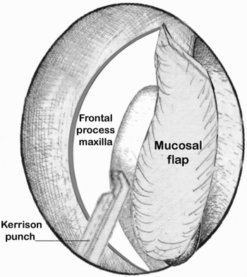
An osteotomy was performed with straight, 2 mm Smith–Kerrison punch forceps (Figure 3). An angled punch was used to remove bone at the upper limit of the sac. Sometimes, removal of thick bone from the frontal process of the maxilla was required, for which a drill with 1–2 mm cutting burr was used (Otodrill; Saeyang Company, Marathon with straight hand Piece-nsk; Japan). In a few cases, a chisel and hammer were used for removal of thick bone. The complete anteroposterior extent of the medial wall of the sac was exposed. Lacrimal bone was removed with a Freer's elevator or ball probe. At this point, it was important to meticulously locate and remove all small bone fragments.
Osteotomy performed with Smith–Kerrison punch forceps.

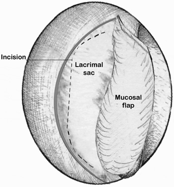
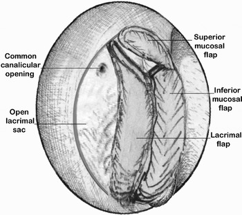
A small amount of xylocaine with adrenaline was infiltrated into the medial wall of the sac, in order to obtain local anaesthesia of the sac wall and to improve haemostasis during incision and fashioning of the mucosal flap. The lacrimal sac was incised vertically on the anteromedial aspect of the sac using a sickle knife or ophthalmic keratome; pus, mucopus or mucus usually flowed from the sac. Two horizontal incisions were made at the superior and inferior limit of the sac (Figure 4), and a U-shaped posterior based flap was created and folded posteriorly. The nasal mucosal flap was trimmed in such a way as to create a small superior flap, which covered the raw bone left superiorly over the upper limit of the sac, and a larger inferior flap, which lay in apposition with the folded posterior lacrimal sac flap (Figure 5). The rhinostomy was further enlarged by removing the remaining lacrimal sac wall anteriorly. The anterior limit of the rhinostomy and osteotomy was left to heal by secondary intention, as nasal mucosa usually remained close to the rhinostomy.
The exposed lacrimal sac, showing the vertical incision and horizontal extension used to create a posteriorly based flap.

The lacrimal sac flap rolled posteriorly to come into apposition with the inferior nasal mucosal flap. A superior nasal mucosal flap covers the exposed bone superiorly.

If there was any doubt regarding correct identification of the sac, then a transcanalicular lacrimal probe was inserted through the inferior canaliculus to identify the medial wall of the sac, or external pressure was exerted on the lateral wall just below the medial canthus area, causing the sac to protrude medially in the nasal cavity.
Stoma patency was checked by syringing with saline solution, and a full lacrimal sac wash was performed. The entire procedure was performed using a 0º, 4 mm endoscope. The sac interior was visualised with a 30º, 4 mm endoscope. The surgical site was packed with a small piece of Merocel (Medtronic Xomed, Jacksonville, U.S.A) to hold the flap in position and to ensure haemostasis.
Post-operative care and follow up
Patients were discharged uneventfully the day after surgery, with oral antibiotics for one week. The Merocel was usually removed after 48 hours, and saline nasal drops four to five times a day were advised to avoid crust formation. Antibiotic eye drops were advised four times a day for three to four weeks in order to ensure continuous flow through the lacrimal system. Patients were advised to avoid nose-blowing for four to seven days, so as to avoid nasal haemorrhage and orbital emphysema.
Patients were followed up weekly for a month. Endoscopic visualisation of the nasal cavity was performed in order to remove crusts and granulations (if any) and to check the patency of the newly created ostium using lacrimal irrigation. Subsequent follow up was at monthly intervals for three months and then every three months for two years; this included symptom evaluation (i.e. checking for subjective improvement in eye watering) and endoscopic assessment of the newly created ostium, in order to check for adhesion formation and restenosis.
Results
`Of the 226 patients, 179 were female (79 per cent) and 47 male (21 per cent). The youngest patient was an eight-year-old girl and the eldest a 74-year-old woman. The main presenting complaint was epiphora (95 per cent); this included cases presenting with purulent discharge at the medial canthus (21 per cent), mucocele (13 per cent) and lacrimal fistula (3 per cent).
Septoplasty was performed in 36 per cent of cases. A high Deviated Nasal Septum (DNS) adjacent to the anterior end of the middle turbinate was removed endoscopically through a Killian's incision. The anterior cartilage was kept intact. In 18 per cent of cases, endoscopic sinus surgery was performed for chronic sinusitis and nasal polyposis. In 10 per cent of cases, conchoplasty and turbinoplasty were performed to improve access and to avoid post-operative synechiae formation.
Patients were followed up for six to 24 months after their operation. Eight patients who were immediately lost to follow up were excluded from the series. Overall, the primary success rate of the procedure was 92 per cent, in terms of subjective improvement in eye watering and endoscopic visualisation of a patent rhinostomy on lacrimal irrigation.
Since cases of functional obstruction and canalicular obstruction were excluded from the study group, the results obtained were exclusively for anatomical obstruction of the nasolacrimal duct.
In 8 per cent of cases, DCR scarring and ostium fibrosis were noted. These were cases in which the technique had been difficult to perform properly, due to poor visibility (due to bleeding) or to anatomical abnormality (such as an excessively thick frontal process of the maxilla or a prominent uncinate).
The complications and difficulties observed during surgery were as follows. Five patients with known hypertension were taken to surgery after controlling their blood pressure, but peri-operative haemostasis was difficult because of the surgeon's guarded use of adrenaline. In cases of acute dacryocystitis, nasal mucosal inflammation led to significant haemorrhage during incision of the lacrimal sac wall. Six cases had excessively thick bone in the frontal process of the maxilla, which required more extensive drilling. In five cases, the lacrimal sac was difficult to locate; three due to anatomical abnormality of the uncinate process, and two to an inadequate osteotomy. In two cases, orbital fat prolapsed when the mucoperiosteal flap was raised posteriorly and the recessed uncinate process and lamina papyracea were inadvertently injured; this event was managed without complication.
Post-operatively, minor delayed complications included: granuloma formation at the anterior lip of the rhinostomy (seven cases), removed endoscopically; synechiae between the middle turbinate and lateral wall (three cases), released during follow up visits; excessive crusting (eight cases); and a periorbital saline collection along the inferior lid plus emphysema, noted during syringing in one case, due to creation of a false track during canaliculi probing.
Discussion
Epiphora is an annoying symptom which is embarrassing to the patient both socially and functionally. Epiphora resulting from obstruction of the nasolacrimal duct has two widely accepted treatment modalities: external and endoscopic DCR. Since Toti'sReference Toti18 original description of DCR in 1904, external incision has been used for relief of lacrimal obstruction. The success rate of external DCR has improved to the present day with few modifications, and may be up to 90–95 per cent in the hands of a trained oculoplastic surgeon.Reference Hartikainen, Antila, Varpula, Puukka, Seppa and Grenman17 The advent of endoscopic instrumentation for nasal and sinus surgical procedures has prompted renewed interest in endoscopic transnasal DCR.Reference McDonogh and Meiring3, Reference Metson19 The endoscopic approach not only avoids an external incision but also enhances the surgeon's ability to identify and correct common intranasal causes of DCR failure, including adhesions, an enlarged middle turbinate and ethmoid sinus disease.Reference Metson, Kennedy, Bolger and Zinreich20
The main advantage of external DCR is visualisation of the anatomy, allowing precise removal of the bone in the lacrimal fossa and exact anastomosis of the nasal mucosa and lacrimal sac wall. Endoscopic surgeons should have a good knowledge of the anatomy of the lacrimal sac and duct within the nose, in order to obtain optimum results comparable to those of external DCR.
A study by Wormald et al.,Reference Wormald, Kew and Van Hasselt21 based on computed tomography (CT) dacryocystograms and CT scans, showed that the mean height of the lacrimal sac was 8.8 mm above the middle turbinate insertion and 4.1 mm below it. The average measurement of the lacrimal sac, on CT dacryocystogram, was 5.3 mm above the common canaliculus and 7.7 mm below it. These study findings confirm that the sac may be adequately exposed during DCR by removal of sufficient bone and mucosa above the anterior insertion of the middle turbinate. A cadaver-based study, by Raut et al.,Reference Raut, Yung and Logan22 revealed that the posteromedial aspect of the lower lacrimal sac and upper duct is covered by the extremely thin lacrimal bone (average thickness 0.057 mm), which was consistently found to lie immediately anterior to the uncinate process in the middle meatus, thus constituting a ‘surgical window’ (average size 2.5 × 7.2 mm). The lower part of the lacrimal sac and the upper part of the nasolacrimal duct can be easily assessed from within the nose by following this anatomical approach. In our study, we found that adequate exposure of the lacrimal sac on its anteromedial wall could be achieved by removing the bone of the frontal process of the maxilla anterior and superior to the attachment of the middle turbinate, with added removal of thin lacrimal bone in the posteromedial aspect of the lower sac, thus creating a large surgical window. Sufficient bone removal inferior to the level of the sac–duct junction may help prevent accumulation of debris within the sac.Reference Chastain, Cooper and Sindwani23
Another study of the clinical anatomy of the lacrimal sac fossa, by Zhang et al.,Reference Zhang, Wang, Wang, Shan and Wang24 revealed that the proportion of the frontal portion of the maxillary bone is bigger than the lacrimal bone. This favours removal of the frontal process of the maxillary bone for complete exposure of the sac.
Tsirbas and WormaldReference Tsirbas and Wormald25 stated that the key to successful endoscopic DCR is to fully expose the lacrimal sac and marsupialise it into the lateral nasal wall, with the nasal and lacrimal mucosa in apposition, allowing healing by primary intention rather than the formation of granulation tissue, reducing the risk of closure of the sac opening into the nose. Our technique involved creation of a large bony ostium and a large posterior flap at the medial sac wall, reflecting it posteriorly in apposition with the nasal mucosal flap. A superior nasal mucosal flap is also created to cover the exposed bones superiorly on the lateral nasal wall. The remaining small anterior flap of lacrimal sac is punched out to create a large opening. The initial nasal mucosa incision is tailored in such a way that, after removing the bone for osteotomy, no exposed bone is left anteriorly. We have tried to minimise the exposed bone anteriorly by placing the mucosal incision line almost at the edge of the predicted bony ostium.
Using this technique, we achieved a success rate of 92 per cent. The results obtained in this series relate exclusively to anatomical obstruction of the nasolacrimal duct; therefore, our success rate is attributable not only to the surgical technique but also to the case selection (revision cases, cases of functional obstruction and canalicular obstruction were excluded from the series). A greater success rate (89 per cent) has been reported for the lacrimal sac flap technique than for conventional endonasal DCR in which the entire medial sac wall is excised.Reference Yuen, Lam, Tse, Chan, Wong and Chan26 Endonasal DCR has also been reported to be quicker than the traditional external approach, equivalently successful and preferred by patients.Reference Dolman27
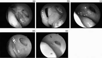
It has been observed that creation of a lacrimal and nasal mucosal flap results in primary intention healing with minimal risk of granulation tissue formation and therefore minimal shrinkage of the post-operative DCR ostium. Mann and WormaldReference Mann and Wormald28 proposed that the DCR ostium shrinks a small but significant amount in the first four weeks after surgery and then stabilises. Following use of their mucosal flap technique, they observed a 10.1 mm DCR ostium at four months and a 9.8 mm ostium at six months. In our study, the rate of surgical success (in terms of symptomatic relief and anatomical patency) was 92 per cent, and a 6–8 mm rhinostomy was observed in 80 per cent of cases at six months' follow up (Figure 6).
Post-operative pictures of the marsupialised lacrimal sac at (a) 15 days, (b) 30 days, (c) three months, (d) six months and (e) one year.

We did not use a silicone stent or mitomycin application in any of our cases. Neither silicone stenting nor the application of mitomycin C is routinely indicated. Laser-assisted techniques do not currently appear to improve results. In our view, appropriate post-operative care is essential to prevent endonasal synechiae and subsequent recurrences. Kong et al. Reference Kong, Kim and Kong7 reported granulation at the internal nasal opening in approximately 50 per cent of patients at eight weeks post-operatively.
Endonasal laser DCR is not the procedure of first preference, due to its high cost, long operation time and less satisfactory results.Reference Onerci29 It may be an alternative in cases with a tendency to bleeding. Lasers (holmium:YAG and Nd:YAG) are helpful but not essential. Kong et al. found that the Otodrill or microrongeur was more effective than the holmium:YAG laser in removing thick bone, and also less painful for patients.Reference Kong, Kim and Kong7 The inferior results achieved with laser DCR may be due to the size of the ostia created; small ostia created by laser DCR have been found to have patency rates of only 64 to 70 per cent.Reference Hartikainen, Antila, Varpula, Puukka, Seppa and Grenman17, Reference Hehar, Jones, Sadiq and Downes30
• This study involved a prospective series of 226 consecutive endoscopic transnasal dacryocystorhinostomy procedures
• The technique involved the creation of nasal mucosal and large posterior lacrimal flaps at the medial lacrimal sac wall
• A large bony ostium was also created
• The procedure achieved a 92 per cent success rate, in terms of symptom relief and anatomical patency
Endoscopic DCR has many advantages over external DCR (e.g. avoidance of facial scarring, of disruption of lacrimal sac pump action from the orbicularis oculi muscle, and of division of the medial canthal ligament). However, this technique does not have the same success rate as external DCR.Reference Hartikainen, Antila, Varpula, Puukka, Seppa and Grenman17 We support the proposal of WormaldReference Wormald10 that a large bony ostium and complete lacrimal sac exposure are important for achieving both a patent post-operative ostium and a result comparable to external DCR.
Endoscopic transnasal DCR is less traumatic, quicker and cosmetically more convenient, and has a lower complication rate, minimal morbidity and a success rate comparable to traditional external DCR.Reference Cokkeser, Evereklioglu and Er31 With an appropriate operative technique and in experienced hands, the success rate of endonasal DCR is comparable to that of the classical external approach. The major advantages of the endonasal approach are its short operation time, low complication rate and minimal patient morbidity.Reference Keerl and Weber32 The endoscopic approach provides excellent visualisation and management of intranasal structures, and it may be associated with improved outcome, considering that intranasal synechiae and improper rhinostomy site placement are common causes of failure of external DCR.Reference Metson, Kennedy, Bolger and Zinreich20
The most common causes of failure of endonasal DCR are false localisation of the lacrimal sac, granulation tissue formation, retained bony spicules, inadequate removal of the medial sac wall, and synechiae between the lateral wall and the middle turbinate.Reference Onerci, Orhan, Ogretmenoglu and Irkec33
Conclusion
Endoscopic transnasal DCR is indeed a valid alternative to the traditional extranasal procedure, once exclusively the domain of the ophthalmologist. However, this technique does require a certain expertise in endoscopic surgery.
A wide surgical window is fashioned by: judicious incision of the nasal mucosa on the lateral nasal wall (avoiding exposed bones); adequate removal of bone; creating a flap of lacrimal sac mucosa and approximating it to the nasal mucosa; and regular post-operative endoscopic follow up to remove crusts, synechiae and granulations. Creation of a wide surgical window helps ensure a success rate comparable to that of the external procedure.
In most cases, sophisticated equipment is not required and the procedure can be performed with just a few, routinely used endoscopic sinus surgery instruments.
We have modified the nasal mucosal incision on the lateral nasal wall, from 8–10 mm above and in front of the axilla of the middle turbinate (used in earlier cases in this series), to a nasal mucosa incision 8 mm above and 6 mm in front of the axilla of the middle turbinate, so as not to leave any exposed bone anteriorly after bony osteotomy.