Introduction
Obstructive sleep apnoea (OSA) is estimated to occur in 24 per cent of men and 9 per cent of women, and when it coincides with sleepiness it is called OSA syndrome.Reference Young, Palta, Dempsey, Skatrud, Weber and Badr1 Obstructive sleep apnoea is associated with hypertension, obesity, insulin resistance, cardiac arrhythmias and mortality.Reference He, Kryger, Zorick, Conway and Roth2–Reference Punjabi, Shahar, Redline, Gottlieb, Givelber and Resnick4
Various methods have been used to evaluate the airway of affected individuals; these include physical examinations, flexible fibre-optic nasopharyngoscopy, lateral cephalometry, computed tomography (CT) and magnetic resonance imaging.Reference Schendel and Hatcher5–Reference Barrera, Holbrook, Santos and Popelka7 An increasingly popular modality for assessing the airway is cone beam CT.Reference Schendel and Hatcher5 Changes in the size of the upper airway associated with a supine versus an upright position have been examined using lateral cephalometry for OSA patients and cone beam CT for non-OSA patients;Reference Bharadwaj, Ravikumar and Krishnaswamy8–Reference Ono, Lowe, Ferguson and Fleetham13 however, as yet there have been no cone beam CT studies comparing these position-related changes in OSA patients. Automated three-dimensional (3D) airway analysis of cone beam CT scans can be performed using programs such as 3dMD (Atlanta, Georgia, USA). Such programs evaluate the size of the airway, calculating total airway volume and individual cross-sectional area measurements.Reference Schendel and Hatcher5
Materials and methods
Literature search
A search of Medline, Scopus and the Cochrane Database of Systematic Reviews was performed to identify previous studies on upright and supine imaging in OSA patients. There were no studies identified in the literature that assessed the airway of OSA patients in upright and supine positions using cone beam CT imaging.
Subjects and procedure
The Stanford institutional review board was contacted, and written approval was obtained prior to initiating this study. Patients who presented and were treated at the Stanford Hospital and Clinics for adult OSA were recruited for this retrospective chart review.
The study comprised adult patients with OSA, confirmed by polysomnography, who underwent cone beam CT (whilst awake) in upright and supine positions. Children, non-OSA patients, and OSA patients who had not undergone both upright and supine cone beam CT imaging were excluded from the study.
During the cone beam CT procedure, care was taken to ensure uniformity of airway positioning. Specifically, the patients remained in the standard position, called ‘the natural head position’. Patients were instructed on how to keep their head in the same flexion–extension position when in both the upright and supine positions. Additionally, to help avoid motion artefacts, all patients were instructed not to swallow, and to take a normal breath in and then hold their breath for the duration of the cone beam CT scan, which takes less than 10 seconds.
Demographic, radiographic (Digital Imaging and Communication in Medicine (‘DICOM’) files) and polysomnographic data were collected and analysed.
Data analysis
The Digital Imaging and Communication in Medicine files for the cone beam CT images for each patient, for both upright and supine positions, were obtained. Measurements and analysis of the airway were conducted using the 3dMD program.
For consistency and reproducibility, the upper airway was defined with the use of bony landmarks: the upper aspect was defined as the inferior border of the posterior nasal spine, and the lower aspect was defined as the inferior border of the third cervical vertebra. The total upper airway volume, the posterior nasal spine, uvula tip, retrolingual and tongue base cross-sectional areas, and the site of the minimum cross-sectional area (of the overall airway), were all marked manually using the 3dMD software. Volume and area measurements were subsequently calculated by the program. The region from the posterior nasal spine to the uvula tip was also considered as the retropalatal region. The retrolingual area was defined as the cross-sectional area at the level of the superior aspect of the third cervical vertebra. The base of the tongue was defined as the inferior aspect of the third cervical vertebra.
Microsoft Excel 2013 spreadsheets were used to collate the data. Quantitative data analysis was performed using IBM SPSS Statistics Data Editor software, version 20.0 (Armonk, New York, USA). The paired samples t-test was utilised to calculate: the paired differences of the means, standard deviations, standard errors of the means, 95 per cent confidence intervals (lower and upper), t-test values, degrees of freedom and two-tailed significance values. A two-tailed p value of less than 0.05 was considered statistically significant.
Results
The chart review identified 9 OSA patients who had undergone upright and supine cone beam CT in the previous 36 months. Because of considerable motion artefacts, data for four of the patients had to be excluded from the study. The remaining five patients were all were males, diagnosed with moderate to severe OSA. Mean age was 35.0 ± 9.3 years, mean body mass index was 28.1 ± 2.7 kg/m2 and mean apnoea–hypopnoea index was 39.3 ± 23.0 per hour. Physical examination findings, apnoea–hypopnoea indices, lowest oxygen saturation values and Epworth Sleepiness Scale results are listed in Table I.
Patients' characteristics

*Tonsil size classification based on Friedman Tonsil Position score. Pt no = patient number; y = years; BMI = body mass index; AHI = apnoea–hypopnoea index; LSAT = lowest oxygen saturation; ESS = Epworth Sleepiness Scale; FTP = Friedman Tongue Position; Dent occl = dental occlusion; n/a = not applicable; WNL = within normal limits; C1 = class 1 dental occlusion (i.e. the mesiobuccal cusp of the permanent maxillary first molar was in occlusion with the buccal groove of the permanent mandibular first molar); TA = adenotonsillectomy; turb = turbinate reduction; T = tonsillectomy; UPPP = uvulopalatopharyngoplasty; SD = standard deviation
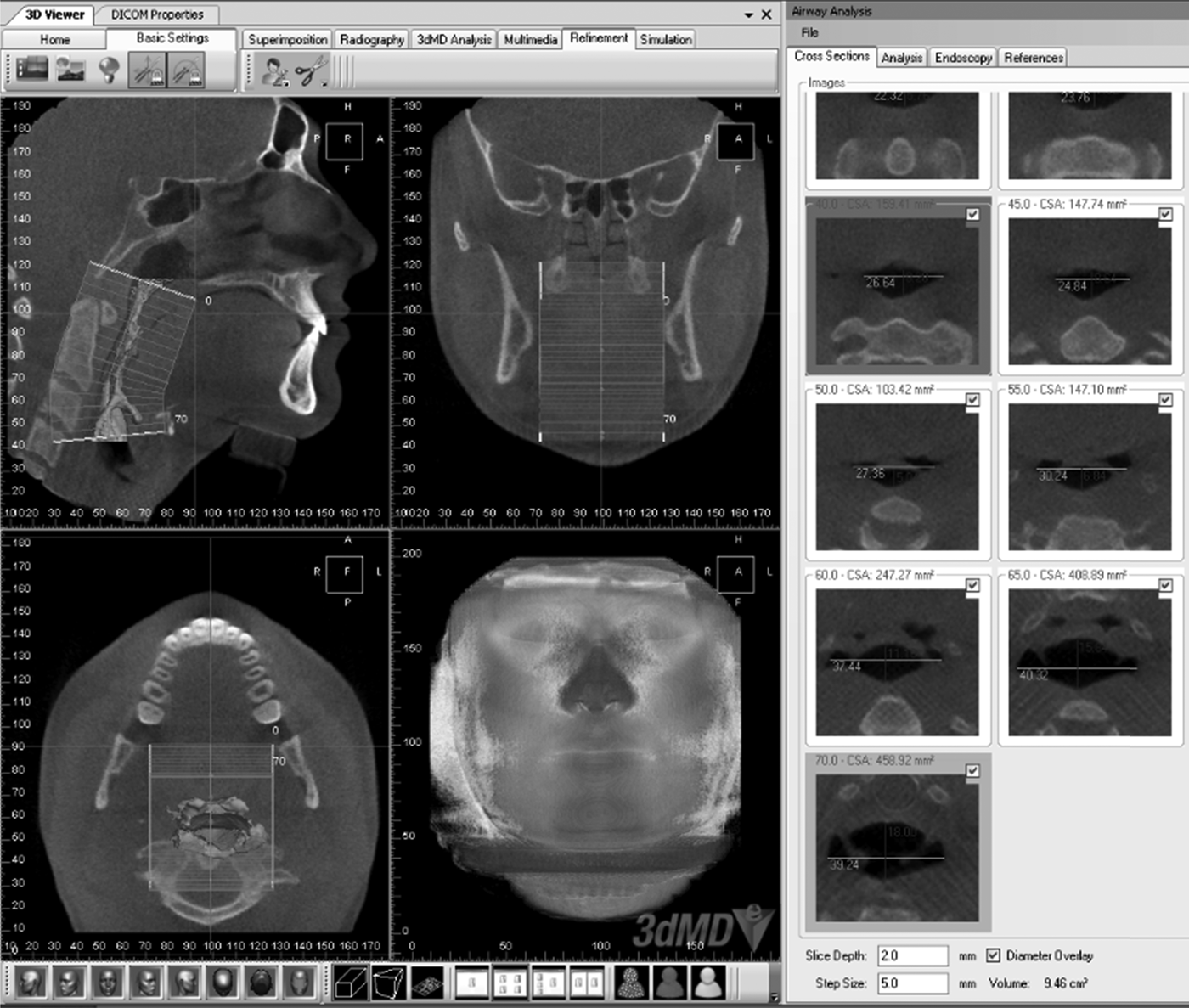
The Digital Imaging and Communication in Medicine images were loaded into the 3dMD program. The airway volume measurements were calculated based on user-identified upper and lower anatomical limits (the user defined the boundaries and the software calculated the airway volume based on these boundaries); an example is shown in Figure 1.
The 3dMD program provides sagittal, coronal and axial views of the airway (left side of figure), and cross-sectional measurements of the airway in the axial dimension (right side of figure). H = head; P = posterior; R = right; A = anterior; F = feet; L = left
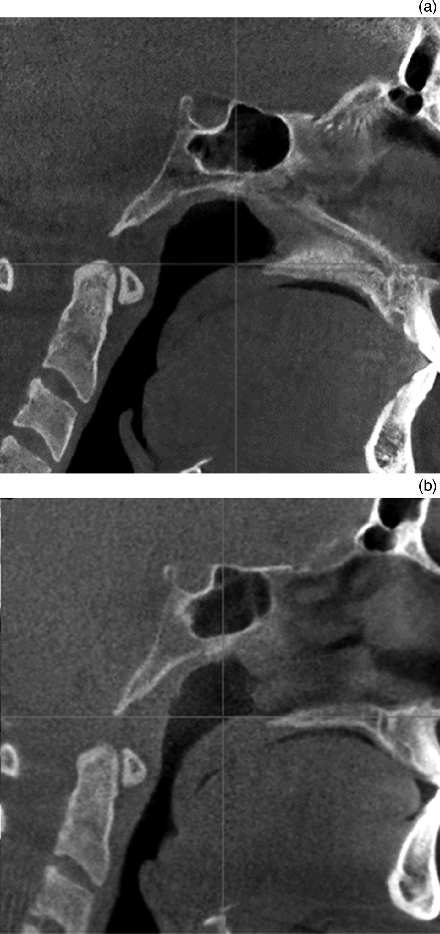
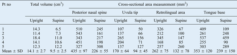
The airway was smaller when patients were in a supine compared with an upright position, as reflected by decreases in the following airway measurements: total upper airway volume, 14.1 ± 2.7 cm3 decreased to 9.5 ± 2.5 cm3 (p = 0.033); posterior nasal spine cross-sectional area, 435 ± 97 mm2 decreased to 226 ± 55 mm2 (p = 0.029); uvula tip cross-sectional area, 170 ± 64 mm2 decreased to 94 ± 45 mm2 (p = 0.024); retrolingual cross-sectional area, 262 ± 75 mm2 decreased to 132 ± 78 mm2 (p = 0.047); base of tongue cross-sectional area, 353 ± 120 mm2 decreased to 239 ± 150 mm2 (p = 0.104); and the site of the minimum cross-sectional area (of the overall airway), 124 ± 29 mm2 decreased to 30 ± 5 mm2 (p = 0.025). Figure 2 demonstrates an example of the sagittal appearance of the airway in the upright versus supine position. The paired t-test values for the differences between the upright and supine position upper airway measurements are listed in Tables II–IV.
Sagittal views of the airway in patient number 3 in an upright (a) and a supine (b) position. Note the retro-displacement of the base of tongue, palate and epiglottis with obliteration of the pre-epiglottic space. Additionally, the tip of the tongue in (a) is abutting the central incisors, whilst in (b) it is posteriorly displaced.
Upright and supine position upper airway measurements
Pt no = patient number; SD = standard deviation
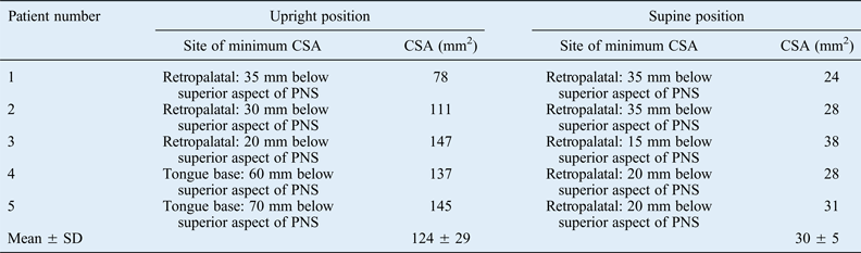
Site of minimum cross-sectional area findings
CSA = cross-sectional area; PNS = posterior nasal spine; SD = standard deviation
Upright versus supine position cone beam CT measurement differences
The paired t-test results revealed significant differences between upright and supine position measurements for all airway parameters except tongue base cross-sectional area (CSA). CT = computed tomography; MD = mean difference; SD = standard deviation; SEM = standard error of the mean; CI = confidence interval; df = degrees of freedom
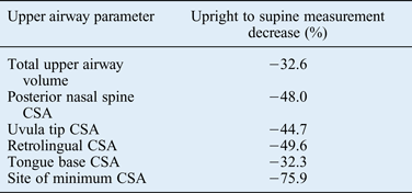
The differences between the upright and supine position measurements reached statistical significance (p < 0.05) for total upper airway volume and for cross-sectional area at all the measured sites except for the tongue base (p = 0.104). The percentage decreases (associated with a supine versus an upright position) are presented in Table V. Overall, total airway volume decreased by 32.6 per cent and cross-sectional area measurements decreased by 32.3–75.9 per cent.
Percentage changes in upper airway measurements
CSA = cross-sectional area
Discussion
This study revealed that the airway of OSA patients was smaller when the patients were in a supine compared with an upright position. The difference between the upright and the supine position measurements of upper airway volume were significant; total upper airway volume decreased by 32.6 per cent when patients were in the supine position. The site of the minimum cross-sectional area of the overall upper airway demonstrated the most significant decrease in size, decreasing by an average of 75.9 per cent. The effect of gravity and tissue laxity are likely to be significant contributors to the change in upper airway size.
The findings of this study are consistent with previous research, which has demonstrated that the retropalatal region is the region of the overall airway with the minimum cross-sectional area. In the upright position, the site of the minimum cross-sectional area for patients 4 and 5 was the tongue base subsite; however, for all other patients, the site of the minimum cross-sectional area was the retropalatal region (between the posterior nasal spine and uvula tip). In the supine position, the site of the minimum cross-sectional area, for all patients, was the retropalatal region. This may be due to the palate being more susceptible to gravity in the supine position.
Interestingly, the patient who had previously undergone a tonsillectomy and uvulopalatopharyngoplasty (patient number 5) demonstrated minimal position-related changes in all of the sites measured, with total volume measurements of 12.3 cm3 in the upright position and 12.2 cm3 in the supine position. The tonsillectomy and uvulopalatopharyngoplasty would explain the lack of changes at the level of the posterior nasal spine and the uvula tip, but do not clearly explain the lack of changes at the retrolingual or tongue base subsites.
As there were no control subjects in our study, we searched the literature for studies on airway CT changes associated with a supine versus an upright position in non-OSA patients. Two studies were identified. In the first study, conducted on 15 healthy volunteers, Van Holsbeke and colleagues compared CT data for upright versus supine position cross-sectional areas.Reference Van Holsbeke, Verhulst, Vos, De Backer, Vinchurkar and Verdonck12 The upper airway regions measured in that study correspond well to our study (top = retropalatal, central = retrolingual and bottom = base of tongue). Based on the data provided in their article, the percentage changes for supine compared with upright position measurements were: a 41.2 per cent decrease in the retropalatal region, a 8.9 per cent decrease in the retrolingual region and a 13.4 per cent decrease in the tongue base region.Reference Van Holsbeke, Verhulst, Vos, De Backer, Vinchurkar and Verdonck12 Our study demonstrated the following percentage changes for supine compared with upright position measurements: a 44.7–48 per cent decrease in the posterior retropalatal region, a 49.6 per cent decrease in the retrolingual region and a 32.3 per cent decrease in the tongue base region. Compared with the non-OSA patients,Reference Van Holsbeke, Verhulst, Vos, De Backer, Vinchurkar and Verdonck12 the retropalatal region decreased slightly more in the OSA patients; however, the retrolingual and tongue base cross-sectional areas decreased significantly more in the OSA patients. However, it is difficult to draw reliable conclusions as Van Holsbeke et al. reported median values and our study examined the mean values, making comparison difficult.
The second study evaluating position-related changes in airway CT measurements in non-OSA patients is by Sutthiprapaporn and colleagues.Reference Sutthiprapaporn, Tanimoto, Ohtsuka, Nagasaki, Iida and Katsumata10 In their study of seven patients, they assessed the smallest cross-sectional area in the oropharynx, posterior to the soft palate (corresponding to the retropalatal or posterior nasal spine-uvula tip in our study). Their study revealed a decrease in the smallest cross-sectional area of 35.3 per cent. In our study, this measurement decreased by 44.7–48 per cent. No other cross-sectional area measurements were reported in their study.Reference Sutthiprapaporn, Tanimoto, Ohtsuka, Nagasaki, Iida and Katsumata10
There are some limitations to this study. First, our study consisted of only five patients. A thorough review of our electronic medical records of the previous 36 months indicated that 38 patients underwent imaging of the airway (mostly upright cone beam CT scans). However, only nine of those patients underwent cone beam CT in both the upright and supine position, and had Digital Imaging and Communication in Medicine images available. Because of significant motion artefacts, data for four of the nine patients had to be excluded from the study. Despite there being only five patients, there were statistically significant position-related differences in total airway volume and in the measured cross-sectional areas (posterior nasal spine, uvula tip and retrolingual cross-sectional areas, and the site of the minimum cross-sectional area of the overall airway; the decrease in tongue base cross-sectional area was not significant). Second, the patients were all awake during testing, which is a limitation to most assessments of the airway (aside from drug-induced sleep endoscopy or sleep magnetic resonance imaging). Because the patients were awake, the pharyngeal and tongue tone were maintained, and the dynamic changes seen in sleeping patients (snoring, flow limitation, rapid eye movement atonia) were not reproduced. Nonetheless, there were significant decreases in total volume and most of the cross-sectional area measurements evaluated. Third, patients are exposed to radiation during cone beam CT imaging, and although it is only a small amount of radiation, it may be a concern to some patients. One study found that despite cone beam CT imaging having doses which are, on order of magnitude, less than conventional CT, cone beam CT imaging emits a much higher amount of radiation than standard dental imaging.Reference Roberts, Drage, Davies and Thomas14
• Obstructive sleep apnoea (OSA) can be challenging to treat surgically; imaging modalities are often used to target sites of obstruction
• Imaging modalities used to evaluate the airway include lateral cephalography, computed tomography (CT) and magnetic resonance imaging
• Cone beam CT imaging of awake patients in upright and supine positions demonstrates the effect of gravity on narrowing the airways
• The airway of OSA patients was smaller when patients were in a supine compared with an upright position
To our knowledge, this is the first study evaluating the upper airway of OSA patients in upright and supine positions utilising cone beam CT. The previous two studies described above compared position-related airway changes in non-OSA patients. Cone beam CT imaging with 3D reconstruction is a newer modality used to assess airway changes and it provides another tool for evaluating the upper airway. Further research is needed to evaluate cone beam CT as a tool for the assessment of OSA patients.
Conclusion
This paper, which describes a case series of OSA patients who underwent cone beam CT imaging, demonstrated that the airway was smaller when patients were in a supine compared with an upright position, as reflected by statistically significant decreases in total volume and cross-sectional area measurements.