INTRODUCTION
Tsetse-transmitted trypanosomes cause both human and livestock diseases in sub-Saharan Africa and the presence of infected tsetse flies in a region severely affects livestock health and productivity. Assessment of the trypanosomiasis challenge posed by a population of tsetse flies requires accurate identification of the trypanosomes present and several molecular methods have been developed for this purpose. We have previously used 2 generic PCR-based methods, ITS-1 (internal transcribed spacer 1) PCR and FFLB (fluorescent fragment length barcoding), to identify tsetse midgut infections in Tanzania (Adams et al. Reference Adams, Malele, Msangi and Gibson2006, Reference Adams, Hamilton, Malele and Gibson2008; Hamilton et al. Reference Hamilton, Adams, Malele and Gibson2008). Both methods rely on detection of species-specific size polymorphisms in regions of the ribosomal RNA (rRNA) locus that can be amplified using primers complementary to flanking conserved sequences. In the tsetse fly, the trypanosome species found in the midgut do not include the major cattle pathogen, Trypanosoma (Duttonella) vivax, as its entire developmental cycle is completed within the proboscis and adjacent cibarial pump (Jefferies et al. Reference Jefferies, Helfrich and Molyneux1987). Here, we have used FFLB to accurately identify proboscis infections in order to assess the prevalence of this trypanosome.
T. vivax is a major pathogen of ruminants both in Africa and in South America; it is transmitted cyclically by tsetse flies and mechanically by biting flies such as tabanids. South American T. vivax strains have been shown to have close genetic similarity to West African strains (Fasogbon et al. Reference Fasogbon, Knowles and Gardiner1990; Cortez et al. Reference Cortez, Ventura, Rodrigues, Batista, Paiva, Anez, Machado, Gibson and Teixeira2006), in agreement with the historical evidence that T. vivax was imported into the New World in cattle from West Africa (Hoare, Reference Hoare1972). Differences in pathogenicity are recognized between East and West African T. vivax strains, the West African strains being generally regarded as more pathogenic to cattle (Stephen, Reference Stephen1986). Nevertheless, there are also reports of a severe haemorrhagic disease caused by T. vivax in East Africa (Gardiner, Reference Gardiner1989). In South America, most T. vivax infections are chronic and asymptomatic, with rare outbreaks of severe disease (Osorio et al. Reference Osorio, Madruga, Desquesnes, Soares, Ribeiro and da Costa2008).
The study of genetic diversity in T. vivax has been hindered by the difficulty of growing most T. vivax strains in laboratory rodents or culture, but wider sampling is now possible through PCR-based methods, for example Duffy et al. (Reference Duffy, Morrison, Black, Pinchbeck, Christley, Schoenefeld, Tait, Turner and MacLeod2009) investigated the population structure of T. vivax in The Gambia by microsatellite analysis of blood samples from infected livestock. While these West African field samples showed a clonal population structure, the single Kenyan sample used for comparison was divergent with unique alleles at half the microsatellite loci examined. This finding is echoed by several other studies suggesting genetic divergence of East and West African strains of T. vivax and significant diversity within T. vivax in East Africa. Firstly, isoenzyme and DNA fingerprinting studies showed that while West African, Ugandan and South American isolates were similar, Kenyan isolates, including some known to cause the haemorrhagic syndrome, were very divergent (Fasogbon et al. Reference Fasogbon, Knowles and Gardiner1990; Dirie et al. Reference Dirie, Murphy and Gardiner1993a, Reference Dirie, Otte, Thatthi and Gardinerb). Secondly, while West African, Ugandan and South American T. vivax isolates hybridized with the 180 bp satellite DNA repeat from a Nigerian T. vivax (Dickin and Gibson, Reference Dickin and Gibson1989), it did not hybridize with four isolates of T. vivax from Kenya (Dirie et al. Reference Dirie, Otte, Thatthi and Gardiner1993b). It has now become apparent from molecular epidemiological studies that the PCR test based on this sequence fails to pick up many T. vivax infections, particularly in East Africa (Gibson, Reference Gibson2009). In Tanzania, further investigation of some of the non-identified trypanosomes from infected tsetse proboscides revealed a T. vivax-related trypanosome with 86% sequence identity in a 300 bp fragment of the18S rRNA gene to the Nigerian reference isolate, T. vivax ILDat 1.2 (Y486) (Malele et al. Reference Malele, Craske, Knight, Ferris, Njiru, Hamilton, Lehane, Lehane and Gibson2003). Cortez et al. (Reference Cortez, Ventura, Rodrigues, Batista, Paiva, Anez, Machado, Gibson and Teixeira2006) found that the Kenyan T. vivax strain, IL 3905, was distinct from West African and South American T. vivax by comparison of 18S rRNA (V7-V8 region), ITS and 5·8S rRNA gene sequences. A further T. vivax isolate (TviMzNy) from an infected wild antelope (nyala, Tragelaphus angasi) in Mozambique was inoculated into a goat, allowing examination of morphology and pathology, in addition to molecular data (Rodrigues et al. Reference Rodrigues, Neves, Garcia, Viola, Marcili, Da Silva, Sigauque, Batista, Paiva and Teixeira2008). Sequence analysis of the 18S rRNA gene and ITS regions revealed this trypanosome to be most closely related to T. vivax, yet distinct from the strains found by Malele et al. (Reference Malele, Craske, Knight, Ferris, Njiru, Hamilton, Lehane, Lehane and Gibson2003) and Cortez et al. (Reference Cortez, Ventura, Rodrigues, Batista, Paiva, Anez, Machado, Gibson and Teixeira2006). In summary, these studies show high levels of diversity among T. vivax isolates from East Africa, compared to high levels of similarity between West African and South American strains of T. vivax.
Proboscis-only infections in tsetse were traditionally assumed to be T. vivax (Lloyd and Johnson, Reference Lloyd and Johnson1924) before the advent of accurate methods of identification by PCR (Masiga et al. Reference Masiga, Smyth, Hayes, Bromidge and Gibson1992; Masake et al. Reference Masake, Majiwa, Moloo, Makau, Njuguna, Maina, Kabata, OleMoiYoi and Nantulya1997; Morlais et al. Reference Morlais, Ravel, Grebaut, Dumas and Cuny2001). Although the ability of PCR tests to identify all T. vivax strains has gradually improved, identification of proboscis infections in the field remains problematic, because of the low numbers of trypanosomes present and the unquantified level of genetic variability (Morlais et al. Reference Morlais, Ravel, Grebaut, Dumas and Cuny2001; Malele et al. Reference Malele, Craske, Knight, Ferris, Njiru, Hamilton, Lehane, Lehane and Gibson2003; Njiru et al. Reference Njiru, Makumi, Okoth, Ndungu and Gibson2004). In this paper, we have used the generic, highly sensitive and accurate FFLB technique to identify the trypanosome species present in the infected proboscides of wild-caught tsetse in Tanzania. While we were unable to detect T. vivax with an identical FFLB profile to reference isolates from West Africa, instead unique FFLB profiles led to the discovery of 2 new genotypes of T. vivax trypanosomes, both different from the East African T. vivax genotypes described by Cortez et al. (Reference Cortez, Ventura, Rodrigues, Batista, Paiva, Anez, Machado, Gibson and Teixeira2006) and Rodrigues et al. (Reference Rodrigues, Neves, Garcia, Viola, Marcili, Da Silva, Sigauque, Batista, Paiva and Teixeira2008).
MATERIALS AND METHODS
Tsetse sampling
Field work in Tanzania was carried out in collaboration with the Tsetse and Trypanosomiasis Research Institute (TTRI), Tanga, Tanzania. Glossina pallidipes were collected from Msubugwe (June 2006 and September 2007), a forest reserve about 70km south of Tanga, and G. swynnertoni from Tarangire National Park (July 2006); both field sites were described previously (Adams et al. Reference Adams, Hamilton, Malele and Gibson2008). Tsetse were dissected and the midgut and proboscis of each fly were examined for the presence of trypanosomes by light microscopy; infected midguts and proboscides were retained for further analysis.
DNA preparation from tsetse
Trypanosome-positive tsetse proboscides were placed into 50 μl of ethanol in the field and DNA was subsequently extracted using the ammonium acetate precipitation method (Bruford et al. Reference Bruford, Hanotte, Brookfield, Burke and Hoelzel1998). Briefly, samples were digested in Digsol buffer (50 mm Tris, 20 mm EDTA, 117 mm NaCl and 1% SDS w/v) with Proteinase K (final concentration 10 mg/ml) at 55°C for 3 h, and then precipitated using ammonium acetate at a final concentration of 2·5 m, followed by ethanol precipitation and washing with 70% ethanol. Pellets were air-dried and DNA was re-suspended in 20 μl of sterile water. The processing and analysis of infected midguts have been described elsewhere (Adams et al. Reference Adams, Hamilton, Malele and Gibson2008).
DNA preparation from animal blood
DNA samples from 2 other East African T. vivax-like trypanosomes isolated in Mozambique (Rodrigues et al. Reference Rodrigues, Neves, Garcia, Viola, Marcili, Da Silva, Sigauque, Batista, Paiva and Teixeira2008; Cortez et al. Reference Cortez, Rodrigues, Garcia, Neves, Batista, Bengaly, Paiva and Teixeira2009), as well as 2 Brazilian T. vivax isolates described by Cortez et al. (Reference Cortez, Ventura, Rodrigues, Batista, Paiva, Anez, Machado, Gibson and Teixeira2006) and Rodrigues et al. (Reference Rodrigues, Neves, Garcia, Viola, Marcili, Da Silva, Sigauque, Batista, Paiva and Teixeira2008) were used for comparison; DNAs were extracted from blood samples of trypanosome-positive cattle and nyala antelope as described in these references. DNA samples from cattle in Burkina Faso and Cameroon were extracted from buffy coat samples spotted onto filter paper; other DNA samples were made by extraction of DNA from cryopreserved infected host blood.
Fluorescent Fragment Length Barcoding
FFLB analysis was carried out as described previously (Hamilton et al. Reference Hamilton, Adams, Malele and Gibson2008). A total of 4 primer sets were used (2 sets within the 18S rRNA gene and 2 within the 28S α rRNA gene) to create a barcode for each sample with fluorescently labelled tags. These barcodes were then compared to those of other African tsetse-transmitted trypanosomes obtained in previous studies (Hamilton et al. Reference Hamilton, Adams, Malele and Gibson2008, Reference Hamilton, Adams, Njiokou, Gibson, Cuny and Herder2009).
Whole genome amplification
Several proboscis samples did not contain enough DNA for FFLB and sequence analysis. These samples were subjected to whole genome amplification (REPLI-g Ultrafast mini kit, Qiagen) according to the manufacturer's instructions, using 1 μl of template DNA. The resultant DNA was diluted 1:25 and 3 μl was used as template per PCR.
Sequencing and phylogenetic analysis
An approximately 800 bp region of the glycosomal glyceraldehyde phosphate dehydrogenase gene (gGAPDH) was amplified by nested PCR and sequenced as described previously (Adams et al. Reference Adams, Hamilton, Malele and Gibson2008; Hamilton et al. Reference Hamilton, Adams, Malele and Gibson2008) using primers G1 (Hannaert et al. Reference Hannaert, Opperdoes and Michels1998), G3, G4a and G5 (Hamilton et al. Reference Hamilton, Stevens, Gaunt, Gidley and Gibson2004). The high-fidelity enzyme Phusion (New England BioLabs) was used to generate PCR products for direct sequencing. Consensus sequences were created using AutoAssembler version 2.0 (ABI). Sequences were added to an existing alignment for gGAPDH sequences (Accession no. ALIGN_001078) (Hamilton et al. Reference Hamilton, Gibson and Stevens2007), which includes sequences from all available tsetse-transmitted trypanosomes. Sequence data were also obtained from The Sanger Institute website at http://www.sanger.ac.uk/Projects/T_vivax/. From this alignment, 72 trypanosomes and 21 other trypanosomatids were selected for analysis, including representatives of all the major clades and lineages. The alignment was analysed by maximum likelihood (ML), maximum parsimony (MP) and neighbour-joining, using ML distances, with PAUP* version 4.0b10 (Swofford, Reference Swofford2003). A distance matrix between the T. vivax sequences was created using PAUP, based on 766 bp of sequence; shorter sequences were not included.
RESULTS
Tsetse dissection
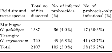
A total of 2107 tsetse proboscides from Glossina pallidipes (Msubugwe) and G. swynnertoni (Tarangire) were examined for the presence of trypanosomes (Table 1). Infection was found in 105 proboscides (5·0%), with the highest infection rate in G. swynnertoni from Tarangire; a large proportion of infections (55·2%) were proboscis only, particularly in G. swynnertoni (83·7%) (Table 1). Midgut infection rates for these fly populations of 6·0% and 6·7% were recorded for G. pallidipes at Msubugwe in 2006 and 2007 respectively, and 4·6% for G. swynnertoni at Tarangire in 2006. Trypanosomes identified in the midgut by ITS-1 PCR and/or FFLB were T. brucei, T. simiae, T. simiae Tsavo, T. godfreyi, T. congolense savannah, T. congolense Kilifi and T. brucei-like (Adams, Reference Adams2008; Adams et al. Reference Adams, Hamilton, Malele and Gibson2008; Hamilton et al. Reference Hamilton, Adams, Malele and Gibson2008).
Proboscis infection rates in two tsetse species in Tanzania
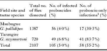
1 No. of infected proboscides without an accompanying midgut infection as a percentage of proboscis infections.
Identification of proboscis infections
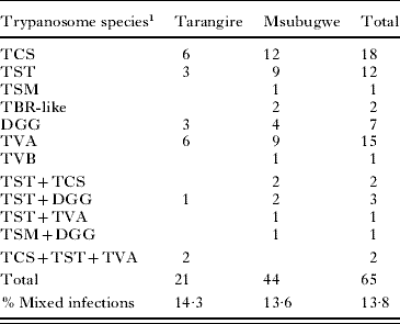
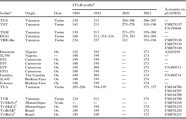
Trypanosomes present in proboscis samples were identified using FFLB. A total of 65 of the 105 infected proboscides (61·9%) were identified; 9 (13·8%) were mixed infections, 7 of which were double infections and 2 triple infections (Table 2). Seven different FFLB profiles were recorded from the proboscides (Table 3), 5 of which had been detected previously in midgut samples of the same flies (T. simiae, T. simiae Tsavo, T. godfreyi, T. congolense savannah and T. brucei-like) (Adams, Reference Adams2008; Adams et al. Reference Adams, Hamilton, Malele and Gibson2008; Hamilton et al. Reference Hamilton, Adams, Malele and Gibson2008). The remaining 2 FFLB profiles were new, although they resembled that of the reference T. vivax from Nigeria, strain Desowitz (Table 3); primer set 24S1 had been found previously not to work for T. vivax (Hamilton et al. Reference Hamilton, Adams, Malele and Gibson2008). A total of 18 flies from both field sites carried T. vivax A, sometimes in mixed infections with other trypanosomes; not all samples gave a full FFLB profile, presumably due to insufficient DNA template. The T. vivax B profile was distinctive (Table 3) and found only in a single fly at Msubugwe (Table 2). Overall, a total of 19 of 65 (29%) infected proboscides contained either T. vivax A or B. Most of these were proboscis-only infections, except where there was co-infection with trypanosomes of subgenus Nannomonas. Although proboscis-only infections are normally assumed to be T. vivax, we found that 14 such infections consisted of trypanosomes of subgenus Nannomonas (T. congolense or T. godfreyi).
Species of trypanosome identified in tsetse proboscis samples in two field sites
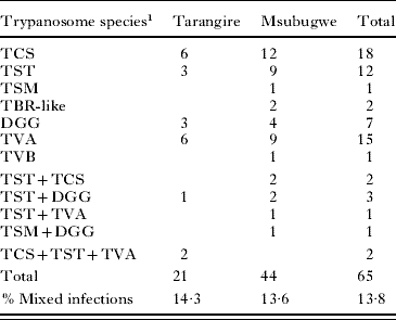
1 Number of single, double and triple infections is shown. TCS, T. congolense savannah; TST, T. simiae Tsavo; TSM, T. simiae; TBR, T. brucei; DGG, T. godfreyi; TVA, T. vivax genotype A; TVB, T. vivax genotype B.
FFLB profiles and Accession numbers for gGAPDH sequences
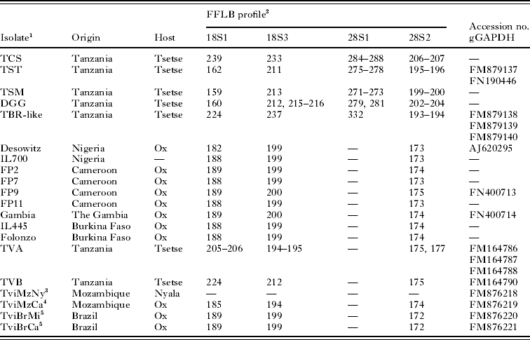
1 TCS, T. congolense savannah; TST, T. simiae Tsavo; TSM, T. simiae; TBR, T. brucei; DGG, T. godfreyi; TVA, T. vivax genotype A; TVB, T. vivax genotype B.
2 Dash indicates that no peak was detected.
3 Rodrigues et al. (Reference Rodrigues, Neves, Garcia, Viola, Marcili, Da Silva, Sigauque, Batista, Paiva and Teixeira2008).
4 Cortez et al. (Reference Cortez, Rodrigues, Garcia, Neves, Batista, Bengaly, Paiva and Teixeira2009); FFLB profile gave weak peak for 18S1.
5 Cortez et al. (Reference Cortez, Ventura, Rodrigues, Batista, Paiva, Anez, Machado, Gibson and Teixeira2006); FFLB profiles of both Brazilian samples gave weak peaks.
Comparing the proboscis results with those for the identification of midgut infections obtained previously (Adams, Reference Adams2008), of 47 flies with infection in both the midgut and proboscis, identification of both samples was made in 41 cases (87%), with the majority (36 of 41, 88%) containing the same infection in both compartments. Of the 5 flies with different infections, 2 had T. simiae in the midgut and T. godfreyi in the proboscis, 2 had T. godfreyi in the midgut and T. vivax in the proboscis, and 1 had T. congolense savannah in the midgut and T. simiae Tsavo in the proboscis.
Identification of animal infections
T. vivax samples of various origins were identified using FFLB (Table 3). All samples from West Africa and Brazil produced fragments of 188–189 bp for 18S1, 199–200 bp for 18S3, and 172–175 bp for 28S2. Sizes for the reference T. vivax, Desowitz, were within this range except for 18S1 with the slightly lower value of 182 bp. The Mozambiquan sample, TviMzCa, gave values outside the range for both 18S1 and 3; unfortunately, the other Mozambiquan sample from a nyala, TviMzNy, failed to amplify. None of the T. vivax samples produced a specific fragment with primer set 28S1, leading to the conclusion that non-amplification of this region is by default diagnostic for T. vivax. The primer sequences match exactly the database sequence of the 28Sα rRNA gene from T. vivax ILDat 1.2 (Accession no. U22316) and should yield a specific fragment of 252 bp. The problem may lie in the GC-rich nature of the rRNA locus in T. vivax compared to other trypanosomes (Haag et al. Reference Haag, O'Huigin and Overath1998). For example, in ILDat 1·2 the 252 bp 28S1 FFLB region is 72·6% GC, whereas the corresponding 290 bp region from T. brucei is 53·1% GC. This is also true for the 28S2 region though, where the respective GC contents are 71·6% and 56·5%. However, considering that FFLB fragments were generally more problematic to amplify in T. vivax than other trypanosomes, modification of protocols to favour amplification of GC-rich areas may improve overall FFLB detection of T. vivax.
Phylogenetic analysis
To further characterize trypanosomes with the new FFLB profiles, gGAPDH gene sequences were obtained by PCR amplification; Accession numbers are given in Table 3. Only a few proboscis samples contained sufficient DNA to produce suitable fragments for sequencing. Twelve samples from 2006, representing T. vivax profile A, were sequenced, but only 4 produced high quality sequences. The gGAPDH gene could not be amplified from another 6 proboscis samples even after whole genome amplification (WGA), presumably due to low quality or quantity of DNA. A further gGAPDH sequence was obtained from the sole sample with the B genotype after WGA. For comparison, gGAPDH sequences were also obtained from the 2 T. vivax samples from Mozambique (TviMzNy, TviMzCa), 2 Brazilian T. vivax samples (TviBrMi, TviBrCa), and 1 T. vivax sample each from Cameroon and The Gambia (Table 3). These gGAPDH sequences were compared with those from the Nigerian T. vivax strains Desowitz (Accession no. AJ620295) and ILDat 1.2 (Y486) from the T. vivax genome sequencing project (http://www.sanger.ac.uk/Projects/T_vivax/), and also with 2 database sequences for T. vivax of unknown origin (Accession nos. AF053744, AF047500).
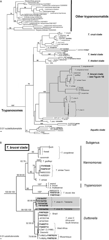
Phylogenetic trees constructed from the gGAPDH sequence data, such as the maximum likelihood (ML) tree (Fig. 1), show T. vivax to fall within, but on the periphery of the T. brucei clade of African tsetse-transmitted trypanosomes, as found previously for both 18S rRNA and gGAPDH sequence data (Haag et al. Reference Haag, O'Huigin and Overath1998; Stevens et al. Reference Stevens, Noyes, Dover and Gibson1999; Hamilton et al. Reference Hamilton, Stevens, Gaunt, Gidley and Gibson2004). With the addition of more sequences in this study, T. vivax is now seen to belong to a subclade of the T. brucei clade (Fig. 1B). This subclade is equivalent to subgenus Duttonella, the subgenus defined by development restricted to the tsetse proboscis (Hoare, Reference Hoare1972), and contains 3 distinct lineages: a main group of T. vivax (hereafter referred to as T. vivax C) containing isolates from Africa and South America, and T. vivax A and B from Tanzania.
Maximum likelihood gGAPDH gene tree. (A) Full tree containing 72 trypanosome taxa representing all known clades, and rooted using 21 other diverse trypanosomatids. (B) Detail showing the Trypanosoma brucei clade only. The tree is based on an alignment of 766 characters, all of which (412 parsimony-informative) were included in the analysis. The model of nucleotide substitution for maximum likelihood (ML) and ML distance analysis was GTR+I+G with estimated nucleotide base frequencies; a 4-category gamma distribution was used. −Ln=14963·52480. Single values at nodes are ML bootstrap values. Multiple values at nodes are in order: ML bootstrap value (100 replicates), MP, ML distance bootstrap values (1000 replicates). 100*=support values of 100 by all methods. Accession numbers of sequences obtained in this study are shown in bold. For other information on trypanosome sequences see (Hamilton et al. Reference Hamilton, Stevens, Gaunt, Gidley and Gibson2004, Reference Hamilton, Stevens, Gidley, Holz and Gibson2005, Reference Hamilton, Gibson and Stevens2007; Stevens et al. Reference Stevens, Noyes, Dover and Gibson1999); strain information for sequences AF047500 and AF053744 is not available through the GenBank/EMBL database.

T. vivax B is represented by the single sequence obtained after WGA, a technique which may introduce errors into the amplified fragment. We therefore checked the fidelity of the sequence obtained for T. vivax B by analysis of the codon position of each base change; errors introduced during WGA would be predicted to occur at random. However, in pairwise comparisons, almost all differences were at the third codon position; for example, of a total of 31 nucleotide differences between the gGAPDH sequences of T. vivax B and ILDat 1.2, three were at position 1 and 28 at position 3. This indicates that the sequence obtained for T. vivax B after WGA is likely to be correct.
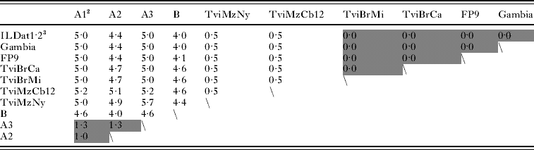
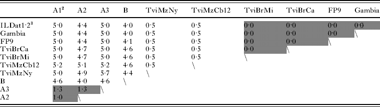
The genetic distances between the 3 subgroups of T. vivax range from 4·0 to 5·7%, differences higher than within T. vivax C (0–0·5%) or within T. vivax A (1·0–1·3%) (Table 4). These within-group distances are similar to those within well-characterized species such as T. brucei (0·4%) or T. cruzi 2·8% (including T. cruzi marinkellei, a trypanosome of bats) (Hamilton et al. Reference Hamilton, Stevens, Gaunt, Gidley and Gibson2004). Together with the genetic differences detected at the rRNA locus by FFLB, these high levels of genetic distance suggest that T. vivax A and B are distinct genetic lineages divergent from T. vivax C, the main group of African and South American T. vivax including the genome strain, ILDat 1.2 (Y486).
Distance matrix for gGAPDH sequences based on an alignment of 766 bp common to all sequencesFootnote 1
1 Intraclade differences are highlighted.
2 T. vivax A sequences: A1=FM164786, A2=FM164787, A3=FM164788.
3 ILDat1.2=Y486. Sequence data obtained from the Sanger Institute website at http://www.sanger.ac.uk/Projects/T_vivax/
T. vivax C, which includes isolates from West Africa (Desowitz, ILDat 1.2 from Nigeria, FP9 from Cameroon and one sample from The Gambia), Mozambique (TviMzNy, TviMzCa) and Brazil (TviBrMi, TviBrCa), received strong to high bootstrap support (ML 79%, MP 99%, MLDist 96%; Fig. 1). As noted above, genetic distances within this group are relatively small (0–0·5%; Table 4). Isolates originating from West Africa and South America form a distinct subclade within T. vivax C, receiving moderate bootstrap support (ML 70%, MP 63%, MLdist 68%), while the 2 Mozambiquan samples, TviMzNy and TviMzCa, fall on the periphery of this subclade and differ from each other by 0·5% (Table 4; Fig. 1). This confirms previous findings on the distinctive nature of these two East African isolates based on phylogenetic analysis of the 18S rRNA and cathepsin L-like genes (Cortez et al. Reference Cortez, Ventura, Rodrigues, Batista, Paiva, Anez, Machado, Gibson and Teixeira2006, Reference Cortez, Rodrigues, Garcia, Neves, Batista, Bengaly, Paiva and Teixeira2009; Rodrigues et al. Reference Rodrigues, Neves, Garcia, Viola, Marcili, Da Silva, Sigauque, Batista, Paiva and Teixeira2008).
The 4 T. vivax A sequences fell in a clade receiving strong bootstrap support (ML 85%, MP 95%, MLDist 99%; Fig. 1). These sequences differ by 4·4–5·7% from those of T. vivax C, but within-group by 1·0–1·3% (Table 4). Thus there is greater variation within T. vivax A than within T. vivax C. The T. vivax B sequence differs from T. vivax A by 4·0–4·6%, and from other T. vivax sequences by 4·0–4·7% (Table 4). Despite its unique genotype, the phylogenetic relationships of T. vivax B with other T. vivax isolates are not resolved; it forms a weakly supported clade with T. vivax C (ML 52%, MP 55%, MLDist 52%; Fig. 1). More samples and sequences from other genes should help clarify this issue.
We also obtained the gGAPDH sequences of other trypanosome species found in tsetse from Tanzania for comparison (Accession nos. T. simiae Tsavo FM879137, FN190446; T. brucei-like FM879138, FM879139, FM879140). For T. simiae Tsavo, both sequences were identical and, including 2 other sequences (both from Tanzania: AM503353, AJ620291), the within-group genetic distance was 1·2%. Two of the sequences of the T. brucei-like trypanosomes were identical to each other and to the gGAPDH sequence previously obtained from a midgut isolate of this trypanosome AM503075 (Hamilton et al. Reference Hamilton, Adams, Malele and Gibson2008); the third sequence FM879140 showed 1 bp difference, giving an overall within-group genetic difference of 0·1%. This confirms that reliable sequence data can be obtained from tsetse samples and provides further illustrative examples of intraspecific genetic distances. These sequences were also included in the phylogenetic analysis (Fig. 1B).
DISCUSSION
Trypanosome infections present in the proboscis are those that pose an immediate threat to livestock bitten by tsetse flies. Our analysis in Tanzania shows that many of these infections contain new trypanosome genotypes that have not been described previously. Of 2 new T. vivax genotypes found here, 1 was widespread, being recorded in 2 tsetse species from 2 geographically distant regions of the country. Another new trypanosome genotype was identified in tsetse proboscides in Msubugwe, having been previously detected in fly midguts in the same region (Adams et al. Reference Adams, Hamilton, Malele and Gibson2008); this trypanosome was shown to be most closely related to T. brucei by phylogenetic analysis and therefore called T. brucei-like (Hamilton et al. Reference Hamilton, Adams, Malele and Gibson2008). The mammalian hosts and pathogenic effects of this trypanosome remain unknown. Similarly, we do not know the mammalian host range or pathogenicity of the new T. vivax genotypes described here. In such a well-known and well-studied disease as animal trypanosomiasis, the finding of putative new pathogenic genotypes is surprising and even alarming. Without knowledge of the pathogenicity and host range of an infectious agent, or of its response to chemotherapy, it is difficult to design a rational control programme.
T. vivax is considered to be one of the most important of the salivarian trypanosomes because of its pathogenicity to cattle and its relatively high infection rates in tsetse. Its developmental cycle is the simplest among the tsetse-transmitted trypanosomes, being completed entirely within the proboscis and adjacent cibarial pump (Jefferies et al. Reference Jefferies, Helfrich and Molyneux1987). This has presumably facilitated its emancipation from tsetse transmission and allowed it to spread beyond the tsetse belt of sub-Saharan Africa and to South America, where its main vectors are biting flies such as tabanids. Molecular characterization studies have shown that New World T. vivax isolates are similar to those from West Africa, but that isolates from East Africa are much more diverse (Dickin and Gibson, Reference Dickin and Gibson1989; Fasogbon et al. Reference Fasogbon, Knowles and Gardiner1990; Dirie et al. Reference Dirie, Murphy and Gardiner1993a, Reference Dirie, Otte, Thatthi and Gardinerb; Masake et al. Reference Masake, Majiwa, Moloo, Makau, Njuguna, Maina, Kabata, OleMoiYoi and Nantulya1997; Cortez et al. Reference Cortez, Ventura, Rodrigues, Batista, Paiva, Anez, Machado, Gibson and Teixeira2006). While the West African/South American T. vivax is clearly recognizable in our FFLB and phylogenetic analyses, East African isolates of T. vivax were very heterogeneous and fell into 3 distinct groups. The relationship of the highly divergent T. vivax lineages found here to other East African T. vivax from Mozambique echoes that of the unique T. vivax zymodeme MID627 from Muhaka on the Kenya coast, which fell outside the cluster of Kenyan T. vivax isolates and also the main group of West African and Ugandan isolates (Fasogbon et al. Reference Fasogbon, Knowles and Gardiner1990). Clearly, much more comprehensive and detailed analysis of T. vivax isolates from East Africa needs to be carried out before the full picture of genetic diversity and its bearing on pathogenicity will be understood.
The gGAPDH gene has proved to be very useful for phylogenetic analysis of trypanosomes, as this short gene sequence is readily amplified from both purified DNA and DNA from field-collected material, as in this study. Furthermore, gGAPDH gene sequences align without gaps, in contrast to 18S rRNA gene sequences, facilitating both intra- and interspecific comparisons. However, gGAPDH gene sequences are not available for all trypanosomes and some T. vivax genotypes have been described as yet only from partial 18S rRNA gene sequences (Malele et al. Reference Malele, Craske, Knight, Ferris, Njiru, Hamilton, Lehane, Lehane and Gibson2003; Rodrigues et al. Reference Rodrigues, Neves, Garcia, Viola, Marcili, Da Silva, Sigauque, Batista, Paiva and Teixeira2008). Unfortunately, we failed to obtain 18S rDNA fragments from T. vivax A and B, even after WGA, although gGAPDH gene fragments amplified readily. This may indicate a specific problem with amplification of the 18S rRNA gene, since it is typically present in higher copy number than the gGAPDH gene in trypanosomes. For example, in mixed proboscis infections containing T. vivax A, the 18S rDNA of the other trypanosome species amplified preferentially. It is known that the GC content of the 18S and 28S rDNAs of T. vivax is high compared to other salivarian trypanosome species (Haag et al. Reference Haag, O'Huigin and Overath1998), and this difference may underlie problems with PCR amplification both to obtain the gene sequence and in the FFLB analysis.
Currently, there is no consensus among biologists on the level of sequence divergence that signifies a species level difference, and this is likely to vary between organisms anyway. The problem is compounded for microbial organisms to which the biological species concept cannot readily be applied. The taxonomy of the salivarian trypanosomes exemplifies this problem, where utilitarian considerations that relate causative organisms to disease have driven taxonomy (Gibson, Reference Gibson2007). Judging by the high levels of genetic distance among T. vivax gGAPDH sequences described here, the highly divergent T. vivax A and B lineages may well represent new species or perhaps belong to one of the little known or forgotten taxa within subgenus Duttonella, such as T. uniforme or T. vivax ellipsiprymni, both previously described in East Africa (Hoare, Reference Hoare1972; Stephen, Reference Stephen1986). It seems likely, from their ubiquity in the tsetse we examined, that T. vivax A and B represent the genotypes of T. vivax circulating in livestock in Tanzania and causing disease. It remains an open question whether the T. brucei-like trypanosome identified from proboscides and midguts of infected tsetse is also a pathogen.
Molecular methods for identification of trypanosome infections in tsetse have once again proved more informative than the simple dissection methodology (Lloyd and Johnson, Reference Lloyd and Johnson1924) relied on in the past. Besides identifying new trypanosome genotypes, the sensitivity and accuracy of the FFLB technique allowed the detection of mixed infections of 2 and even 3 different trypanosomes in the proboscis. FFLB was able to distinguish different trypanosome species occurring in the midgut and proboscis of the same fly, and to show that many proboscis-only infections are due to trypanosomes of subgenus Nannomonas rather than subgenus Duttonella as generally assumed. This reiterates the findings of (Lehane et al. Reference Lehane, Msangi, Whitaker and Lehane2000) and suggests that the prevalence of subgenus Duttonella in tsetse may be lower than estimates based on fly dissection alone.
In conclusion, we have discovered 2 novel genotypes of T. vivax in Tanzania, 1 of which is prevalent in 2 geographically distant areas in different tsetse species. This was achieved through the application of new molecular and phylogenetic approaches to the analysis of the small amounts of trypanosome DNA available from field-collected tsetse proboscides. Together with the other new T. brucei-like trypanosome recently described from Tanzania (Cortez et al. Reference Cortez, Ventura, Rodrigues, Batista, Paiva, Anez, Machado, Gibson and Teixeira2006; Hamilton et al. Reference Hamilton, Adams, Malele and Gibson2008; Rodrigues et al. Reference Rodrigues, Neves, Garcia, Viola, Marcili, Da Silva, Sigauque, Batista, Paiva and Teixeira2008), the discovery of these new trypanosome genotypes demonstrates that current knowledge of genetic diversity within the salivarian trypanosomes is severely limited, despite the pre-eminent importance of these trypanosomes as agents of Nagana. We now urgently need to find out about the pathogenicity and response to chemotherapy of these new trypanosome genotypes, as well as details of their distribution and host range.
We are grateful to L. Neves in Mozambique, F. Paiva and J. S. Batista in Brazil, and all staff members at the TTRI, Tanzania for their hard work and invaluable contribution to field collections. Sequence data from T. vivax and T. congolense were obtained from The Sanger Institute website at http://www.sanger.ac.uk/ and sequencing of these trypanosome genomes was funded by The Wellcome Trust.
FINANCIAL SUPPORT
E. R. A. was funded by a NERC research scholarship. P. B. H. received financial support from DEFRA, the Environment Agency and NERC. Work in Tanzania (E. R. A. and I. I. M.) was supported by NERC, the Genetics Society and the Tanzanian government. Work in Brazil and Mozambique (A. C. R. and M. M. G. T.) was supported by the Brazilian agency CNPq within the PRO-AFRICA programme.