Introduction
Hyaluronidase is widely used to remove cumulus cells from cumulus cell–oocyte complexes (COCs) prior to oocyte cryopreservation, intracytoplasmic sperm injection (ICSI) and DNA injection. During oocyte maturation, cumulus cells synthesize and accumulate hyaluronic acid, a high-molecular-weight glycosaminoglycan that consists of alternate repeats of d-glucuronic acid and N-acetylglucosamine. The hyaluronic acid accumulates within the cumulus oophorus. Hyaluronidase specifically acts on hyaluronic acid, and induces the loss of cumulus cells from COCs by digesting the extracellular matrix that is mainly composed of high-molecular-weight hyaluronic acid in the cumulus cell layer. Although hyaluronidase is widely used to denude oocytes, various detrimental effects associated with this treatment have been reported. In human oocytes, increased rates of degeneration and parthenogenetic development have been observed (Palermo et al., Reference Palermo, Joris, Derde, Camus, Devroey and Van Steirteghem1993), while the rate of oocyte fertilization and the quality of the produced blastocysts decrease after use of hyaluronidase (Taylor et al., Reference Taylor, Elliott, Colturato, Straub, Mitchell-Leef and Nagy2006; Moura et al., Reference Moura, Gurgel, Machado, Marques, Rolim, Lima and Salgueiro2017). In the mouse, treatment of oocytes with hyaluronidase reduces the survivability, fertilization rate and developmental ability (Ishizuka et al., Reference Ishizuka, Takeo, Nakao, Yoshimoto, Hirose, Sakai, Horikoshi, Takeuji, Tsuchiyama and Nakagata2014). The mechanism(s) of these detrimental effects of hyaluronidase is uncertain. Additionally, the effects of hyaluronidase treatment on bovine oocytes have not yet been determined.
The objective of this study was to determine the responses of bovine oocytes to the use of hyaluronidase. Hyaluronidase was used to remove cumulus cells from COCs; embryonic development, the rate of parthenogenetic development, Ca2+ levels, reactive oxygen species (ROS) levels and reduced glutathione (GSH) levels were then determined and compared with those of oocytes not treated with hyaluronidase for cumulus cell removal.
Materials and methods
Ovary collection
Bovine ovaries were obtained from a slaughterhouse; they were kept at 15–20°C and transported to the laboratory in saline solution supplemented with 0.1% antibiotics and antimycotics (AB; No. 15240-062, Gibco, Grand Island; NY, USA). Transportation time from the slaughterhouse to the laboratory was approximately 1 h. The ovaries were held in saline supplemented with 0.3 mM reduced glutathione (GSH; G6013, Sigma-Aldrich, St. Louis, MO, USA) and 1% AB at 15°C for 20 h with shaking (Nagao et al., Reference Nagao, Harada, Yamaguchi, Igarashi and Ooshima2010).
Oocyte collection and in vitro maturation (IVM)
Oocyte collection and IVM of the oocytes were carried out as previously described (Nagao et al., Reference Nagao, Saeki, Hoshi and Kainuma1994). In brief, good quality COCs from 2–6 mm diameter follicles were cultured in 50-µl droplets of modified TCM-199 (m-TCM199; No. 12340, Invitrogen, Carlsbad, CA, USA) with FSH (Antrin, Kyoritsu Seiyaku, Tokyo, Japan) and estradiol-17 β (E2758, Sigma-Aldrich) covered with mineral oil (M8410, Sigma-Aldrich) in a culture dish (Falcon 351007, Becton, Dickinson and Company, Franklin Lakes, NJ, USA) for 22–25 h at 39°C under 5% CO2 in humidified air.
Removal of cumulus cells by hyaluronidase
Hyaluronidase was purchased from Sigma-Aldrich (400–1000 units/mg of hyaluronidase from bovine testes, Type I-S; Sigma-Aldrich). The titre of hyaluronidase (Lot No. SLBC1666V), measured by Sigma-Aldrich, was 631 units/mg. After maturation of COCs, cumulus cells were thoroughly dispersed with or without 0.1% hyaluronidase and removed by vortexing for 7 min.
ICSI, artificial activation, and embryo culture
ICSI was performed as described previously (Ashibe et al., Reference Ashibe, Miyamoto, Kato and Nagao2019) with some modifications. Briefly, frozen–thawed semen was washed and incubated in Brackett and Oliphant medium (BO) (Brackett and Oliphant, Reference Brackett and Oliphant1975) supplemented with 0.05% (w/v) polyvinyl alcohol (PVA; P8136, Sigma-Aldrich) and 1% AB (BO-PVA) for 0.5–3 h at 39°C under 5% CO2 in humidified air. After removal of cumulus cells, denuded oocytes with a visible first polar body were selected and cultured in m-TCM199 for approximately 1 h at 39°C under 5% CO2 and 5% O2 with humidity. Spermatozoa that had been immobilized by applying several piezo pulses (Cell Tram Vario, PiezoXpert, Eppendorf, Germany) were singly injected into each oocyte with a minimal amount of medium. The injected oocytes were cultured in modified synthetic oviduct fluid supplemented with 0.05% (w/v) PVA and 1% AB (SOF-PVA) with BSA (SOF-BSA) (Nagao et al., Reference Nagao, Saeki, Hoshi and Kainuma1994) for 3 h, and treated with 7% ethanol in TCM-199 containing 1 mg/ml polyvinylpyrrolidone (P0930, Sigma Chemical Co.) for 3 min at 39°C under 5% CO2 in air for oocyte activation (Ashibe et al., Reference Ashibe, Miyamoto, Kato and Nagao2019). Cleavage (≥2-cell) and development to the blastocyst stage were examined under a stereomicroscope (×60) at 7 days after sperm injection.
IVF and in vitro culture (IVC) of embryos
IVF and IVC were performed as described previously (Nagao et al., Reference Nagao, Iijima and Saeki2008, Reference Nagao, Harada, Yamaguchi, Igarashi and Ooshima2010) with some modifications. Briefly, denuded oocytes were fertilized with spermatozoa (2 × 10⁶ cells/ml) that had been washed in BO-PVA supplemented with 2 μg/ml heparin (H3393, Sigma-Aldrich) at 39°C under 5% CO2 in humidified air for 18 h. The embryos were then cultured in 50 μl droplets of SOF-BSA (22–50 embryos/drop) at 39°C under 5% CO2 and 5% O2 with humidity. Cleavage (≥ 2-cell) and development to the blastocyst stage was examined under a stereomicroscope (×60 magnification) at 7 days after insemination.
Assessment of parthenogenetic development of ICSI oocytes
Assessment of parthenogenetic development was performed as described previously (Li et al., Reference Li, Hamano, Qian, Funauchi, Furudate and Minato1999) with some modifications. Briefly, ICSI embryos at the 2-cell stage at 2 days after fertilization were stained with Hoechst 33342 (B2261, Sigma-Aldrich) in ethanol (1 µg/ml; No. 14033-00, Kanto Chemical, Tokyo, Japan) for 10 min at 4°C and placed on a glass microscope slide in a very small droplet of 50% glycerol. To determine whether a sperm head was present, the embryos were examined using a fluorescence microscope (OLYMPUS, Tokyo) with the appropriate filter (excitation: 352 nm, emission: 455 nm). Parthenogenetic development was identified by the presence of a condensed sperm head in blastomere.
Assessment of intracellular Ca2+, ROS and GSH levels
Ca2+ levels in oocytes were measured using Fluo-4 AM (F311, Dojindo Laboratories, Japan). Denuded oocytes were cultured in SOF-PVA with 5 µM Fluo-4 AM for 1 h. Then, they were placed on the base of a glass dish in SOF-PVA and examined using a fluorescence microscope (OLYMPUS, Tokyo) with the appropriate filter (excitation: 494 nm, emission: 516 nm).
To determine ROS levels, denuded oocytes were cultured in SOF-BSA with 10 µM 2′7′-dichrolorohydrofluorescein diacetate (DCHFDA; STA-342, Cell Biolabs, Inc., USA) for 15 min. The oocytes were then placed on a glass microscope slide in 1× Dulbecco’s phosphate buffered saline (PBS; 142200-075, Gibco, Grand Island, NY, USA) supplemented with 1% (w/v) PVA (PBS-PVA) and images were captured using a cooled charge-coupled device (CCD) camera attached to a fluorescence microscope (BX51, OLYMPUS, Tokyo) with the appropriate filter (excitation: 450–490 nm, emission: 515–565 nm).
GSH levels were measured in denuded oocytes that had been cultured in SOF-PVA with 10 µM cell tracker blue (1266189, Molecular Probes, USA) for 30 min. The oocytes were then placed on a glass microscope slide in PBS-PVA and images were captured using a cooled CCD camera attached to a fluorescence microscope (BX51, OLYMPUS, Tokyo) with the appropriate filter (excitation: 330–385 nm, emission: 420 nm).
Recorded fluorescence images were analyzed using FL10-ASW3.1 (OLYMPUS, Tokyo) to determine the intensity of fluorescence to provide a measure of Ca2+, ROS and GSH levels in each oocyte.
Statistical analysis
The rates of cleavage, blastocyst formation and parthenogenetic development were compared between the two treatment groups using chi-squared tests. The levels of Ca2+, ROS and GSH were compared in the two treatment groups using analysis of variance (ANOVA) with the F-test and t-test. In all experiments, values were considered to be significantly different at P-values < 0.05.
Results and discussion
Hyaluronidase treatment is widely used to remove cumulus cells from COCs and is an essential component of animal and human ARTs such as ICSI. In the present study, the rates of development to the blastocyst stage were significantly lower (P < 0.05) after hyaluronidase treatment compared with the controls: ICSI, 22.4% vs. 36.1%; IVF, 21.2% vs. 30.4% (Fig. 1). Treatment of human oocytes with hyaluronidase has been shown to increase the rate of parthenogenetic development (Palermo et al., Reference Palermo, Joris, Derde, Camus, Devroey and Van Steirteghem1993) and may also decrease the rates of fertilization and blastocyst development (Taylor et al., Reference Taylor, Elliott, Colturato, Straub, Mitchell-Leef and Nagy2006; De Vos et al., Reference De Vos, Van Landuyt, Van Ranst, Vandermonde, D’Haese, Sterckx, Haentjens, Devroey and Van der Elst2008). A similar effect on fertilization and blastocyst development has been reported for mouse oocytes (Ishizuka et al., Reference Ishizuka, Takeo, Nakao, Yoshimoto, Hirose, Sakai, Horikoshi, Takeuji, Tsuchiyama and Nakagata2014). In the present study, we similarly identified a reduction in the rates of blastocyst development after treatment of bovine oocytes with hyaluronidase. Our observations suggested that the use of hyaluronidase to remove cumulus cells on bovine COCs has a negative effect on early embryonic development.
Effect of the use of hyaluronidase to remove cumulus cells from bovine COCs on the developmental competence of bovine oocytes in ICSI (a) and IVF (b). Different letters indicate significant differences (a, b: P < 0.05). n = number of embryos cultured. Data are from 5–8 repeat experiments. −: Non-treated group; +: Hyaluronidase treatment group.

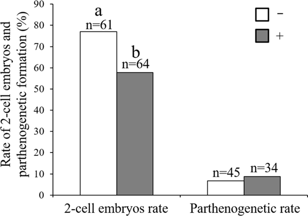
The rate of 2-cell embryos after ICSI was significantly lower at 57.8% (37/64) in the hyaluronidase treatment group compared with 77.0% (47/61) in the control group. There was significant difference in the rate of 2-cell embryos in Fig. 2 (P = 0.022), but not in Fig. 1(a) (ICSI, 62.4% vs. 75.9%, P = 0.057; IVF, 67.6% vs. 70.0%, P = 0.540). The rate of 2-cell embryos was evaluated at day 2 after ICSI in Fig. 2, as it becomes more difficult to detect the sperm head at the embryo development stage. Conversely, the rate of cleavage was evaluated at day 7 in Fig. 1(a), to measure the blastocyst rate more accurately without the influence of the environment outside the incubator. Therefore, we speculated that the zygotes received some negative effects when using hyaluronidase to remove cumulus cells, so the first cleavage may be delayed in the hyaluronidase treatment group. Moreover, the oolemma and cytoplasmic membrane are physically broken to inject spermatozoa when following the ICSI protocol, which may generate result in high stress on the oocytes. Therefore, the detrimental effect of hyaluronidase may be further emphasized in ICSI embryos. The balance of intracellular components such as Ca2+ may be disrupted more severely by ICSI compared with IVF. In this study we used only one batch of hyaluronidase, but reductions in the rates of cleavage and blastocyst development were also observed during treatment with different batches of hyaluronidase in our pre-experiment (data not shown). These results suggested that hyaluronidase may have a common adverse effect regardless of batch.
Effect of the use of hyaluronidase to remove cumulus cells from bovine COCs on parthenogenetic development after ICSI. Different letters indicate significant differences (a, b: P < 0.05). n (2-cell embryos rate) = number of embryos cultured. n (parthenogenetic rate) = number of 2-cell embryos. Data are from three repeat experiments. Parthenogenetic development was identified by the presence of a condensed sperm head in a blastomere. –: Non-treated group; +: Hyaluronidase treatment group.

As mentioned above, it was reported that hyaluronidase treatment of human COCs increased the rate of parthenogenetic development (Palermo et al., Reference Palermo, Joris, Derde, Camus, Devroey and Van Steirteghem1993). We compared the rates of parthenogenetic development in surviving bovine embryos at day 2 after ICSI, but found no significant difference in parthenogenetic development rates: 8.8% (3/34) after hyaluronidase treatment compared with 6.7% (3/45) in the non-treated group (Fig. 2). The mechanism of this reduction in cleavage after hyaluronidase treatment of oocyte is not clear, although it has been reported that hyaluronidase induces morphological changes in mouse oocytes (Ishizuka et al., Reference Ishizuka, Takeo, Nakao, Yoshimoto, Hirose, Sakai, Horikoshi, Takeuji, Tsuchiyama and Nakagata2014). In the present study, we did not observe any morphological changes after hyaluronidase treatment, and fertilization by IVF was normal after hyaluronidase treatment of oocytes (data not shown). Moreover, in the non-treated hyaluronidase group there was an increased rate of blastocyst development, although the time lengths for vortexing and experimental exposure were same in both groups. These results suggested that hyaluronidase reduced the cleavage rate in bovine oocyte by qualitative factors that do not affect fertility or parthenogenesis.
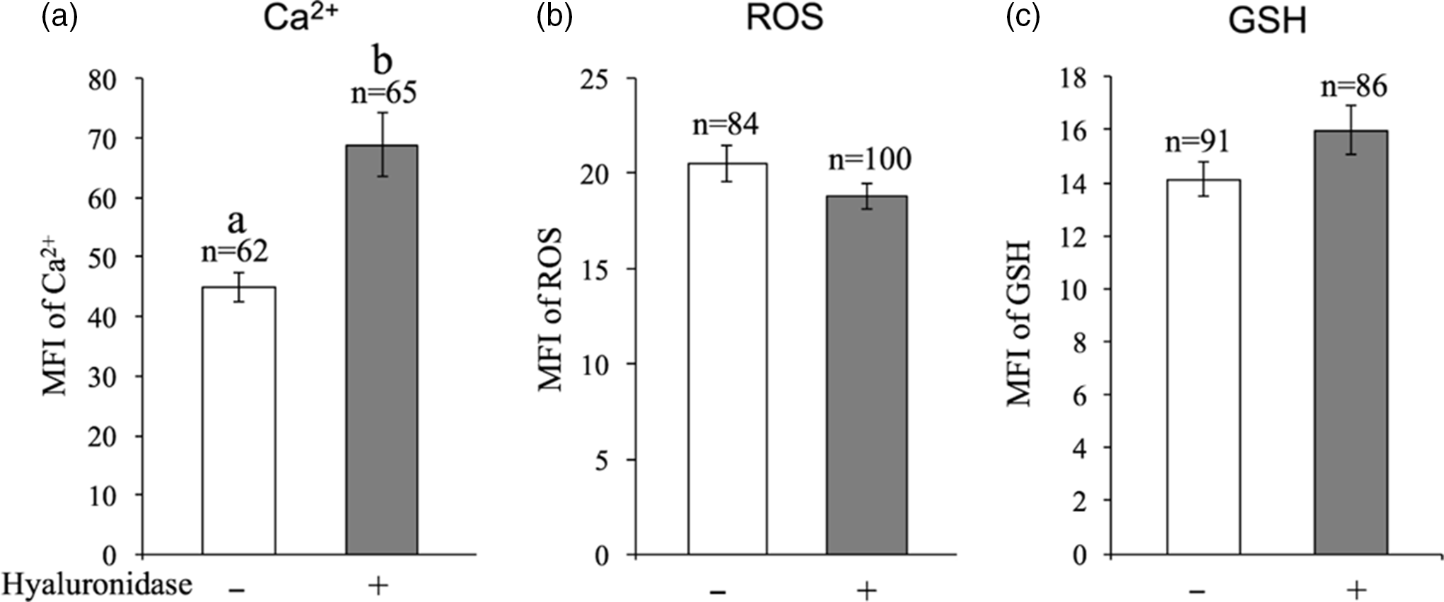
Next, we investigated potential qualitative changes to oocytes induced by hyaluronidase treatment. Analysis of Ca2+ levels showed a significantly higher mean intensity of fluorescence in the hyaluronidase treatment group, indicating higher Ca2+ levels: hyaluronidase group, 68.8 ± 5.3; control group, 45.0 ± 2.5 (P < 0.05; Fig. 3a). Ca2+ has complex functions in biological systems and is intrinsic to many aspects of cellular metabolism (Heilbrunn, Reference Heilbrunn1940); Ca2+ acts as a signalling molecule that plays important roles in the regulation of various physiological cellular processes, including secretion, contraction, proliferation, development, gene expression and programmed cell death (Berridge et al., Reference Berridge, Lipp and Bootman2000; Bandyopadhyay et al., Reference Bandyopadhyay, Lee, Lee, Lee, Yu, Jee, Cho, Jung, Lee, Zannoni, Singson, Kim, Koo and Ahnn2002). In the animal kingdom, an intracellular Ca2+ increase in oocytes is an important step for successful fertilization and further development (Kashir et al., Reference Kashir, Deguchi, Jones, Coward and Stricker2013). However, dimethyl sulfoxide (DMSO)-mediated cytotoxicity during bovine oocyte vitrification is associated with Ca2+ release from the endoplasmic reticulum into the cytoplasm, resulting in increased Ca2+ levels in oocytes and a reduced rate of subsequent blastocyst development (Wang et al., Reference Wang, Hao, Li, Zhao, Wang, Yan, Du, Wang, Liu, Pang, Zhu and Zhao2017). Therefore, the increase in Ca2+ levels observed here after treatment of oocytes with hyaluronidase may have reduced the rate of blastocyst development. Also, in our pre-experiment, Ca2+ levels in oocytes increased depending on the concentration of hyaluronidase (0 mg/ml, 0.5 mg/ml, 0.1 mg/ml, 10 mg/ml). As the molecular mass of hyaluronidase is 60 kDa, it cannot pass through the cell membrane, therefore the increase in Ca2+ levels might be associated with plasma membrane function, and not endoplasmic reticulum stress as induced by DMSO (Kang et al., Reference Kang, Das, Gurunathan, Park, Song, Park and Kim2017). Hyaluronidase may induce dysfunction in Ca2+ channels on the plasma membrane, and then cause defective excretion of Ca2+ from the cells or Ca2+ inflow into the cytoplasm from outside the cells. It has been reported that an increase in Ca2+ levels in the physiological range triggers the generation of ROS in rat oocytes (Prasad et al., Reference Prasad, Tiwari, Tripathi, Pandey and Chaube2015). Martín-Romero et al. (Reference Martín-Romero, Miguel-Lasobras, Domínguez-Arroyo, González-Carrera and Alvarez2008) reported that H2O2 can trigger an increase in Ca2+ levels in human oocytes. Therefore, we analyzed the intracellular levels of ROS and GSH, an intracellular modulator of ROS related to intracellular Ca2+ levels; we found no significant differences in the levels of ROS and GSH between the hyaluronidase treatment group and the control group: ROS, 18.8 ± 0.7 vs. 20.5 ± 0.9; GSH, 16.0 ± 0.9 vs. 14.1 ± 0.7 (Fig. 3b, c).
Effect of treatment with hyaluronidase to remove cumulus cells from bovine COCs on the levels of intracellular Ca2+ (a), intracellular ROS (b), and GSH of oocytes (c). The levels of each metric are shown as mean fluorescence intensities (MFI). Different letters indicate significant differences (a, b: P < 0.05). n = number of oocytes examined. Data shown are from 3–5 replicate experiments. Error bars represent the standard error of the mean. –: Non-treated group; +: Hyaluronidase treatment group.

In conclusion, the present study demonstrated that hyaluronidase could be a detrimental factor for the developmental competence of bovine oocytes, as has previously been reported for mouse and human oocytes. With regard to the potential mechanism of this detrimental effect, we showed that hyaluronidase might trigger an increase in Ca2+ levels in oocytes due to a functional change in Ca2+ channels in the cell membrane. This increase of Ca2+ levels in the oocytes resulted in a decrease in the rate of the embryonic development. We concluded that oocyte manipulation without hyaluronidase might be an effective means of improving mammalian ARTs.
Acknowledgements
We thank T. Masafumi of Obihiro University for invaluable advice. The authors would like to thank Genetics Hokkaido Co. Ltd for providing the frozen semen, and Chikusei Meat Center and Nakao Chikusan Co. Ltd for providing the ovaries used in this study.
Financial support
This research received no specific grant from any funding agency, commercial, or not-for-profit sectors.
Conflicts of interest
None.
Ethical standards
Not applicable.