Introduction
Achalasia is a motility disorder of the lower oesophagus, characterised by absent peristalsis in the body of the oesophagus, increased lower oesophageal sphincter pressure and failure of sphincter relaxation during swallowing.Reference Francis and Katzka1 It is caused by idiopathic degeneration of the myenteric plexus.Reference Francis and Katzka1 It results in dysphagia for liquids and solids, and possible regurgitation.Reference Francis and Katzka1 The incidence in British children under the age of 15 years old is 1 per million.Reference Mayberry and Mayell2 A PubMed database search for achalasia and stridor revealed case reports of achalasia presenting acutely with respiratory distress in adults, but there were no reports of such cases in children.Reference Miyamoto, Konda, Matsui, Sawada, Ikeda and Watanabe3, Reference Doshi, Aw, Costa, Cohen and Som4
Case report
A 12-year-old Afro-Caribbean girl presented to the ENT clinic with an 18-month history of dysphagia and noisy breathing when consuming both solids and liquids. Sometimes the dysphagia was total, and she occasionally regurgitated solids. She did not have any symptoms to suggest she was aspirating and her voice was normal. Her weight was normal for her age, but she had only gained 2 kg in the last year.
The patient had been born at term by caesarean section. She was healthy post-delivery and did not need intubation. She has asthma, which is well controlled with inhalers. When she was two years old, she underwent adenotonsillectomy for obstructive sleep apnoea, with a successful outcome. She does not suffer from recurrent chest infections. She lives with her parents who are both healthy.
Her symptoms had been investigated by the gastroenterology team. Upper gastrointestinal endoscopy showed normal appearances of the oesophagus, stomach and duodenum. There was no narrowing at the lower end of the oesophagus. Biopsy results were normal. A 48-hour pH monitoring study, conducted to investigate the occurrence of reflux, was normal.
On examination in the ENT clinic, the patient's oral cavity was normal, as were her lower cranial nerves. Flexible laryngoscopy showed a slightly retroverted epiglottis, but otherwise the tongue base, hypopharynx and larynx all appeared normal.
A fibre-optic endoscopic evaluation of swallowing was performed with an orange squash drink. Initially, the swallowing seemed normal, with no coughing or choking. However, after repeated swallowing, biphasic stridor occurred, which seemed to be arising from the subglottis or trachea. There was no malacic collapse at the supraglottic level. It was thought that this could be due to a laryngeal cleft or a tiny tracheoesophageal fistula.
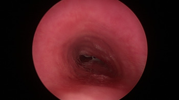
Microlaryngoscopy, bronchoscopy and upper oesophagoscopy were performed. Microlaryngoscopy showed a normal larynx, with no evidence of either a tracheoesophageal fistula or a laryngeal cleft. Bronchoscopy showed flattening of the cartilage rings from posterior compression caused by redundant oesophageal mucosa, which resulted in tracheomalacia of the distal trachea by up to 75 per cent (Figure 1). Rigid oesophagoscopy to 26 cm showed a dilated oesophagus with normal mucosa. The lower sphincter was not seen, but the upper sphincter was normal.
Bronchoscopy image showing distal tracheomalacia caused by flattened cartilage rings from posterior compression, which was a result of redundant oesophageal mucosa.

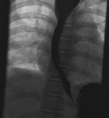
Videofluoroscopy showed a degree of achalasia with delayed passage of contrast into the stomach (Figure 2). Achalasia was confirmed by oesophageal manometry, which demonstrated incomplete relaxation of the lower oesophageal sphincter and absent peristalsis, both of which are diagnostic features of the condition.
Videofluoroscopy image showing delayed passage of contrast from the oesophagus into the stomach.

Following a Heller's myotomy performed by the paediatric surgeons, the patient's symptoms improved considerably.
Discussion
This is the first reported case in the English literature of childhood stridor caused by achalasia. The accompanying oesophageal dilatation caused extrinsic compression of the tracheal cartilages, leading to tracheomalacia. Stagnating food in the oesophagus compressed her trachea producing biphasic stridor.
There are case reports of achalasia in adults presenting with acute respiratory distress and stridor from tracheal compression by massive oesophageal dilatation.Reference Miyamoto, Konda, Matsui, Sawada, Ikeda and Watanabe3, Reference Doshi, Aw, Costa, Cohen and Som4 Decompression of the oesophagus with a nasogastric tube can be lifesaving.Reference Doshi, Aw, Costa, Cohen and Som4 Chest X-ray will show dilatation of the upper oesophagus in these patients.
• This is the first paediatric case in the English literature of achalasia presenting with stridor
• Microlaryngoscopy, bronchoscopy and upper oesophagoscopy showed oesophageal dilatation, which caused extrinsic compression of tracheal cartilage, leading to tracheomalacia
• Achalasia was confirmed with oesophageal manometry and successfully treated with Heller's myotomy
• Achalasia can present acutely in adults, with respiratory distress and stridor
• Decompression of the oesophagus with a nasogastric tube can be life saving
Although stridor caused by achalasia is extremely rare in children, the condition is important to recognise as it is amenable to surgical treatment. It can present to gastroenterologists with dysphagia, or to ENT surgeons and respiratory physicians with breathing difficulties. Stridor has many causes in children, but if it occurs after eating then achalasia should be considered.