

Introduction
As common bacterial pathogens in both mammals and birds, Clostridium perfringens is associated with necrotic enteritis in poultry and pigs (Songer, Reference Songer1996; Timbermont et al., Reference Timbermont, Haesebrouck, Ducatelle and Van Immerseel2011), Staphylococcus aureus with skin and soft tissue infections, mastitis, and several other diseases (Nagase et al., Reference Nagase, Sasaki, Yamashita, Shimizu, Wakita, Kitai and Kawa2002; Duijkeren et al., Reference Duijkeren, Ikawaty, Broekhuizen-Stins, Jansen, Spalburg, de Neeling, Allaart, Van Nes, Wagenaar and Fluit2008), while Escherichia coli is mostly associated with enteritis, especially post weaning diarrhoea in piglets (Noamani et al., Reference Noamani, Fairbrother and Gyles2003; Fairbrother and Gyles, Reference Fairbrother, Gyles, Straw, Zimmerman, D'Allaire and Taylor2006). Infections in animals by these organisms are usually treated with antibiotics. Concern about antimicrobial resistance and ban of antibiotic growth promoters in the European Union have increased the interest in finding natural alternatives to control the proliferation of pathogenic bacteria in animals and birds (Bartolo and Sall, Reference Bartolo and Sall2015; Cariou et al., Reference Cariou, Fadel and Rothstein2015; Huyghebaert et al., Reference Huyghebaert, Ducatelle and Van Immerseel2011; Kettunen et al., Reference Kettunen, Vuorenmaa, Rinttila, Gronberg, Valkonen and Apajalahti2015; Tellez et al., Reference Tellez, Latorre and Hargis2015).
Resin from coniferous trees has been used as a traditional medicine for centuries in Scandinavian countries (Kettunen et al., Reference Kettunen, Vuorenmaa, Rinttila, Gronberg, Valkonen and Apajalahti2015). It contains resin acids, for example, abietic, dehydroabietic, neoabietic, isopimaric, levopimaric and palustric acids; and these compounds have antimicrobial, antifungal and antiparasitic activity (San Feliciano et al., Reference San Feliciano, Gordaliza, Salinero and Miguel del Corral1993; Rubio et al., Reference Rubio, Calderón, Flores, Castroa and Céspedes2005; Savluchinske-Feio et al., Reference Savluchinske-Feio, Curto, Gigante and Roseiro2006). Resin acids are known to be more effective against Gram-positive bacteria (San Feliciano et al., Reference San Feliciano, Gordaliza, Salinero and Miguel del Corral1993). Natural resin acids with enriched composition (RAC) has been shown to improve the performance of broiler chickens in recent studies (Kettunen et al., Reference Kettunen, Vuorenmaa, Rinttila, Gronberg, Valkonen and Apajalahti2015, Reference Kettunen, Eerden, Lipinski, Rinttila, Valkonen and Vuorenmaa2017).
A resin acids-based product (RAP) is a newly introduced feed ingredient containing naturally occurring free resin acids. Limited data is available in the literature on the effects of resin acids to a wider variety of bacterial pathogens infecting mammals and birds. Hence, the present study aimed to evaluate the inhibitory activity of a natural RAP at different concentrations to Cl. perfringens, S. aureus, and E. coli O149 isolated from different mammals and birds using various in vitro techniques.
Materials and Methods
Bacteria
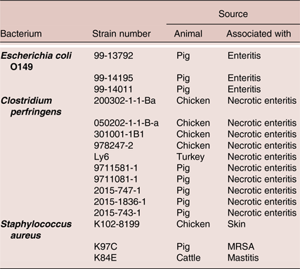
Ten strains of C. perfringens (Nauerby et al., Reference Nauerby, Pedersen and Madsen2003; Lyhs et al., Reference Lyhs, Perko-Makela, Kallio, Brockmann, Heinikainen, Tuuri and Pedersen2013), three strains of E. coli O149, and three of S. aureus isolated from different animals were used in the inhibition study (Table 1). All strains were clinical isolates that were characterised and preserved by the National Veterinary Institute, Denmark.
The list of bacteria along with their sources used in the inhibition study.

MRSA, Methicillin-Resistant Staphylococcus aureus.
Composition of the test product
The resin acids-based product (RAP) was tested for inhibitory activity against the bacterial pathogens. The product consists of 50% of tall oil fatty acids and resin acids (Progres, Hankkija Oy/Suomen Rehu, Hyvinkää, Finland), which were absorbed into 50% of silicate carrier (Tixosil 38X). The final product contained 4% natural resin acids from Scots pine (Pinus sylvestris) and Norwegian spruce (Picea abies). The resin acid fraction consisted abietic acid (33%), dehydroabietic acid (18%), pimaric acid (14%) and other minor resin acids (35%). The product contained 5% water, 35% crude fat and 55% ash. As feed additives, it contained E551a synthetic silicic acid (500 g/kg) as an anti-caking agent.
Target concentrations of the test product, and pH determination
Inhibitory bio-activity of the RAP was tested against 0.01%, 0.1% and 0.5%, and again 0.5%, 1% and 5% resin concentration of the product, including carrier substance, in brain heart infusion (BHI) broth. However, the actual concentrations of the final product were 0.02%, 0.2% and 1.0%, and 1%, 2% and 10%, respectively as only 50% of the product was resin. The pH of each concentration of the product was determined before starting the inhibition study and the pH ranged from 7.32 to 7.39.
Inoculum, inoculation and incubation
Growth curves were prepared on the basis of optical density at 600 nm wavelength (OD600nm) of brain heart infusion (BHI) broth cultures for one strain from each of the three different bacterial species as representatives. To make a final inoculum for each strain, 60 µl of broth cultures stored overnight was added into 60 ml BHI in a 300-ml conical flask, and then the covered flask was incubated at 37 °C up to the stationary phase. Thereafter, inoculations of 100 µl (containing approximately 108 CFU/ml) of the respective strains into 200 ml bottles were done at the pre-stationary phase, i.e. at 7 and 12 hours in C. perfringens and E. coli O149, respectively, and at stationary phase at 9 h for S. aureus. The cultures were incubated at 37 °C in order to run the inhibition study later at different time points. For the incubation of C. perfringens, an anaerobic cabinet (Modular atmosphere controlled system, du Scientific®, Copenhagen, Denmark) was used.
Methods
Bacteriological culture on solid medium were done on blood agar base (CM 55; Oxoid, Basingstoke, UK) added 5% citrated bovine blood, and incubated at 37 °C. The anaerobic cabinet was used for incubation of C. perfringens. Any growth in the respective plates resembling E. coli, C. perfringens and S. aureus was identified on the basis of colony morphology.
Two different methods were used- a 10-fold broth dilution method (BDM), and an agar diffusion method (ADM). The BDM was applied to three strains of each of the bacterial species, where the three strains of C. perfringens were 200302-1-1-Ba, Ly6 and 9711581-1. Samples were taken from the respective bottles after 2, 4 or 5, 8, and 24 h incubation, where after, 10-fold dilutions were made and plated onto blood agar plates. Counts were expressed as colony forming units per ml (CFU/ml) as log10. The BDM was performed against 0.01%, 0.1% and 0.5% concentrations of the product. The ADM was run on E. coli 99-13792, Cl. perfringens 200302-1-1-Ba and S. aureus K97C, first using 0.01%, 0.1% and 0.5% concentrations of the product. Subsequently, all strains (ten C. perfringens, three S. aureus and three E. coli O149 strains) were tested with ADM against 0.5%, 1% and 5%. For the ADM, test strains were spread on the surface of blood agar plates, where after, 5 mm Φ holes were plugged in the agar. Eighteen μl of solution of the resin based product were added to the wells, and after incubation at 37 °C for 24 h, the plates were read for inhibitions zone surrounding the wells. As a negative control, BHI was used with the specific bacterial strain without product in BDM, and only BHI in ADM.
Statistical analysis
GraphPad Prism software (Version 5, GraphPad Software, San Diego California USA) was used for data analyses. Two-Way Analysis of Variance was conducted and Bonferroni post tests for multiple comparisons were followed. Outcomes from data analysis were considered significant at P < 0.05 and 95% Confidence intervals.
Results and discussion
C. perfringens
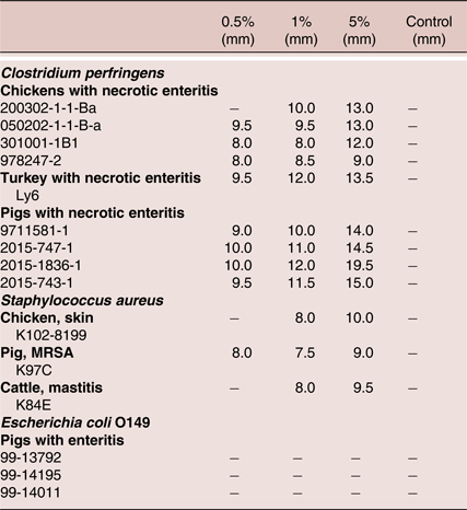
For the BDM, no C. perfringens was found at any of the tested concentrations of the RAP (Table 2), indicating efficient inhibition of growth even at 0.01%. In ADM, narrow inhibition zones developed around the product concentration of 0.5% (inhibitions zones: 8 to 10 mm), 1% (8 to 12.0 mm), and 5% (9.0 to 19.5 mm) when performed on all 10 strains (Table 3). Kettunen et al. (Reference Kettunen, Eerden, Lipinski, Rinttila, Valkonen and Vuorenmaa2017) demonstrated the significant reduction of Lactobacillus spp. and tended to lessen the loads of Bifidobacterium spp. and the entire numbers of eubacteria in broiler chickens using RAC. San Feliciano et al. (Reference San Feliciano, Gordaliza, Salinero and Miguel del Corral1993) suggested that the resin acids perform their antimicrobial activity through interacting with phospholipid membranes of gram-positive bacteria. It has been hypothesised that the antibacterial impact of resin acids is connected either to the bacterial cell wall solubilisation (Soderberg et al., Reference Söderberg, Johansson and Gref1996) or changed proton gradients of the cell membrane (Jokinen and Sipponen, Reference Jokinen and Sipponen2016). It is interesting that other plant-derived products have been described to have an inhibitory activity against C. perfringens, such as tannic acid and derivatives extracted from chestnut trees (Bartolo and Sall, Reference Bartolo and Sall2015).
The concentrations of bacteria in different concentrations of the RAP following 10-fold dilution method. Concentrations of bacteria are expressed in log10 of colony forming unit per ml.

RAP, resin acids-based product (Progres, Hankkija Oy/Suomen Rehu, Hyvinkää, Finland). 0.01%, 0.1% and 0.5%, the product concentrations tested. h, hour. NE, necrotic enteritis.# 0, no bacteria detected.* Significant difference. There is significant difference with control (P < 0.05). There are also significant differences between 0.1% and 0.01% at 8 h in S. aureus strain K102-8199, between 0.5% and 0.01% or 0.1% at each time point in strain K97C, and between 0.5% and 0.01% at 24 h in strain K84E; between 0.5% and 0.01% or 0.1% at 4 h in E. coli O149 strain 99-14195, and between 0.5% and 0.01% at 4 h in strain 99-14011 (P < 0.05).
Inhibition zone diameters evolved against different concentrations of the RAP in agar diffusion method.

RAP, resin acids-based product (Progres, Hankkija Oy/Suomen Rehu, Hyvinkää, Finland).
−, absence of zone of inhibition. 0.5%, 1% and 5%, the product concentrations tested. mm, the zone of inhibition is expressed in mm.
S. aureus
From the results using BDM, 0.5% resin completely prevented the growth of all S. aureus strains (Table 2). A concentration of 0.1% inhibited the growth of all strains, although not all figures were statistically significant. The concentrations of the bacteria were significantly lower in 0.1% compared to their corresponding controls at 5 and 8 h for strain K102-8199, and at 5 h in MRSA strain K97C (Table 2). The findings indicated an inhibition of S. aureus but required higher concentrations than needed for the inhibition of C. perfringens. In ADM, inhibition zones evolved around the product concentration of 1% (7.5 to 8 mm) and 5% (9 to 10 mm) in all S. aureus isolates (Table 3), and it evolved only around 0.5% (8 mm) in the MRSA strain. Antimicrobial activity of resin acids to S. aureus has been reported by Savluchinske-Feio et al. (Reference Savluchinske-Feio, Gigante, Roseiro and Marcelo-Curto1999). Smith et al. (Reference Smith, Williamson, Zloh and Gibbons2005) reported sensitivity of MRSA to resin acids, however, these findings indicated a lower impact than on C. perfringens.
E. coli O149
No inhibitory effect was found for strain 99-13792, and for strain 99-14195 and 99-14011 there was only a significant effect after 4 h at 0.5% level (Table 2). Antimicrobial activity of resin acids to E. coli have been shown by Savluchinske-Feio et al. (Reference Savluchinske-Feio, Gigante, Roseiro and Marcelo-Curto1999). However, no strain of E. coli O149 displayed inhibition zones at any concentration of the product using the ADM. The tentative reason for this could be that cell wall properties of gram-negative bacteria may prevent the resins entering into the cells, although this needs to be clarified. Thus, our results suggest that resins are considerably less inhibitory to E. coli compared to C. perfringens.
Comparing the pathogens, C. perfringens was inhibited at lower concentration (0.01%) of the RAP, and S. aureus were inhibited at higher concentrations (0.1, 0.5, 1 and 5%), while E. coli O149 were inhibited only at higher concentration (0.5%), as demonstrated by the CFU counts. The three different bacteria were inhibited at early time points, only requiring 4 to 5 h, in higher concentrations, where, C. perfringens were inhibited at 5 h and at lower concentrations using BDM.
Conclusions
In conclusion, RAP can inhibit the growth of C. perfringens, whereas E. coli O149 and S. aureus are comparatively less sensitive to the RAP. Further investigation is needed to study the exact mode of action at the pharmacokinetics and pharmacodynamics properties, especially regarding penetration of cell membranes by RAP.
Acknowledgements
The authors would like to thank Suomen Rehu, Finland for funding this work.
Declaration of interest
None.