Images in congenital cardiac disease
A 34-year-old woman presented with a foetal echocardiogram at 32 weeks’ gestation after suspicion of an interrupted inferior vena cava. The aortic arch was not well visualised, given the maternal body size. Delivery occurred at 32 6/7 weeks’ gestation with postnatal diagnosis of a right-sided congenital diaphragmatic hernia and tracheoesophageal fistula. The initial echocardiogram demonstrated a left aortic arch with a hypoplastic transverse arch and left-sided patent ductus arteriosus. At 1 week of life, repeat echocardiogram showed coarctation of the aorta with an arch-branching pattern, suggesting a double aortic arch.
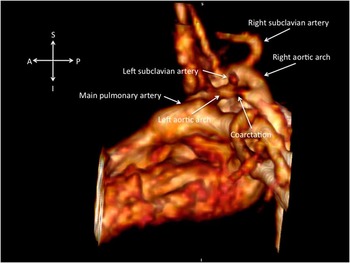
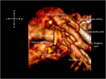
Cardiac magnetic resonance was carried out and demonstrated a dominant right arch that gave rise to the right common carotid artery and right subclavian artery. The hypoplastic left arch gave rise to the left common carotid artery and left subclavian artery with discrete narrowing of the segment between the left subclavian artery and the descending aorta. There was also an interrupted inferior vena cava with an azygous continuation to the right superior caval vein. At 4 months of age, the infant underwent successful repair of the vascular ring with division and ligation of the left arch (Figs 1 and 2).
Left lateral view of a volume-rendered gadolinium-enhanced 3D magnetic resonance angiogram demonstrating the coarctation of the hypoplastic left aortic arch with a discrete narrowing seen distal to the takeoff of the left subclavian artery. The right aortic arch is substantially larger with no evidence of obstruction. These images were obtained on a non-sedated patient free-breathing on room air.

Superior view of a volume-rendered gadolinium-enhanced 3D magnetic resonance angiogram demonstrating the vascular ring. Again, a discrete coratctaion is noted distal to the left subclavian artery. Note the very dilated azygous vein to the right superior caval vein just rightward of the right aortic arch.

Congenital diaphragmatic hernia is a life-threatening disease affecting 1/3000 live births. Its association with congenital heart disease has been reported as 11–21%Reference Wynn, Krishnan and Aspelund 1 ; this is the first reported case with a double aortic arch. Given the maternal morbid obesity and a postnatal finding of congenital diaphragmatic hernia, use of cardiac magnetic resonance in this patient was crucial in accurately delineating the cardiac anatomy.
Acknowledgements
None.
Financial Support
This research received no specific grant from any funding agency, commercial or not-for-profit sectors.
Conflicts of Interest
None.