The bi-directional cavo-pulmonary anastomosis, or Glenn anastomosis, is an intermediate step in the multi-stage palliation of single ventricle physiology in which the superior caval vein is sutured to the right pulmonary artery. Late thrombotic events in patients with a history of Glenn operation are rare and are more commonly seen as long-term complications of the Fontan palliation.Reference Imanaka, Takamoto, Murakami and Kaneko 1 – Reference Idorn, Jensen and Juul 3 At present, there are no standard recommendations for anti-thrombotic therapy after a Glenn anastomosis.Reference Monagle, Chan and Goldenberg 4 We report a case of a superior caval vein thrombus in an adult with Glenn anastomosis and left pulmonary artery atresia.
Case report
A 28-year-old woman with history of tricuspid atresia type II-B and left pulmonary artery atresia presented with progressive lower extremity and facial oedema refractory to diuretic therapy. Her past medical history consisted of a right-sided modified Blalock–Taussig shunt as a neonate and a left-sided modified Blalock–Taussig shunt at 1 year of age in an effort to re-establish flow to the left lung. At the age of 4, her left-sided modified Blalock–Taussig shunt was taken down and an attempt to repair her left pulmonary artery was undertaken; however, her left pulmonary artery became atretic. At 5 years of age, she underwent a completion Fontan operation to the right lung that was complicated by acute failure and subsequent low cardiac output syndrome. Therefore, the Fontan anastomosis was emergently taken down and she was left with a single-lung “Classic Glenn” anastomosis to the right pulmonary artery.
Despite her unusual physiology, she fared reasonably well as an outpatient at our Adult Congenital Heart Disease clinic until she began to have worsening bilateral lower extremity oedema. Although she was initially managed with increased diuretic therapy, her symptoms worsened.
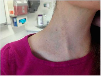
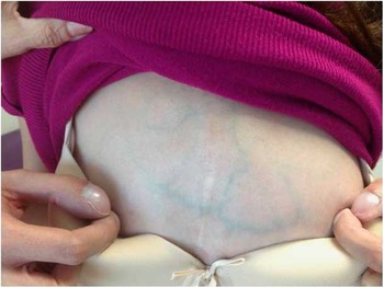
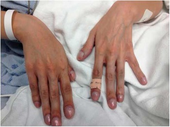
She complained of facial fullness with worsening desaturation to 67%, and she sought an urgent clinic visit. On physical examination, she was tachycardic, hypotensive, and more desaturated than her baseline. She had facial plethora, prominent neck veins, engorged subcutaneous chest wall veins, and swollen, cyanotic hands (Figs 1–3).
Distended jugular vein.

Engorged subcutaneous chest wall veins and finger clubbing.

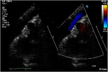
Echocardiographic evidence of the SVC thrombus with and without doppler ultrasound. SVC=superior caval vein.

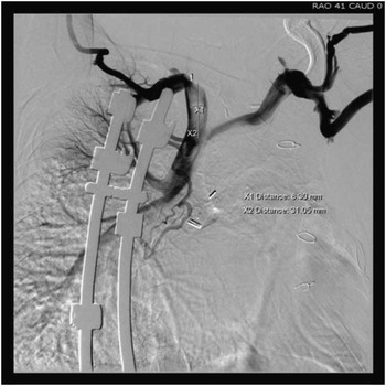
Echocardiography showed a large 9×3 mm nearly occlusive thrombus within the superior caval vein near the bifurcation of the left and right innominate vein (Fig 4a and b). The left ventricle appeared dilated with mildly depressed systolic function (calculated ejection fraction of 43%) in the presence of significant mitral regurgitation. She received one dose of enoxaparin (1 mg/kg) subcutaneously at the clinic. She was on an oral contraceptive on admission and it was discontinued. Her haemoglobin and haematocrit levels were 18.4 g/dl and 62.8%, respectively. An emergent right subclavian venogram was performed to confirm the location and size of the thrombus (Fig 5). Given the potential for complete occlusion of her only pulmonary artery or distal embolisation through the Glenn to the single functional lung, the patient was treated as having a potentially life-threatening pulmonary embolus and was given tissue plasminogen activator for thrombolysis (0.5 mg/kg/hour over 24 hours without bolus) and was later started on a heparin drip (20 units/kg/hour) as an inpatient. Her facial and upper extremity swelling greatly improved after thrombolytic therapy and her saturation returned to her baseline of 87%. Her lower extremity oedema entirely resolved by hospital discharge. She was later sent home on warfarin and was instructed to discontinue her oral contraceptive medication. As this was her first thrombotic event, and her oral contraceptive pills were a likely culprit, we did not pursue a hypercoaguable work-up at the time. Follow-up echocardiogram 1 month later showed complete resolution of the clot (Fig 6).
( a and b ) Emergent venogram obtained demonstrating occlusion due to thrombus (arrow).

Echocardiographic evidence of clot resolution 4 weeks later.

Fluoroscopic image illustrating superior caval vein thrombus near the Glenn anastamosis site.

Discussion
This is the first report of a superior caval vein thrombus in a patient with a single-lung or Classic Glenn anastomosis. Owing to her single pulmonary artery physiology, the superior caval vein thrombus was addressed as a pulmonary embolus and considered potentially fatal. Early or late thrombosis is not commonly seen after Classic or Bi-directional Glenn procedures and is more commonly seen after Fontan palliation. The incidence of thrombus formation after Fontan is highly variable, ranging from 8–49% in retrospective cohorts.Reference Idorn, Jensen and Juul 3 , Reference Khairy, Fernandes and Mayer 5 – Reference Johnson, Lindsay and Day 7 Phasic, non-pulsatile blood flow, artificial mural surfaces, and abnormalities in both pro-coagulant and anti-coagulant factors such as Factor VIII, X, prothrombin, antithrombin, and proteins C and S are likely responsible for the rates of thrombosis in Fontan patients.Reference Jahangiri, Kreutzer, Zurakowski, Bacha and Jonas 8 , Reference Cromme-Dijkhuis, Henkens, Bijleveld, Hillege, Bom and van der Meer 9 Furthermore, ex-vivo data suggest increased platelet reactivity and aggregation in Glenn patients, despite their low platelet counts.Reference Ravn, Hjortdal and Stenbog 10 Of note, Odegard et alReference Odegard, McGowan and DiNardo 11 demonstrated that similar haemodynamic abnormalities precede the Fontan and are present in patients after the Glenn procedure when compared with age-matched controls; however, outcomes analysis of 227 patients who underwent Glenn procedures did not report any thrombotic events.Reference Alsoufi, Manlhiot and Awan 12 Our patient was unusual as she was left with a Classic Glenn anastomosis for nearly 25 years, as opposed to most patients who usually have a Glenn for <5 years. She was, therefore, in a chronically low-venous flow state – passive venous return – and developed physiological changes from cyanosis such as secondary polycythaemia. These two states may have contributed to her thrombus formation compounded by her oral contraceptive medications.
Forbes et alReference Forbes, Rosenthal, Reul, Ott and Feltes 13 described a case series of six patients who developed life-threatening superior caval vein or pulmonary thrombosis following a Glenn, and they identified the following risk factors: bilateral superior caval vein, female sex, older age, decreased pulmonary artery size, increased right atrial pressure, increased systemic ventricular end-diastolic pressure, prolonged aortic cross-clamp time, poor post-operative ventricular function, and elevated post-operative superior caval vein pressure. Among them, one patient, who developed a thrombus following endovascular stent implantation for severe left pulmonary artery stenosis, was treated with balloon dilation of the thrombus and streptokinase.Reference Forbes, Rosenthal, Reul, Ott and Feltes 13 Our patient’s single-lung physiology was a major factor in deciding whether to use thrombolytic therapy or to proceed with thrombectomy. Catheter or surgical thrombectomy are typically reserved for cases of massive pulmonary embolus with cardiac arrest, cardiogenic shock with peripheral hypotension, or echocardiographic findings indicative of right ventricular overload and/or pulmonary hypertension,Reference Kasper, Konstantinides and Geibel 14 none of which our patient demonstrated. Before success with superior caval vein, transcatheter thrombectomy has been reported in a child with Fontan palliation and confluent pulmonary arteries.Reference Gossett, Rocchini and Armstrong 15 Although distal embolisation can theoretically occur even with systemic thrombolytic therapy, we believed that the risk was much greater with transcatheter thrombectomy from mechanical dislodgement. Given the potential catastrophic consequences in our patient with only a single functional lung, we opted for the less-invasive approach and elected to use thrombolytics followed by anticoagulation.
Conclusion
Following Glenn procedures, patients demonstrate an imbalance in pro- and anti-coagulant factors that can lead to thrombus formation exacerbated by traditional risk factors such as oral contraceptive drugs. The complexity of each patient’s physiology necessitates an individualised approach when weighing the risks and benefits between thrombolytic therapy and thrombectomy. We report successful thrombolytic administration in a high-risk Classic Glenn patient with left pulmonary artery atresia who developed a superior caval vein thrombus.
Acknowledgement
The authors acknowledge Allison Mahlstedt, RPAC, for her assistance in the preparation of this manuscript.
Financial Support
This research received no specific grant from any funding agency, commercial, or not-for-profit sectors.
Conflicts of Interest
None.
Ethical Standards
No human or animal experimentation was involved.