Introduction
Cystic echinococcosis (CE) is a parasitic disease of humans and some animals, caused by the metacestode of the tapeworm Echinococcus granulosus. Humans may be infected accidentally through ingestion of vegetables or any other foodstuffs contaminated by the ova of the parasite. The infection can also be acquired by direct contact with the definitive hosts, carnivorous animals, having adult E. granulosus in their gut (Pakala et al., Reference Pakala, Molina and Wu2016; Thompson, Reference Thompson2017). CE has a worldwide distribution and is a major public health problem in some regions (Craig et al., Reference Craig, McManus and Lightowlers2007).
The phenotypic variations in E. granulosus were described in the early 1970s. The development and maturation of E. granulosus from sheep origin in culture media have been demonstrated, whereas isolates of the parasite from horses failed to develop or mature (McManus, Reference McManus2013). Ten genotypes of E. granulosus have been described by mitochondrial DNA sequence. Based on phenotypic and genetic characteristics, that which was formerly known as a single species E. granulosus sensu lato with ten genotypes is now a cluster of species: E. granulosus sensu stricto G1–G3 (G1, sheep strain; G2, Tasmanian sheep strain; G3, buffalo strain) (Romig et al., Reference Romig, Ebi and Wassermann2015), E. equinus (G4, horse strain), E. ortleppi (G5, cattle strain), E. intermedius (G6, camel strain; G7, pig strain), E. canadensis (G8 and G10, cervid strains) and E. felidis (Thompson, Reference Thompson2017).
The metacestode of E. granulosus sensu lato, which forms in intermediate hosts, is a complex structure covered outwardly by a laminated layer and a germinal layer. A host-derived layer, the fibrous layer, encircles the hydatid cyst (Tamarozzi et al., Reference Tamarozzi, Mariconti, Neumayr and Brunetti2016; Thompson, Reference Thompson2017). The laminated layer is a non-cellular multi-laminated structure, which is synthesized initially by the developing hexacanth larva and then by the germinal layer. It is made up of glycosylated glycoproteins and is crucial in the metacestode's immune evasion in the intermediate host (Díaz et al., Reference Díaz, Casaravilla, Allen, Sim and Ferreira2011a, Reference Díaz, Casaravilla, Irigoín, Lin, Previato and Ferreirab; Tamarozzi et al., Reference Tamarozzi, Mariconti, Neumayr and Brunetti2016).
Untreated hydatid cysts generally pass through several stages, active to inactive (Tamarozzi et al., Reference Tamarozzi, Mariconti, Neumayr and Brunetti2016), or tend to be stable over time (Frider et al., Reference Frider, Larrieu and Odriozola1999). Two essential immunological mechanisms – immune evasion and immune modulation – are highlighted for the often long-lasting and asymptomatic CE. There are also some degrees of protective immunity to reinfection (Lightowlers, Reference Lightowlers2010; Zhang et al., Reference Zhang, Wen, Li, Lin and McManus2012; Tamarozzi et al., Reference Tamarozzi, Mariconti, Neumayr and Brunetti2016). Both humoral and cellular immune responses are involved against hydatid cysts in intermediate hosts (Kharebov et al., Reference Kharebov, Nahmias and El-On1997), yet 30–40% of patients with CE are antibody-negative. This suggests that the proliferation and activity of B cells may be inhibited and/or regulated by E. granulosus antigens. It is not known how these antigens target and regulate the immune mechanisms of the host (Zhang et al., Reference Zhang, Wen, Li, Lin and McManus2012).
There are variations in the interaction of intermediate hosts and hydatid cysts, and the causes of these variations are not completely known. However, the parasite genotype and host-related factors are suggested to be involved. Furthermore, it has been demonstrated that in naturally infected sheep, antibody response may be accompanied by a cellular inflammatory response involving neutrophils, lymphocytes, eosinophils and macrophages (Rickard & Williams, Reference Rickard and Williams1982). The same cellular immune response has also been reported for experimental secondary infections in mice (Rogan et al., Reference Rogan, Bodell and Craig2015).
Most of our knowledge about the immunology of CE is based on experimental studies and observations in animals, yet we know little about the local cellular immune response in human infections. Studies on human subjects usually represent only single-point observations and we do not really know much about how immune response varies within humans (Riganò et al., Reference Riganò, Profumo, Buttari, Delunardo, Ortona, Margutti, Teggi and Siracusano2004; Rogan et al., Reference Rogan, Bodell and Craig2015). We have even less information about the local immune response to hydatid cysts, especially regarding the lymphocyte subtypes involved in local responses. There have been three studies on the local proliferation of different subtypes of lymphocytes in hydatid cysts: one on ovine CE (Vismarra et al., Reference Vismarra, Mangia and Passeri2015), one on bovine cases (Sakamoto & Cabrera, Reference Sakamoto and Cabrera2003) and one on hydatid cysts of the liver in humans (Vatankhah et al., Reference Vatankhah, Halász, Piurkó, Barbai, Rásó and Tímár2015). Thus in the present study we aimed to evaluate and explain the local immune cell infiltration pattern using CD3 as pan T cell, CD19 as pan B cell, CD4 as T helper cell, CD8 as cytotoxic T cell (CTL), CD56/NCAM as natural killer (NK) cell, CD68 as macrophage, Foxp3 (forkhead box P3) as regulatory T cell (Treg) and Ki-67 as proliferation biomarkers in human CE. The similarities in antigens of hydatid cysts and some cancers have been reported (Darani & Yousefi, Reference Darani and Yousefi2012), and thus we also investigated the pattern of cellular immune response to hydatid cysts using the above-mentioned biomarkers, and compared it with samples of hepatocellular carcinoma (HC) and chronic hepatitis (CH).
Materials and methods
Sample collection and fixation
We collected 44 tissue samples from livers, five from lungs and one from a kidney, from surgically removed human hydatid cysts (containing the fibrous layer and the host-tissue reaction site) from patients referred to Al-Zahra Hospital, Isfahan, Iran, during April 2015–March 2017. The isolated hydatid cysts belonged to E. granulosus G1 (43 samples) and G3 (3 samples), and E. intermedius G6 (4 samples). The genotypes of the collected samples have been defined previously. The ultrasound features of the samples were also defined by the method described by the World Health Organization Informal Working Group on Echinococcosis (WHO-IGWE) (Wuestenberg et al., Reference Wuestenberg, Gruener, Oeztuerk, Mason, Haenle, Graeter, Akinli, Kern and Kratzer2014). Samples from paraffin-embedded blocks of four hepatocellular carcinoma and four non-autoimmune chronic hepatitis tissues were included in the study for comparison. The tissue samples were cut into small pieces and kept in 10% natural buffered formalin for 48–72 hours. The paraffin-embedded blocks were prepared from the collected tissues and stored in a cool, dry place until examination. The fertility of the hydatid cysts was also determined.
Slide preparation
Using a microtome, 4 μm sections were prepared, placed on Superfrost Plus positively charged slides (Thermo Scientific, Waltham, Massachusetts, USA) and incubated at 37°C overnight. Routine eosin and haematoxylin staining was also performed for histopathological analysis of the eosinophils.
Immunohistochemistry (IHC)
1. De-waxing and hydrating
The tissue sections were deparaffinized in three changes of xylene for 5 minutes each and then hydrated through a graded series of ethanol (100, 96, 70, 50%) and deionized water, each for 5 minutes. The hydrated tissue sections were incubated in phosphate-buffered saline (PBS), pH 7.0 (0.02 mol/l sodium phosphate buffer, 0.15 mol/l NaCl; Dako, Denmark), for 5 minutes.
2. Antigen retrieval
A target retrieval solution, Tris/EDTA buffer, pH 9 (Dako, Denmark), was used for heat-mediated antigen retrieval. The slides were then immersed in the 1X solution, heated to the boiling threshold (95°C) and kept at this temperature for 30 minutes (the buffer was boiled very slowly), and then cooled for 15 minutes at room temperature (RT).
3. Endogenous peroxidase blocking
After the antigen retrieval, the slides were incubated in wash buffer for 5 minutes (0.05 mol/l Tris/HCl, 0.15 mol/l NaCl, 0.05% Tween 20, pH 7.6; Dako, Denmark). To block endogenous peroxidase the slides were incubated in 3% hydrogen peroxide (H2O2 30%; Merck, Germany) for 10 minutes, washed for 1 minute in two changes of deionized water to remove excess H2O2, and then incubated in PBS for 5 minutes.
4. Incubation with the primary antibody
Water-repelling circles were drawn around the tissue sections using a Dako Pen (Dako, Denmark), and primary antibodies were added to the sections. The following biomarkers were used: polyclonal rabbit anti-human CD3 (Dako, Denmark), monoclonal mouse anti-human CD4 (clone 4B12; Dako, Denmark), monoclonal mouse anti-human CD8 (clone C8/144B; Dako, Denmark), monoclonal mouse anti-human CD19 (clone LE-CD19; Dako, Denmark), mouse monoclonal anti-human CD56/NCAM-1 (clone 123C3.D5; Diagnostic Biosystems, USA), monoclonal mouse anti-human CD68 (clone KP1; Dako, Denmark), monoclonal mouse anti-human Foxp3 (clone 236A/E7; eBioscience, USA) and monoclonal mouse anti-human Ki-67 (clone MIB-1; Dako, Denmark). Except for anti-human Foxp3, all antibodies were prediluted (ready to use) and incubated for 20 minutes in a humidity chamber. Anti-human Foxp3 was diluted in TTBS (Tris-buffered saline or TBS and Tween 20), optimized in 5 μg/ml concentration and incubated for 120 minutes in a humidity chamber. After incubation with the primary antibody, the slides were washed in two changes of wash buffer for 7 minutes each and then washed once in PBS for 7 minutes.
5. Secondary antibody and visualization
EnVision Detection System, Mouse/Rabbit, Peroxidase/DAB (3,3′-diaminobenzidine) (Dako, Denmark) was used for visualization (in the ready-to-use/prediluted format). The slides were incubated with the secondary antibody for 30 minutes. The washing step was repeated as previously described. DAB was diluted (25 μl DAB in 1000 μl of DAB buffer) and used fresh. Slides were incubated with the diluted DAB for 5 minutes at RT to develop the enzymatic reaction. Then slides were washed with water, stained with haematoxylin for 1–2 minutes, dehydrated, cleared in xylene and mounted in Entellan (Merck Millipore, USA).
Negative and positive controls
Negative and positive control tissues were stained with each IHC run. Normal human tonsil tissue was used as the positive control for all the biomarkers studied. Biliary epithelial layer and Kupffer cells in the liver were also used as additional positive controls for CD56 and CD68, respectively (see supplementary fig. S5). The same slides incubated with PBS instead of the primary antibody were used as the negative controls.
Data analysis
Slides were studied under a microscope, and micrographs were taken from the stained tissue sections using 40X objective, considering all the microscopic fields with immune cell infiltration. Then the positive and negative inflammatory cells for CD4, CD8, CD3, CD19, CD56, CD68, Ki-67 and Foxp3 were counted using ImageJ software (https://imagej.nih.gov/ij/). The mean numbers of cells counted were then reported as a ratio of the inflammatory cells, using the following formula: positive cells/total cells × 100. The observatory data were reported as seen, and the counted cells were compared in groups and analysed by one-way analysis of variance (ANOVA), regression and Student's t-tests using SPSS v. 22.0 (IBM Corp., Armonk, USA).
Results
In this investigation we analysed 50 tissue samples of isolates of human hydatid cysts (44 from livers, 5 from lungs and one from kidney). To compare the inflammatory cell distribution in the host-tissue reaction site of liver hydatid cysts with other diseases, four samples from patients with hepatocellular carcinoma and four from patients with chronic hepatitis were also studied. The mean age of patients with hydatid cysts, hepatocellular carcinoma and chronic hepatitis was 40 ± 14, 65 ± 13 and 26 ± 12 years, respectively. All hydatid cysts belonged to the CE2 category and were also fertile.
Considering the biomarkers, there were positive cells for CD3, CD19, CD8, CD4, CD68 and Ki-67 in all cases, yet in some there were no positive cells for CD56 (seven CE patients) and Foxp3 (one CE and one chronic hepatitis), and there were no eosinophils in non-CE samples. The studied cells were arranged in distinct ways in the various organs, as described below.
Pattern of inflammatory cell distribution in the host-tissue reaction site of liver hydatid cysts
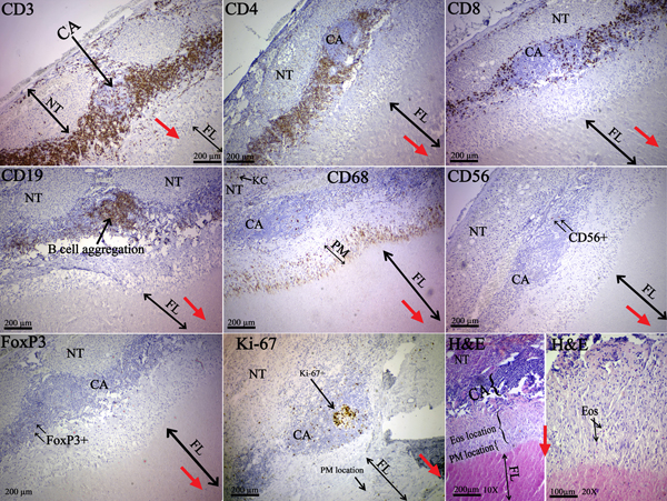
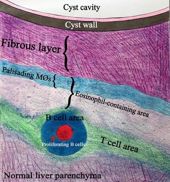
In the host-tissue reaction site of liver hydatid cysts, a general pattern of inflammatory cell distribution was observed. This pattern consisted outwardly of a pack of the fibrous element (adjacent to the laminated layer of the parasite), a layer of palisading macrophages, an eosinophil-containing layer, a layer of aggregated lymphocytes, and normal host tissue (fig. 1; supplementary figs S1 and S2). In the aggregated lymphocyte layer, CD3+ T cells, CD4+ T helper cells, and CD19+ B cells were dominant (table 1). However, Foxp3+ regulatory T cells (Tregs) were not aggregated and there were few CD56+ natural killer (NK) cells. The CD8+ CTL population was less dense than the CD4+ Th cell population and was scattered all around the inflammation site. CD68+ macrophages were mostly around the aggregation site, with a few located inside the site, and were cut into the fibrous layer as palisading macrophages connecting fibrous tissue to the inflammatory site (fig. 1). The fibrous layer was thick, separating the inflammatory site from the parasite (mean thickness 551 ± 224 μm). The Ki-67+ proliferating cells tended to cluster in the core of lymphocyte aggregation where CD19+ B cells were localized (fig. 1, micrographs CD19 and Ki-67), and there were very few in the palisading macrophage location. This pattern of immune cells around the liver hydatid cyst was consistent among nearly all individuals.
The studied cell types in liver hydatid cysts. The red arrows point towards the location of the parasite. A thick fibrous layer separates the parasite from the host parenchyma, and a considerable number of eosinophils and macrophages are visible at the border of the fibrous layer and the inflammatory site (H&E and CD68). B-cell clusters are mostly present within the granulomas. Ki-67+ cells tend to cluster at the centre of the granuloma (micrograph Ki-67), where the B-cell follicle is present. The observed B cells inside the granuloma may indicate germinal centres. There are few proliferating cells in the area where palisading macrophages are present (CD68 and Ki-67). All of the micrographs are of the same individual. NT, normal tissue; CA, immune cell aggregation; FL, fibrous layer; PM, palisading macrophages; Eos, eosinophil; KC, Kupffer cell. CD3 (10X), CD4 (10X), CD8 (10X), CD19 (10X), CD68 (10X), CD56 (10X), Foxp3 (10X), Ki-67 (10X), and H&E (10X and 20X).

Mean numbers of cells in liver tissue samples infected with cystic echinococcosis, hepatocellular carcinoma or chronic hepatitis; numbers for CD3, CD4, CD8, CD19, CD56, Foxp3 and Ki67 are relative numbers (0–100) of positive cells per all immune cells, and numbers for CD68 and eosinophils are numbers per high-power microscopic field. The ratios of Foxp3/CD4, CD4/CD8 and CD3/CD19 are also shown.

No significant numbers of CD56+ cells were detected.
Pattern of inflammatory cell distribution in the host-tissue reaction site of lung hydatid cysts
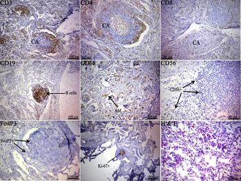
No host-derived fibrous capsule was observed in lung hydatid cysts. Inflammatory cells were observed in most areas of the lung samples, and mostly encompassed all the cells mentioned above in relation to the liver. CD19+ B cells mostly clustered at the core of the immune cell aggregation (fig. 2; supplementary fig. S3), and CD3+ T cells were around and inside the aggregated and non-aggregated sites. As in the liver, a significant number of CD4+ Th cells were observed quite close together, whereas the CD8+ CTLs were less populous and were scattered all around the inflammation site. The CD68+ macrophage arrangement in lungs differed from that in the liver. They were scattered all around the immune cell aggregated sites, with a few inside the sites, and were present in a considerable number inside the alveoli and small airways. CD56+ NK cells were rare, and present in low numbers in the host-tissue reaction site of four out of five samples of lung hydatid cyst. Eosinophils were predominantly outside the lymphocyte aggregation sites and were present in considerable number in all the lung tissues. Similar to the liver samples, the Ki-67+ proliferating cells were observed in most of the same locations as the CD19+ cells. The Foxp3+ Tregs were sparsely scattered in the inflammatory sites (supplementary fig. S3). The aggregation of immune cells around the lung hydatid cyst was almost the same in nearly all individuals.
The studied cell types in a human subject with a lung hydatid cyst. There was no apparent fibrous layer, and massive inflammatory cell aggregation all over the tissue, thus the location of the hydatid cyst could not be determined. No palisading macrophages are visible. Considerable numbers of eosinophils are visible around the immune cell aggregation sites (H&E). All of the micrographs are of the same individual. CA, immune cell aggregation; Eos, eosinophil; AM, alveolar macrophages. CD3 (10X), CD4 (10X), CD8 (10X), CD19 (10X), CD68 (20X), CD56 (20X), Foxp3 (20X), Ki-67 (10X), and H&E (40X).

Pattern of inflammatory cell distribution in the host-tissue reaction site of a kidney hydatid cyst
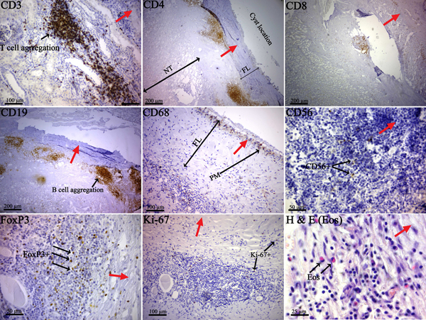
Only one hydatid cyst of the kidney was studied, and the results showed similarity between hydatid cysts of the liver and kidney. As observed in the liver cysts, the kidney cyst had a fibrous layer. Unlike in the liver, the palisading macrophages in the kidney cyst were lined closer to the cyst (fig. 3). The inflammatory cell infiltration was similar to the liver.
The studied cell types in a human subject with a kidney hydatid cyst. The red arrows point towards the location of the parasite. There is a fibrous layer between the cyst and the host-tissue reaction site, separating the cyst from normal tissue. As in the liver cysts, the palisading macrophages (PM) and eosinophils are located between the immune cell aggregation site and the cyst. Lymphocyte proliferation (Ki-67) is not as pronounced as in the lung and liver samples; however, there are few Ki-67+ cells in the location where palisading macrophages are present (micrographs Ki-67 and CD68). The micrographs are all from the single individual studied. NT, normal tissue; FL, fibrous layer; PM, palisading macrophages; Eos, eosinophil. CD3 (20X), CD4 (10X), CD8 (10X), CD19 (10X), CD68 (20X), CD56 (40X), Foxp3 (40X), Ki-67 (20X), and H&E (40X).

Pattern of inflammatory cell distribution in patients with hepatocellular carcinoma and chronic hepatitis
HC and CH tissue samples were studied and compared to the inflammatory site of liver hydatid cysts only. The immune cell infiltration was not as dense or intense as in the host-tissue reaction site of hydatid cysts. Considering the studied cells, the arrangement in HC and CH was visually similar to that in CE except that in non-CE samples there was no eosinophil, or clustering of Ki-67+ cells as in liver and lung hydatid cysts. The distinct pattern of cell layers in liver hydatid cysts was limited to the host-tissue reaction site of patients with CE and was not seen in HC and CH cases. CD56+ NK cells were rare, and present in low numbers in all the CH and HC cases (supplementary fig. S4).
Quantitative analysis
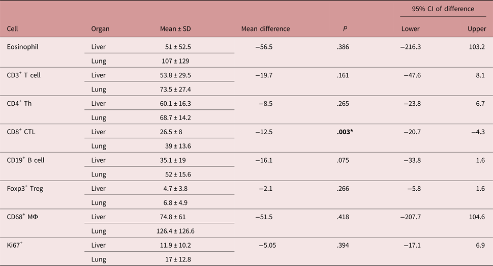
The mean positive cells for CD3, CD4, CD8, CD19, CD56, Foxp3 and Ki67 are presented as a ratio of the inflammatory cells, and CD68+ macrophages and eosinophils are presented as number per high-power microscopic field. The mean numbers of cells are available in table 1.
Multiple comparisons of the mean positive cells showed no significant difference between CH, HC and liver hydatid cyst inflammatory sites (supplementary table S1). There was no significant difference between the number of eosinophils, CD3+ T cells, CD4+ Th cells, CD8+ CTLs, CD19+ B cells, CD56+ NK cells, CD68+ macrophages and Foxp3+ Tregs with the genotype of the parasite in CE cases (supplementary table S2). Considering the cell numbers in different organs of the patients with CE, the CD8+ CTL population was larger in the lungs compared to the liver (P = 0.003) (table 2). There was no significant difference in the mean cell count between males and females.
Mean numbers of cells studied in liver and lung tissue from humans with cystic echinococcosis.

*Significant at P < 0.05
No significant numbers of CD56+ cells were detected.
MΦ, macrophage
In CE patients, the numbers of CD19+ B cells were significantly and positively correlated with the numbers of Ki-67+ proliferating cells (R2 = 0.183, P = 0.032). This phenomenon is also true for CD4+ Th cells with CD19+ B cells (R2 = 0.153, P = 0.005), and CD8+ CTLs with Foxp3+ Tregs (R2 = 0.81, P = 0.045). However, the numbers of Ki-67+ proliferating cells were significantly and negatively correlated with the age of the patient (R2 = 0.165, P = 0.044) (supplementary fig. S5 and table S3). No other significant correlation was observed in the other studied diseases.
Discussion
In this investigation a pattern of immune cell aggregation in the host-tissue reaction sites of human isolates of the hepatic hydatid cysts was observed (summarized in fig. 4). The local immune response against hydatid cysts in humans is barely known. The inflammatory cell aggregation and composition around the hydatid lesion in humans may be driven by specific mechanisms that are likely to be specific to the affected organ and may be activated directly in response to parasite antigens; however, this has not been completely described (Vatankhah et al., Reference Vatankhah, Halász, Piurkó, Barbai, Rásó and Tímár2015). In this study, localization of the immune cell infiltration was described and analysed using IHC. Given the inadequate numbers of G3 and G6 genotypes, they were excluded from further analysis and discussion. Further investigations with a higher number of genotypes other than G1 are recommended.
Diagram of the observed immune cells and their approximate location, based on the findings of this study.

The results showed a predominant CD3+ T cell population, followed by CD19+ B cells in the local host reaction sites in hydatid cysts. The CD4+ Th cell population was 2.3 times larger than that of CD8+ CTL. In general the CD3+ T cell population was observed to be 2.1 times larger than that of CD19+ B cells in CE cases. Foxp3+ cells comprised c. 8% of CD4+ T helper cells. In agreement with our work, a higher T cell population in the host-tissue reaction site of hydatid cysts was previously reported for humans and sheep infected with CE (Vatankhah et al., Reference Vatankhah, Halász, Piurkó, Barbai, Rásó and Tímár2015; Vismarra et al., Reference Vismarra, Mangia and Passeri2015). However, Vatankhah et al. (Reference Vatankhah, Halász, Piurkó, Barbai, Rásó and Tímár2015) reported different CD8+ CTL populations in patients with CE, CH and steatosis (SH), and proposed it as a prognostic value in CD8-dependent T cell activity in some chronic liver inflammations with different etiology (Vatankhah et al., Reference Vatankhah, Halász, Piurkó, Barbai, Rásó and Tímár2015), whereas such difference was not observed in this study.
B cells are important immune cells that produce immunoglobulin; they are also regarded as antigen-presenting cells (APCs) and are important in humoral immune response (LeBien & Tedder, Reference LeBien and Tedder2008). In this study a considerable proportion of inflammatory cells were CD19+ B cells, and among these, plasma cells (not counted) were also observed in histopathological observation, which shows the importance of CD19+ B cells in the local immune response in CE patients. Only a fraction of people with CE (10–91.7%.) show positive levels of antibodies against hydatid cysts, using commercially available immunoassay kits (Zhang et al., Reference Zhang, Wen, Li, Lin and McManus2012). Moreover, most of the proliferating cells tend to cluster in the B cell area in CE patients, and the numbers of CD19+ B cells were significantly and positively correlated with the numbers of Ki-67+ cells, indicating that B cell proliferation is active in the area. A similar phenomenon was reported in the granulomas of Mycobacterium tuberculosis infection in non-human primates (Phuah et al., Reference Phuah, Mattila, Lin and Flynn2012). It has been reported that the B cells at the centre of mycobacterial granuloma are probably suggestive of germinal centres (Ulrichs et al., Reference Ulrichs, Kosmiadi, Trusov, Jörg, Pradl, Titukhina, Mishenko, Gushina and Kaufmann2004; Zhang et al., Reference Zhang, Wang and Graner2011; Phuah et al., Reference Phuah, Mattila, Lin and Flynn2012). Considering the clustering and proliferation of B cells, our results show similarities between mycobacterial granuloma and hydatid cyst host reaction. However, there is no similar study indicating the mentioned results in human hydatid cysts. Furthermore, the localization of B and Ki-67+ cells is reminiscent of their clustering in normal human tonsil tissue (supplementary fig. S6). The presence of CD19+ B cells in great numbers, and considering their proliferation in the host-tissue reaction site of human hydatid cysts, shows a probable strong humoral response against the parasite, but the reason that the immunological diagnosis of CE is not sensitive and specific is not completely clear.
Eosinophils were observed in considerable numbers only in CE patients. Eosinophils have been considered as the end-stage cells involved in host protection against multicellular parasites. These leukocytes are now known to be involved in the initiation and even propagation of various inflammatory responses, as well as innate and adaptive immunity modulation (Rothenberg & Hogan, Reference Rothenberg and Hogan2006).
A large number of macrophages were observed in all of the studied CE patients, and notably the macrophages were seen as the first-line combat against hydatid cysts. CD68+ macrophages were also observed inside and around the lymphocyte aggregation site, and in almost all cases they lined like a guard just behind the fibrous capsule (the palisading macrophages). Considering these cells as important APCs, they may have a significant role in processing and presenting the E. granulosus antigens to the other immune cells, such as T cells, which are also present in great number in the lesion site.
In this study, CD56+ NK cells were the least populous. In fact, NK cells were defined as effector lymphocytes of innate immunity with cytolytic functions. NK cells are now known to express activating and inhibitory receptors that are calibrated for self-tolerance while having efficacy against infections such as viral infections and tumours. It is suggested that NK cells can also be a form of immunologic memory for antigens, thus exerting complex biological functions that are properties of innate and also adaptive immunity (Vivier et al., Reference Vivier, Raulet, Moretta, Caligiuri, Zitvogel, Lanier, Yokoyama and Ugolini2011). It was previously recommended that CD56+ NK cells be investigated in patients with CE (Vatankhah et al., Reference Vatankhah, Halász, Piurkó, Barbai, Rásó and Tímár2015) and we have done so comprehensively in this study. The results showed that in the local inflammatory cell aggregation sites of hydatid cysts the CD56+ NK cells comprised the smallest proportion of the inflammatory cells (0.6%). The highest quantity of these cells was observed in hepatic tumours (1.2%). Moreover, CD56+ NK cells were absent in seven CE patients. The role of CD3+CD56+ NK T cells in the hydatid-induced local immune response is presumed (Vatankhah et al., Reference Vatankhah, Halász, Piurkó, Barbai, Rásó and Tímár2015). Our findings do not support this hypothesis; however, the function of these cells was not studied in the present work and we recommend further investigation of the function of NK cells in the immune response against hydatid cysts.
APCs are a group of cells of the immune system that mediate the immune response by processing and presenting antigens for identification by T cells. Macrophages, B cells, dendritic cells and Langerhans cells are examples of APCs (Hamilos, Reference Hamilos1989). In this study, macrophages were lined behind the fibrous capsule of hydatid cysts and were in a considerable quantity. They may be the most important APCs in the immune response to established human hydatid cysts.
Foxp3 protein is an accepted marker for Tregs. These cells are a population of lymphocytes that regulate and suppress over-responses from other immune cells. Tregs are able to control a variety of other immune cell subsets, including activated effector cells, and inhibit APCs, B cells, NK cells and innate immunity (Gliwiński et al., Reference Gliwiński, Iwaszkiewicz-Grześ and Trzonkowski2017). Foxp3+ Tregs were absent in one CE sample, yet 5.05% of inflammatory cells of 49 CE patients were Foxp3+ Tregs, which is a considerable number. Studies have shown that E. granulosus modulates anti-parasite immune responses in human hosts and this may be the reason the parasite can persist for a long time in intermediate hosts. Furthermore, the probable immune-suppressing role of TGF-β and Tregs has been shown in E. multilocularis infection in animal models (Siracusano et al., Reference Siracusano, Delunardo, Teggi and Ortona2012). Based on the results of the present study, Tregs may play an immunomodulatory role in human CE. Vatankhah et al. (Reference Vatankhah, Halász, Piurkó, Barbai, Rásó and Tímár2015) reported almost similar results regarding Foxp3 expressing Tregs in CE (Vatankhah et al., Reference Vatankhah, Halász, Piurkó, Barbai, Rásó and Tímár2015). It is accepted in the literature that the Th2 response probably favours the establishment of the parasite in an intermediate host, as opposed to a parasite-killing Th1 response. However, often a mixed Th1/Th2 response is present in infected intermediate hosts, and regulatory mechanisms may control the Th1 and Th2 parasite-killing effector mechanisms. For example, Tregs have been reported to increase locally in the first 5 days post protoscolex injection in an animal model (Mourglia-Ettlin et al., Reference Mourglia-Ettlin, Marqués, Chabalgoity and Dematteis2011; Tamarozzi et al., Reference Tamarozzi, Mariconti, Neumayr and Brunetti2016). Echinococcus multilocularis-infected mice with Treg depletion have been shown to exhibit a lower parasite load and an enhanced Th1/Th17 polarization (Wang et al., Reference Wang, Müller, Lin, Siffert, Vuitton, Wen and Gottstein2017).
Vatankhah et al. (Reference Vatankhah, Halász, Piurkó, Barbai, Rásó and Tímár2015) observed a significant difference in the distribution of CD1a expressing dendritic cells (between CE and CH, and CE and SH), α-SMA expressing myofibroblasts (between CE and CH, and CE and SH) and CD68 expressing macrophages (between CE and CH, and SH and CH). Despite the larger sample size in the present study, a significant difference was not observed in the studied cells (CD3, CD4, CD8, CD19, CD56, CD68, Foxp3 expressing cells) in the three diseases studied. The CD1a and α-SMA markers were not used in this study.
Vismarra et al. (Reference Vismarra, Mangia and Passeri2015) studied the lymphocyte populations surrounding established ovine hydatid cysts using the IHC method. They reported a higher number of CD3+ T cells compared to CD79+ B cells, in agreement with the findings of this study on human CE. Similarly, they also reported a fraction of infiltrating lymphocytes as Foxp3+ Tregs and suggested that established ovine cysts may be protected from aggressive immune response through the suppressive effect of Foxp3+ Tregs (Vismarra et al., Reference Vismarra, Mangia and Passeri2015).
Similar to our findings, Sakamoto & Cabrera (Reference Sakamoto and Cabrera2003) and Díaz et al. (Reference Diaz, Willis and Sim2000) reported a kind of macrophage epithelioid cell layer in bovine hydatid cysts (Rickard & Williams, Reference Rickard and Williams1982; Sakamoto & Cabrera, Reference Sakamoto and Cabrera2003). Sakamoto & Cabrera (Reference Sakamoto and Cabrera2003) also showed predominant CD8+ CTLs in most cases of progressive hydatid cysts. In these cases they observed a relatively small number of CD4+ Th cells. The eosinophil-mediated destruction of the laminated layer was also reported in regressive and involuted hydatid cysts (Sakamoto & Cabrera, Reference Sakamoto and Cabrera2003). Considering human CE, the infiltrating immune cell populations, especially T helpers and CTLs, are different from those in cattle. In humans, hydatid cysts persist for a long time, whereas in cattle, hydatid cysts are mostly reported to be sterile or degenerated and calcified (Rinaldi et al., Reference Rinaldi, Maurelli, Veneziano, Capuano, Perugini and Cringoli2008). In the present study, in CE patients the numbers of Foxp3+ Tregs were significantly and positively correlated with the numbers of CD8+ CTL.
In conclusion, we have shown a distinct pattern of inflammatory cells localized in the host-tissue reaction site of human hydatid cysts. The suppressive effects of regulatory T cells and a considerable number of proliferating B cells may be the cause of the persistence of hydatid cysts in human cases for a long period. Moreover, NK cells are present in low numbers in the host-tissue reaction site of human hydatid cysts. Further studies on the functions of these immune cells in human CE are recommended, using a greater number of different genotypes and other organs than the liver to figure out the possible role of each of these cells in the durability of the disease in humans, and possible immunotherapeutic strategies.
Supplementary material
To view supplementary material for this article, please visit https://doi.org/10.1017/S0022149X1800024X
Acknowledgments
The authors would like to acknowledge the Vice-chancellor of Research and Technology, Isfahan University of Medical Sciences, Isfahan, Iran, for the approval of this study. Fig. 4 was kindly produced by Maryam Sajadian. This paper is derived from a PhD thesis in parasitology (No. 394952).
Financial support
This study was supported financially by the Vice-chancellor of Research and Technology, Isfahan University of Medical Sciences, Isfahan, Iran.
Conflict of interest
None.
Ethical standards
This study was approved by the ethical committee of Isfahan University of Medical Sciences (No. 394952). No tissue sample was taken deliberately from patients for this study, and the samples used were derived from those sent to the histopathology lab.