Percutaneous closure of the persistently patent arterial duct is now well established for infants, older children, or adults.Reference Butera, De Rosa and Chessa1 To date, however, to the best of our knowledge, it has not been reported in neonates. Symptomatic preterm and term neonates with persistent patency of the arterial duct nonetheless, are known to have increased morbidity and mortality, albeit that closure of the duct in this group of patients is usually achieved pharmacologically by administration of inhibitors of cyclooxygenase,Reference Heymann, Rudolph and Silverman2 or by surgery. We report here successful transcatheter closure of a persistently patent arterial duct, this being achieved in a critically ill newborn with neonatal lupus erythematosus and multiorgan system failure, in whom the usual treatments were contraindicatied.
Case report
A male infant diagnosed prenatally with complete atrioventricular block was born at 38 weeks of gestation by elective caesarean section. His weight at birth was 2660 grams, with Apgar scores of 7, 9, and 9 at 1, 5, and 10 minutes, respectively. Physical examination was normal except for bradycardia at 75 beats per minute. An electrocardiogram confirmed the presence of complete atrioventricular block. On the 3rd day of life, he presented with poor peripheral perfusion, rectal bleeding, and respiratory distress. We treated this by administering fluids and providing cardiopulmonary support. During the following days, an exchange transfusion was required because of severe haemolytic anaemia, neutropenia, and thrombocytopenia. He became anuric, and continuous veno-venous haemodiafiltration was started on the 8th day of life. He was referred to our hospital 13 days after birth. On admission to our hospital, there was abdominal distension, with air in the peritoneal cavity. An urgent laparotomy revealed a caecal perforation, so an ileostomy was performed. Echocardiographic assessment on the same day showed a persistently patent arterial duct, with a narrowest diameter of 6 mm. There was also a left-to-right shunt, with pulmonary hypertension. He required significant inotropic support, with his brain natriuretic peptide measured at 3500 ng/ml. Respiratory support was needed, and provided using a high frequency oscillator. Cerebral ultrasound revealed a minor intraventricular bleed. On the 15th day of life, he developed multiorgan failure, with coagulopathy and thrombocytopaenia. Surgical closure of the duct was deemed to carry a very high risk, so written consent was obtained from the parents to attempt therapeutic catheterization.
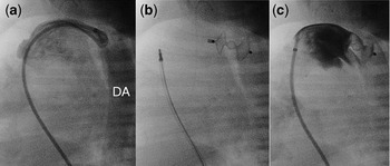
In the cardiac catheterization laboratory, under general anaesthesia, we inserted a 4 French introducer into the right femoral vein. We were unable to access the femoral artery because of excessive bleeding. The mean pulmonary arterial pressure was similar to that in the descending aorta. A pulmonary angiogram (Fig. 1a) showed the patent duct to have a diameter of 3.9 millimetres, and to be 5 millimetres in length. We opted to close the duct using the Amplatzer ductal occluder II. The device, from AGA Medical Corporation, Golden Valley, Minnesota, is made of a nitinol wire mesh, with 2 retention disks of 9 millimetres, and a central connecting part of 3 millimetres diameter. To insert the device, we replaced the 4 French multipurpose catheter used to make the angiogram with a 4 French Amplatzer delivery catheter, which was advanced across the duct into the descending aorta, permitting placement of the device. When judged to be appropriately positioned, having made an angiogram through the side port of the delivery catheter, we released the device (Fig. 1b). After 5 minutes, a repeated angiogram showed almost complete occlusion of the patent duct (Fig. 1c). The fluoroscopic time was 7 minutes.
The pulmonary angiogram in lateral projection (a) shows a patent arterial duct of 3.9 millimetres diameter. Note that the contrast opacifies the descending aorta (DA). Figure 1b shows the situation immediately alter release of the Amplatzer Duct Occluder II, and a subsequent pulmonary angiogram (c) shows that the duct is almost completely occluded.

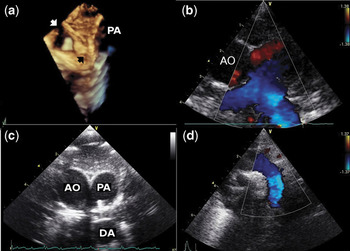
After the procedure, the baby showed marked improvement, re-establishing urinary output. After 1 week, we were able to wean from inotropic and ventilatory support. He remained with abnormal hepatic function, considered to be part of his neonatal lupus erythematosus. We were able to close the ileostomy 4 weeks after insertion of the device, and the baby was discharged in good condition. Echocardiographic follow-up 2 months after the procedure showed complete closure of the duct, with no obstruction to either the left pulmonary artery (Fig. 2b) or the aortic arch (Fig. 2d).
At echocardiography 2 months later, the 3D reconstruction (a) shows the Amplatzer Duct Occluder II (black arrow) to be well adapted to both the pulmonary arterial (PA) and the aortic walls. The white arrow shows the origin of left subclavian artery. The parasternal, short axis view (b) shows no obstruction in the left pulmonary artery, while cranial tilting (c) shows the occluder to be well positioned between the pulmonary artery and the descending aorta (DA). The enlargement of the ascending aorta (AO) is the consequence of the complete atrioventricular block. The suprasternal long axis view (d) shows normal flow in the aortic arch.

Discussion
Although patency of the arterial duct is common in preterm neonates, in some neonates the persistently patent ducts need to be closed at term.Reference Ly, Hawes, Whyte, Teixeira and McNamara3, Reference Takami, Yoda and Kawakami4 Our patient had multiorgan failure related to its persistent patency, with surgical or pharmacological closure contraindicated because of cerebral bleeding, acute renal failure, and profound thrombocytopaenia. To the best of our knowledge, percutaneous ductal closure has not previously been reported in neonates. Closure was achieved in an infant of 3 months weighing less than 3 kilograms using an Amplatzer Vascular Plug, produced by AGA Medical Corporation, Golden Valley, Minnesota, prefilled with Gianturco embolization coils.Reference Glatz, Petit and Gillespie5 The subsequent period, however, was complicated mainly by respiratory problems, and the infant died at 6 months of age.
A series of 18 small infants with symptomatic ducts closed with the initial Amplatzer occluder has been reported,Reference Butera, De Rosa and Chessa1 the smallest child weighing 4.5 kilograms. In all cases, occlusion proved successful with no obstruction of either the left pulmonary artery or the aorta, albeit that 1 instance of acute femoral arterial thrombosis required treatment with urokinase. In these earlier experiences, the investigators placed and delivered the device from the pulmonary artery, using arterial access to define the anatomy of the patent arterial duct, and to evaluate the position of the device before and after delivery.Reference Masura, Walsh and Thanopoulous6 In our case, since we were unable to obtain arterial access, we defined the ductal anatomy with an angiogram made from the pulmonary artery. An angiogram in anteroposterior projection would have been desirable to rule out any encroachment of the left disc into the aorta, but transthoracic echocardiography performed during the procedure helped us to rule out such aortic obstruction. The currently available device proved adequate for our patient, but our experience suggests that the future availability of smaller devices might permit successful percutaneous closure in even smaller patients.
Our experience, in a critically ill neonate, was straightforward and effective, indicating that percutaneous closure should always be considered for those critically ill patients not amenable either to surgical or pharmacological closure.