Introduction
The subfamily Sigmodontinae comprises the single southern experiment of the Cricetidae, a mostly Holarctic group of rodents (Pardiñas et al., Reference Pardiñas, Myers, León-Paniagua, Ordóñez Garza, Cook, Kryštufek, Haslauer, Bradley, Shenbrot, Patton, Wilson, Lacher and Mittermeier2017a). With more than 10 tribes and 430 valid species, one of each three living Neotropical mammals belongs to the Sigmodontinae (Patton et al., Reference Patton, Pardiñas and D'Elía2015). However, understanding of this amazing clade is hampered by a very poor paleontological record. The oldest remains, referred to latest Miocene or early Pliocene beds, are mostly scraps, and some of them were not assessed taxonomically (Pardiñas et al., Reference Pardiñas, D'Elía and Ortiz2002; Verzi and Montalvo, Reference Verzi and Montalvo2008; Nasif et al., Reference Nasif, Esteban and Ortiz2009; Barbière et al., Reference Barbière, Ortiz, Pardiñas, Agnolin, Lio, Brissón-Egli, Chimento and Novas.2016b). Moreover, they came from central or northern Argentina, leaving the major part of South America free of fossil evidence. This poverty of fossil sigmodontines, plus the use of mitochondrial and nuclear gene sequences in phylogenetic analysis, has led investigators to develop evolutionary scenarios for sigmodontines based exclusively on molecular data (e.g., Parada et al., Reference Parada, Pardiñas, Salazar-Bravo, D'Elía and Palma2013; Salazar-Bravo et al., Reference Salazar-Bravo, Pardiñas and D'Elía2013; Leite et al., Reference Leite, Kolokotronis, Almeida, Werneck, Rogers and Weksler2014; see also Barbière et al., Reference Barbière, Ortiz, Pardiñas, Agnolin, Lio, Brissón-Egli, Chimento and Novas.2016b).
Described about four decades ago and based on a lower jaw fragment, Auliscomys formosus Reig, Reference Reig1978 from the Monte Hermoso Formation (Farola de Monte Hermoso, early Pliocene) is, together with Necromys bonapartei Reig, Reference Reig1978, considered the oldest unambiguously dated South American cricetid (e.g., Reig, Reference Reig1978; Pardiñas and Tonni, Reference Pardiñas and Tonni1998; Prevosti and Pardiñas, Reference Prevosti, Pardiñas, Verzi and Montalvo2009; Cione et al., Reference Cione, Gasparini, Soibelzon, Soibelzon and Tonni2015); other cricetid remains have been proposed as being older than the latter species, but that is in question. Nasif et al. (Reference Nasif, Esteban and Ortiz2009) described a molar fragment assigned to an undetermined sigmodontine from the Andalhuala Formation (Catamarca province, Argentina) at about 7 Ma (late Miocene). Pascual and Herrera (Reference Pascual and Herrera1973) mentioned a mandible of an undetermined cricetid from the ‘Irene Formation’ (Montehermosan or Chapadmalalan of Argentina), but this material, never studied, was lost. More recently, Verzi and Montalvo (Reference Verzi and Montalvo2008) described two cricetid hemimandibles from the upper levels of the Cerro Azul Formation (La Pampa province, Argentina), estimated as late Miocene (5.8–5.7 Myr) based on biostratigraphic inference, but they did not propose a taxonomic assignment beyond the subfamily level for the cricetid.
Due to the fragmentary nature of the holotype, and only specimen known, A. formosus was neither included in taxonomic revisions of the tribe Phyllotini nor revisited from a taxonomic point of view (cf. Braun, Reference Braun1993; Steppan, Reference Steppan1995; Salazar-Bravo et al., Reference Salazar-Bravo, Pardiñas and D'Elía2013). However, despite this paucity of information about A. formosus, it was used to calibrate molecular clocks to explore both the origin of Phyllotini and the tempo and mode of the evolution of the entire radiation of the sigmodontine rodents (e.g., Salazar-Bravo et al., Reference Salazar-Bravo, Dragoo, Tinnin and Yates2001; Pardiñas et al., Reference Pardiñas, D'Elía and Ortiz2002; Parada et al., Reference Parada, Pardiñas, Salazar-Bravo, D'Elía and Palma2013; Salazar-Bravo et al., Reference Salazar-Bravo, Pardiñas and D'Elía2013).
Fossils with established chronology useful to generate evolutionary calibrated hypotheses are scarce in the muroid universe (see Steppan et al., Reference Steppan, Adkins and Anderson2004; Kimura et al., Reference Kimura, Hawkins, McDonough, Jacobs and Flynn2015). Regarding sigmodontine diversification, a first crucial question is whether A. formosus can be allocated confidently within Auliscomys Osgood, Reference Osgood1915, a modern genus restricted today to high-Andean environments. A second question is the quality of evidence used to establish the early Pliocene age of this taxon. Both issues are addressed in this contribution based on new materials obtained in the outcrops of the classic fossiliferous locality Farola de Monte Hermoso (~38.58°S, 61.41°W, Buenos Aires, Argentina; see Reig, Reference Reig1978 and Tomassini et al., Reference Tomassini, Montalvo, Deschamps and Manera2013 for information about geography and stratigraphy of this locality), the type locality of A. formosus. The new data obtained allow us to discuss more deeply the origins of the sigmodontine radiation in South America from the perspective of the fossil record.
Materials and methods
The studied fossils are housed in the following Argentinean collections: Instituto Miguel Lillo, Tucumán, Museo Municipal de Ciencias Naturales “Carlos Darwin” de Punta Alta, Buenos Aires, and Museo de La Plata, La Plata, Buenos Aires. Recent specimens examined for comparison are listed in the Appendix. Craniodental anatomy adopted here follows the concepts and nomenclature described by Carleton and Musser (Reference Carleton and Musser1989) and Reig (Reference Reig1977). Dental measurements were taken with a micrometer eyepiece on a stereomicroscope and are provided in millimeters. The phylogenetic framework regarding the evolution and relationships of phyllotines used in this study is from Salazar-Bravo et al. (Reference Salazar-Bravo, Pardiñas and D'Elía2013) and Pardiñas et al. (Reference Pardiñas, Lessa, Teta, Salazar-Bravo and Câmara2014). The general sigmodontine taxonomy adopted here follows Patton et al. (Reference Patton, Pardiñas and D'Elía2015). Upper and lower molars are represented by upper- and lower-case letters, respectively (M, m), and length and width by L and W.
Until now, the only known published material for A. formosus was the holotype (Fig. 1), a right hemimandible with three molars (Reig, Reference Reig1978). Of the five new specimens studied here and referred to A. formosus, two are hemimandibles, two are maxillaries, and the remaining one is an upper incisor. However, as none of these remains were found together, reference of the maxillaries and incisor to A. formosus must be justified anatomically and functionally. To achieve this, we measured and calculated the mean of the length and width of each upper and lower molar in 16 sigmodontines (14 extant species, two extinct). We then calculated a ratio (R) of length to width for each upper (RM) and lower molar (Rm) and the proportion of the length of each molar compared to the length of the total toothrow (PPM for upper molars and PPm for lower molars). Finally, we calculated for each toothrow the geometric mean of the ratio and proportions previously mentioned; for upper molars: geoU = (RM1 × RM2 × RM3 × PPM1 × PPM2 × PPM3)1/6; for lower molars: geoL = (Rm1 × Rm2 × Rm3 × PPm1 × PPm2 × PPm3)1/6. See Supplementary Data Set 1 for all measurements. These indexes summarize the information of both the general morphology of each molar (the ratios) and the configuration of the toothrow (percentage of each molar in the total length of the toothrow). All these calculations were executed with the software R 3.1.0 (R Core Team 2014). We tested the correlation between geoL and geoU, and a linear model was fitted to predict the values of geoU for A. formosus.
Holotype of Kraglievichimys formosus (Reig, Reference Reig1978; PVL 2397) right hemimandible with m1–3 from Monte Hermoso Formation, Montehermosan, Farola de Monte Hermoso, Buenos Aires Province: (1) labial view; (2) lingual view; (3) occlusal view. Scale bar = 5 mm.

Phylogenetic analysis
To test whether Auliscomys formosus belongs to the genus Auliscomys or needs a new generic assignation, a phylogenetic analysis was performed using the matrix provided by Steppan (Reference Steppan1995). Nonphyllotine rodents were removed from the data. All the extant taxa discussed in the present work are included in the analysis. The extinct taxa Chukimys favaloroi Barbière et al., Reference Barbière, Cruz, Ortiz and Pardiñas2016a, Auliscomys fuscus Quintana, Reference Quintana2002, and Auliscomys osvaldoreigi Quintana, Reference Quintana2002 were not included in the analysis as they may belong to the tribe Reithrodontini (Barbière et al., Reference Barbière, Ortiz and Pardiñas2015, Reference Barbière, Cruz, Ortiz and Pardiñas2016a, Reference Barbière, Ortiz, Pardiñas, Agnolin, Lio, Brissón-Egli, Chimento and Novas.2016b). Readers are invited to refer to Steppan (Reference Steppan1995) for the list of characters used; see Supplementary Data Set 2 for the matrix. The phylogenetic analysis thus comprises 27 taxa, and Abrothrix andina Philippi, Reference Philippi1858 (Chroeomys andinus sensu Steppan, Reference Steppan1995) was used as outgroup. The analyses were performed using maximum parsimony with the TNT software (Goloboff et al., Reference Goloboff, Farris and Nixon2008). We conducted a traditional search with tree bisection reconnection (TBR) using 100 random addition sequences, saving 10 trees per round under equal weight with all characters unordered. A resampling technique using a bootstrap method (standard bootstrap of TNT with 100 replications, using traditional search with TBR, 100 random addition sequences and saving 10 trees per round) was performed. Bremer support was calculated with the same program.
Repositories and institutional abbreviations
Types, figured specimens, and other specimens examined in this study are deposited in the following institutions: CEI, Colección de Egagrópilas del INSUGEO, San Miguel de Tucumán, Argentina; CNP, Colección de Mamíferos del Centro Nacional Patagónico, Puerto Madryn, Argentina; CNP-E, Colección de Material de Egagrópilas y Afines ‘Elio Massoia’ del Centro Nacional Patagónico, Puerto Madryn, Argentina; FMNH, Field Museum of Natural History, Chicago, USA; MACN, Museo Argentino de Ciencias Naturales ‘Bernardino Rivadavia,’ Buenos Aires, Argentina; MD-FM, Museo Municipal de Ciencias Naturales ‘Carlos Darwin’ de Punta Alta, Buenos Aires, Argentina; MLP, Museo de La Plata, La Plata, Argentina; MVZ, Museum of Vertebrate Zoology, University of California, Berkeley, USA; PEO, field number of Pablo E. Ortiz, to be deposited at MACN; PVL, Colección Paleontología de Vertebrados Lillo, Instituto Miguel Lillo, San Miguel de Tucumán, Argentina.
Systematic paleontology
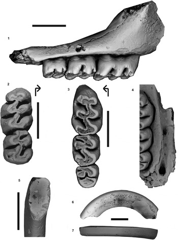
The two new maxillaries (Fig. 2) represent the same taxon based on their similarity in occlusal pattern and measurements. Both are characterized by moderately hypsodont and simplified molars with bileveled occlusal surfaces. These features are in agreement with those observed in the lower molars of the holotype of Auliscomys formosus. The result of the correlation test applied to the variables geoU and geoL shows that they are highly correlated (Pearson's coefficient = 0.94, p value = 9.44e-08). Furthermore, they fit the linear model (see Supplementary Data Set 3). The 95% prediction interval of geoU for A. formosus is between 6.05 and 6.40. The value of geoU calculated for the specimen MLP 48-XII-16-140 (the unique dentary bearing the three molars and thus available for the morphometric approach developed here) is 6.15. From a morphometric point of view, the assignment of this maxillary to A. formosus is supported. Finally, both maxillaries discussed here were found in the same strata and locality where the holotype and the new hemimandibles assigned to A. formosus were retrieved. In addition, the isolated upper incisor can be referred to A. formosus according to its size and general morphology. It is therefore reasonable to conclude these specimens belong to a single taxon, A. formosus.
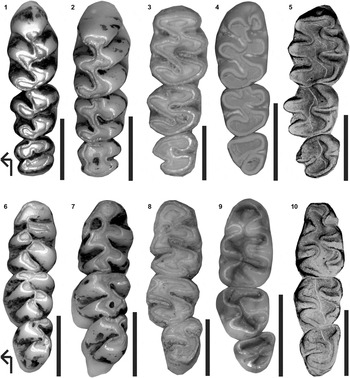
The new material available allows a better comparison of A. formosus (Figs. 1–3) with other phyllotines. A. formosus differs from Calomys Waterhouse, Reference Waterhouse1837, Calassomys Pardiñas et al., 2014, †Chukimys Barbière et al., Reference Barbière, Cruz, Ortiz and Pardiñas2016a, Eligmodontia Cuvier, Reference Cuvier1837, and Salinomys Braun and Mares, Reference Braun and Mares1995 by its greater size and hypsodonty. A. formosus is comparable in size to Andalgalomys Williams and Mares, Reference Williams and Mares1978, the living species of Auliscomys, Galenomys Thomas, Reference Thomas1916, Graomys Thomas, Reference Thomas1916 (except G. edithae Thomas, Reference Thomas1919), Phyllotis Waterhouse, Reference Waterhouse1837 (except the small-bodied forms of P. amicus Thomas, Reference Thomas1900a, P. andium Thomas Reference Thomas1912, and P. gerbillus Thomas, Reference Thomas1900c groups), Loxodontomys, and Tapecomys Anderson and Yates, Reference Anderson and Yates2000 (Figs. 4, 5). Graomys (Fig. 4.1, 4.6) has greater crown height and more planate and prismatic molars than A. formosus. Graomys also differs from A. formosus in the lack of anteromedian flexid on the m1, and its bony palate is clearly larger than in A. formosus. Phyllotis (Fig. 4.2, 4.7) and Tapecomys (Fig. 4.3, 4.8) can be removed from analysis by their clearly more hypsodont, laminated, and planate molars. In addition, these taxa differ from A. formosus by exhibiting a small anteroloph on M2, a reduced posterior lobe on M3, and a more developed anterolabial cingulum (Phyllotis). Although the hypsodonty in Andalgalomys (Fig. 4.4, 4.9) is similar to A. formosus and both taxa have a bileveled occlusal surface, the general morphology of the molars in Andalgalomys is quite distinct from that of A. formosus. Moreover, Andalgalomys differs in other critical ways from A. formosus, such as having a conspicuous posterior palatal foramen that produces a deeply fenestrated palatal region. The morphological differences between A. formosus and Galenomys (Fig. 4.5, 4.10) are also considerable. Indeed, in the latter genus, upper molars exhibit a symmetric pattern with transverse and opposite flexi and cusps that is clearly distinct from the pattern of A. formosus. In addition, the first lower molar in Galenomys exhibits a more reduced anterolabial cingulum, and m3 has a less sigmoid occlusal pattern.
Maxillaries and upper teeth of Kraglievichimys formosus from Monte Hermoso Formation, Montehermosan, Farola de Monte Hermoso, Buenos Aires Province: (1) MLP 48-XII-16-140, right maxillary in lateral view; (2) MLP 77-II-1-20, left M1–2 in occlusal view; (3) MLP 48-XII-16-140, right M1–3 in occlusal view; (4) MLP 48-XII-16-140, detail of the bony palate in ventral view; (5–7) MD-FM-08-53, isolated upper incisor; (5) dentine exposition; (6) lateral view; (7) frontal view. Scale bars = 2 mm. Arrows indicate anterolabial side.

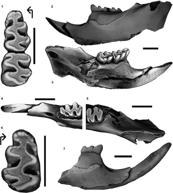
New hemimandibles and lower molars of Kraglievichimys formosus from Monte Hermoso Formation, Montehermosan, Farola de Monte Hermoso, Buenos Aires Province: (1–5) MD-FM-05-10, left hemimandible with m1–2; (1) left m1–2 in occlusal view; (2) left hemimandible in labial view; (3) left hemimandible in lingual view; (4) anterior part of left hemimandible with dentine exposure of incisor; (5) posterior part of left hemimandible, note the narrowing of alveolar toothrow behind m2; (6, 7) MD-FM-05-09, right hemimandible with m1; (6) right m1 in occlusal view; (7) right hemimandible in labial view. Scale bars = 2 mm. Arrows indicate anterolabial side.

Upper (upper row) and lower (lower row) toothrows in occlusal view of various Phyllotini used for comparison: (1, 6) Graomys griseoflavus Waterhouse, Reference Waterhouse1837 (CNP-E 63); (2, 7) Phyllotis xanthopygus Waterhouse, Reference Waterhouse1837 (CNP 5339); (3, 8) Tapecomys primus Anderson and Yates, Reference Anderson and Yates2000 (CNP 830); (4, 9) Andalgalomys olrogi Williams and Mares, 1978 (CNP 2398); (5, 10) Galenomys garleppi Thomas, Reference Thomas1898 (FMNH 53845). Scale bars = 2 mm. Arrows indicate anterolabial side.

All the available material of A. formosus reinforces the hypothesis developed by Reig (Reference Reig1978) that this taxon is more similar to Auliscomys and Loxodontomys (Fig. 5) than to other phyllotines. However, A. formosus exhibits not only a mosaic of characters from both genera but also autapomorphies (Table 1). A clear example of this mosaic can be seen from the lower incisor, which is as narrow as in Auliscomys but also as deep and ungrooved as the upper incisor in Loxodontomys. The presence of an anteromedian flexid on m1 and the low diastema of A. formosus are characteristic of Auliscomys, but a conspicuous capsular projection on the dentary, a masseteric crest that extends up to the anterior border of m1, a bulbous procingulum on M1, and a large posterior lobe on M3 showing an enamel ring in juvenile individuals are characteristics of Loxodontomys. In addition, A. formosus presents unique traits such as a narrowing of the alveolar lower toothrow at m3 and labial extension of the anteroloph on M2.
Comparison of Loxodontomys micropus Waterhouse, Reference Waterhouse1837 (CNP-E 370) and Auliscomys pictus Thomas, Reference Thomas1884 (MVZ 139580): (1–5) Loxodontomys micropus; (1) left hemimandible in labial view; (2) left hemimandible in lingual view; (3) anterior part of left hemimandible; (4) left M1–3 in occlusal view; (5) left m1–3 in occlusal view. (5–9) Auliscomys pictus; (5) left hemimandible in labial view; (6) left hemimandible in lingual view; (7) anterior part of left hemimandible; (8) upper left M1–3 in occlusal view; (9) lower left m1–3 in occlusal view. Scale bars = 2 mm. Arrows indicate anterolabial side.

Comparison of select mandibular and dental characters of Kraglievichimys, Auliscomys Osgood, Reference Osgood1915, and Loxodontomys Osgood, Reference Osgood1947.

The comparisons presented in the receding lead to the conclusion that A. formosus has a unique combination of anatomical features within the phyllotine radiation requiring recognition at the generic level. This conclusion is also sustained by the results of the phylogenetic analysis (Fig. 6). The analysis yielded 1 most parsimonious tree, 335 steps long (CI = 0.34; RI = 0.51). Among the various genera represented by several species, Auliscomys is the only genus recovered as monophyletic. This is noteworthy in the sense that the distinct genera of Phyllotini are mostly retrieved as such in most of the phylogenetic analysis (Fabre et al., Reference Fabre, Hautier, Dimitrov and Douzery2012; Salazar-Bravo et al., Reference Salazar-Bravo, Pardiñas and D'Elía2013; Carrizo and Catalano, Reference Carrizo and Catalano2015; Steppan and Schenk, Reference Steppan and Schenk2017). Nevertheless, the general relationships provided are in agreement with the other phylogenetic analyses. Calomys appears to be a basal member of the tribe (Pardiñas et al., Reference Pardiñas, Lessa, Teta, Salazar-Bravo and Câmara2014). The affinity between Loxodontomys and Tapecomys is also recovered by various phylogenetic analyses (Fabre et al., Reference Fabre, Hautier, Dimitrov and Douzery2012; Carrizo and Catalano, Reference Carrizo and Catalano2015; Steppan and Schenk, Reference Steppan and Schenk2017). Within Phyllotis, Steppan et al. (Reference Steppan, Ramirez, Banbury, Huchon, Pacheco, Walker, Spotorno, Kelt, Lessa, Salazar-Bravo and Patton2007), Fabre et al. (Reference Fabre, Hautier, Dimitrov and Douzery2012), and Steppan and Schenk (Reference Steppan and Schenk2017) have shown, as does our analysis, a closer relationship between P. caprinus Pearson, Reference Pearson1958, P. darwini Waterhouse, Reference Waterhouse1837, P. magister Thomas, Reference Thomas1912, and P. xanthopygus Waterhouse, Reference Waterhouse1837 than between these and the other species of Phyllotis. In this tree A. formosus is retrieved as the sister taxon of Andalgalomys pearsoni Myers, Reference Myers1977 within a clade also grouping Graomys griseoflavus Waterhouse, Reference Waterhouse1837 and Graomys domorum Thomas, Reference Thomas1902. Two synapomorphies support the association of A. formosus and A. pearsoni: an anteromedian flexus on M1 distinct or prominent (character 10) and a ratio of M3 length to alveolar length of molar toothrow between 0.205 and 0.25 (character 32). Nevertheless, the many autapomorphies identified for A. formosus such as the presence of a labial root on M1 (character 4) and two roots on m2 (character 8), the limited development of the anteromedian flexid on m1 (character 17), the anterior masseteric ridge forming a knob slightly below the dorsal edge of the mandible (character 35), the incisive foramina extending posteriorly to the level of paracone and protocone of M1 (character 38), and the mesopterygoid fossa lying between 0 and 1/3 tooth length posterior to M3 (character 32), besides the morphological differences previously mentioned between this taxon and Andalgalomys pearsoni, prevent the assignment of the former to Andalgalomys and thus justify allocation to a new genus, as follows.
Cladogram of one most parsimonious tree of 335 steps long (CI = 0.340; RI = 0.511). Bremer support and bootstrap percentages (above 50%) are indicated above and under each node, respectively.

Family Cricetidae Fischer, Reference Fischer1817
Subfamily Sigmodontinae Wagner, Reference Wagner1843
Tribe Phyllotini Vorontsov, Reference Vorontsov1959
Kraglievichimys new genus
Figures 1–3
Type species
Auliscomys formosus Reig, Reference Reig1978 by monotypy.
Diagnosis
As for the type species by monotypy.
Occurrence
Farola de Monte Hermoso in southwest Buenos Aires Province, Argentina. All the known material comes from the deposits of the Monte Hermoso Formation, middle Pliocene.
Description
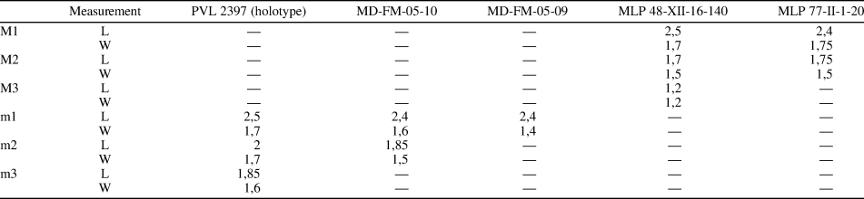
Molars lacking mesoloph or mesolophid, moderately hypsodont, with main cusps with rounded (not prismatic) outer margins and the lingual and labial folds not interpenetrating. Upper and lower molars have terraced crowns; main cusps opposite on upper molars, while cusps are alternate in lower molars and lophs are transverse. See Table 2 for measurements.
Dental measurements (in millimeters) for Kraglievichimys formosus.

The M1 is oval in outline. Labial flexi are curved posteriorly while lingual flexi are nearly transverse. The metaflexus and hypoflexus are alternate and meet at the midline. The procingulum is bulbous and large with the presence of a lingually displaced anteromedian flexus, thus dividing the procingulum into two asymmetric anteroconules, the lingual anteroconule being smaller than the labial one. Hypocone and protocone are similar in size and subtriangular. The mures are large and slightly oblique; the median mure is derived from the anterior arm of the hypocone. Paracone and metacone are oriented anteriorly-posteriorly; a small posteroloph is indicated by a very shallow inflection in the metacone. M1 has four roots, including an accessory labial root under the paracone scarcely visible from the margin of the alveolus.
The M2 is subrectangular in outline. Flexi are alternate and meet at the midline. The hypoflexus is nearly transverse, the metaflexus is strongly curved posteriorly, and the paraflexus is slightly curved posteriorly. This last feature, together with a small protoflexus, defines a labial anteroloph. Hypocone and protocone are subtriangular and similar in size. A small fosette is developed in the hypocone. Mures are oblique, and the median loph is also formed by the anterior arm of the hypocone. Paracone and metacone have the same labial extension. A small enamel projection from the base of the paracone may represent either a small paralophule or an incidental fold in the paracone caused by an anterior penetration of the metaflexus. A small posteroloph is defined by the posteroflexus. M2 has three roots.
The M3, approximately three-quarters of the total length of M2, is rounded in outline and characterized by the fusion of the metaflexus and hypoflexus, creating a bilobed pattern. The anterior lobe is large, with an anteroloph extended up to the labial side; a short paracone connects to the anteroloph. There is a small protoflexus, recognizable as a very shallow inflection, on the anterior side of the strong and subtriangular protocone. The posterior lobe is fan shaped, as large as the anterior lobe. The subtriangular hypocone is smaller than the protocone. The metaloph is transverse and connects to a reduced metacone. A large and extended transverse fossette marks a posteroloph connected to the metacone. The M3 has three roots.
The upper incisor suggests an opisthodont condition (sensu Hershkovitz, Reference Hershkovitz1962); the thick enamel surface lacks grooves and exhibits a straight dentine fissure (sensu Steppan, Reference Steppan1995). Lower incisors in both new hemimandibles are long and narrow, and the anterior surface is smooth.
The posterior part of m1 is wider than the anterior part, giving it a subtriangular outline. The flexi are broad. The procingulum is partially compressed anteroposteriorly, connected to the rest of m1 by a broad, straight anterior murid located in the midline. An anteromedian flexid is displaced lingually and persistent through wear as a slight notch. The anterolabial cingulum is well developed and extended posteriorly, defining an anteriorly directed protoflexid. The protoflexid may become isolated into a fossetid with wear. The median murid is oriented obliquely and joins the protoconid and entoconid. The protoconid and hypoconid are similar in size and subtriangular. The posterolophid is short but well delimited and oriented transversely. The m1 has three roots, including a labial accessory root under the protoconid.
The m2 is subrectangular in outline. The anterolabial cingulum is well developed and closes the protoflexid by joining the anterior base of the protoconid. The median murid is oblique, and the hypoconid and the protoconid are subtriangular. On the posterior edge of the hypoconid, a slight notch marks a posteroloph of similar length as in m1. The molar has two strong roots, one under both the protoconid and metaconid and the other under the hypoconid and entoconid.
The m3 has a sigmoid pattern without evidence of a posterolophid. The m3 is reduced in size compared to m2, both in length and in posterior width, with a subtriangular outline. A slight expansion in the anterior part of the protoconid suggests the presence of an anterolabial cingulum in less-worn specimens. This molar has two roots.
Few details can be surmised from the maxillary and the palate due to the fragmentary nature of the available material. The posterior end of the incisive foramen reaches the protocone of M1. The posterior margin of the body of the zygomatic plate is anterior to the anterior face of M1. The palate is short and broad. It has two distinctive foramina over the palatine bone, a small anterior palatal foramen located on the maxillopalatine suture at the level of the anterior border of the M2, and a posterior palatal pit of moderate size, located at the level of the anterior border of the mesopterygoid fossa. The latter is associated with a groove running anteriorly and flanked externally by the alveolar maxillary process.
The lower jaw is robust, with the height of the horizontal ramus at the level of m1 greater than the diastema length. Although the tip of the diastema is slightly broken, its anterior-most point is below the alveolar plane. The upper and lower masseteric crests converge anteriorly into a ridge below the m1. This ridge extends anteriorly beneath m1. The mental foramen is large and mostly opens dorsally on the diastema, although it can be seen partially from lateral view. The capsular process (incisor capsule) is conspicuous and lies between the coronoid and condyloid processes. Judged by its base, the coronoid process is large. The mandibular foramen is large and ovoid and remains embraced by the mylohyoid line. The retromolar region is moderate in size, restricted to the labial side of m3, and pierced by a number of diminutive foramina.
Etymology
The genus name recognizes the prolific contributions to the knowledge of South American fossil vertebrates by the Argentinean paleontologist Lucas Kraglievich (1886–1932). The epithet is formed from the surname “Kraglievich” + [i] in masculine genitive singular, plus the Latin word mys (= mouse).
Comparisons
Quintana (Reference Quintana2002) described two fossil species of Auliscomys, A. fuscus and A. osvaldoreigi (Fig. 7), both from lowermost Pleistocene deposits of southeastern Buenos Aires province, Argentina. Although it was later suggested that the latter specimens do not belong to Auliscomys, and perhaps not to the Phyllotini but instead to the Reithrodontini (Teta et al., Reference Teta, Pardiñas, Sauthier and D'Elía2009; Barbière et al., Reference Barbière, Ortiz, Pardiñas, Agnolin, Lio, Brissón-Egli, Chimento and Novas.2016b), their original allocation invites comparison with Kraglievichimys. Kraglievichimys is clearly distinct from A. fuscus (Fig. 7.1–7.4) by lacking the spoon-like excavation of the internal face of the condyloid process that characterizes the latter, bearing a conspicuous capsular process (reduced in A. fuscus) and having a larger mental foramen. The masseteric crest is stronger with a more horizontal development in A. fuscus. Furthermore, no evidence of a narrowing of the alveolar row is observed in A. fuscus. As for the teeth, while the lower incisor is narrow in both taxa, the incisor is deeper in Kraglievichimys. Auliscomys fuscus lacks the anterolabial cingulum on m1 and exhibits a square, rather than rectangular, outline (as in Kraglievichimys). From a more general point of view, while both species seem to have the same size judged from measurements, Kraglievichimys displays a more robust mandible.
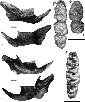
Holotype of Auliscomys fuscus Quintana, Reference Quintana2002 (MACN 19686) from San Andres Formation, Sanandresian, Playa Santa Isabel, Buenos Aires Province and Auliscomys osvaldoreigi Quintana, Reference Quintana2002 (MACN 19691) from San Andres Formation, Sanandresian, Punta San Andres, Buenos Aires Province: (1–4) Auliscomys fuscus; (1) right hemimandible in labial view; (2) right hemimandible in lingual view; (3) right lower m1–3 in occlusal view; (4) left upper M1–2 in occlusal view. (5–7) Auliscomys osvaldoreigi; (5) right hemimandible in labial view; (6) right hemimandible in lingual view; (7) right m1–3 in occlusal view. Scale bars = 2 mm. Arrows indicate anterolabial side.

Auliscomys osvaldoreigi (Fig. 7.5–7.7) also has a slenderer and shallower mandible than Kraglievichimys. The capsular projection in A. osvaldoreigi is as conspicuous as in Kraglievichimys but positioned lower in the mandibular ramus. The masseteric crest in A. osvaldoreigi also develops lower on the ramus and extends farther anteriorly, beyond the anterior border of m1. The mental foramen of A. osvaldoreigi, in contrast to Kraglievichimys, is not visible in lateral view. The diastema is longer and the incisor, although as deep as in Kraglievichimys, is wider. As in A. fuscus, A. osvaldoreigi exhibits a spoon-like excavation of the condyloid process. The m3 is smaller in Kraglievichimys, the m2 is narrower, and the anterolabial cingulum on m1 is not as well developed as in A. osvaldoreigi. Both taxa have a narrowing of the alveolar row.
Although it can be well differentiated from Graomys based on the morphology of the molars (Fig. 4.1, 4.6), Kraglievichimys shares several features with this genus. Both have a well-developed posterolateral palatal pit placed at the level of the posterior border of M3. Nevertheless, while the mesopterygoid fossa reaches this foramen in Kraglievichimys, delimiting a short palate, it remains more posterior in Graomys. Both genera share the presence of a groove in the palate, but the groove is deeper in Graomys. The mental foramen is slightly larger and more lateral in Kraglievichimys.
The shape and size of the mandibular masseteric crest in Kraglievichimys is very similar to that in Phyllotis. The mental foramen in Phyllotis is, as in Kraglievichimys, partially visible in lateral view but much smaller, and in Phyllotis the diastema is short and the capsular process less conspicuous than in Kraglievichimys. The palate is shorter in Kraglievichimys, bearing larger posterolateral foramen. The incisive foramina have a similar posterior extension in both genera but are rounded in shape in Kraglievichimys, whereas they are acuminate in Phyllotis.
The general aspect of the mandible in Kraglievichimys resembles more the robust form of Loxodontomys than the slender form of Auliscomys (Fig. 5). Furthermore, despite differences in size between the known species of Auliscomys, all are smaller than Kraglievichimys, contrary to Loxodontomys, which is similar in size to Kraglievichimys. On the other hand, the proportions of the molars in Kraglievichimys are closer to Auliscomys, both sharing a reduced m3 compared to m2. Both upper and lower second molars in Kraglievichimys are, as in Auliscomys, subrectangular in outline, whereas they are more squared in Loxodontomys.
Remarks
Auliscomys formosus was named by Osvaldo Reig in his doctoral dissertation (Reig, Reference Reig1972, p. 331) and allocated to Phyllotis (Auliscomys) following the taxonomic schema of Hershkovitz (Reference Hershkovitz1962), who favored a largely polytypic Phyllotis. Six years later, Reig (Reference Reig1978, p. 176) published formosus under Auliscomys, adopting the suggestion made by Pearson (Reference Pearson1972) and Pearson and Patton (Reference Pearson and Patton1976) to recognize full generic status of Auliscomys based on karyological grounds. These results forced Reig (Reference Reig1978) to face some taxonomic issues concerning the generic allocation of this fossil species. Indeed, as Reig thought that sigmodontines constituted a group with a long (i.e., well back into the Miocene) and regional (i.e., South American) evolutionary history, he was biased to think that Pliocene fossils should fit into extant genera. The taxonomic paradigm then was to consider genera with much more morphological variation than is accepted today. Thus, as Reig noticed the morphological resemblance between formosus and representatives of the genera Loxodontomys and Auliscomys, Reig (Reference Reig1978, p. 179) wrote, “En un primer análisis, me sentí inclinado a pensar que la especie hermosense estaba más cercanamente relacionada con Phyllotis micropus, único representante del subgénero Loxodontomys (Osgood, Reference Osgood1947), de amplia distribución en Chile y la Patagonia argentina al sur del paralelo 37, y por lo tanto más cercano geográficamente de formosus… Las comparaciones con boliviensis y pictus se realizaron… con mucho escepticismo de que pudiese haber afinidad con especies geográficamente tan distantes y que vivían en hábitats tan especializados” (freely translated: “In a first analysis, I was inclined to think that the Hermosan species was more closely related to Phyllotis micropus, sole representative of the subgenus Loxodontomys [Osgood, Reference Osgood1947], widely distributed in Chile and Argentinean Patagonia south of parallel 37, and therefore geographically closest to formosus… Comparisons with boliviensis and pictus were made… with much skepticism that there could be affinity between such geographically distant species living in such specialized habitats”). In fact, the extant species of Auliscomys have an Andean distribution from central Peru through the Altiplano of Bolivia to northern Argentina and northernmost Chile (Salazar-Bravo, Reference Salazar-Bravo, Patton, Pardiñas and D'Elía2015a). This would have left formosus far from the modern area of distribution of the genus if it were assigned to Auliscomys. However, comparing formosus with Loxodontomys and the three species of Auliscomys, Reig (Reference Reig1978) highlighted a greater similarity between formosus and A. sublimis Thomas, Reference Thomas1900b than between formosus and boliviensis Waterhouse, Reference Waterhouse1846 or pictus Thomas, Reference Thomas1884. This logic represented the basis underlying the hypothesis that Reig (Reference Reig1978) developed to justify the generic allocation of formosus. Here, the new remains, especially with the description of upper molars, allow us to recognize more similarities between formosus and Loxodontomys than Reig (Reference Reig1978) accepted, thus justifying the allocation of formosus to a new genus. In this way, the geographic and environmental incongruence brought up by Reig (Reference Reig1980) in his assignment of formosus to Auliscomys is resolved.
Kraglievichimys formosus (Reig, Reference Reig1978) new combination
Figures 1–3
1978 Auliscomys formosus Reig, p. 178, fig. 5.
Holotype
PVL 2397, a poorly preserved right dentary comprising the molar series and the dorsal portion of the diastema. According to the original label, the material was collected by José Bonaparte and Jorge Zetti in January 1960 in “Acantilado Monte Hermoso; Hermosense Banco N° 3.” These data indicate that the fossil was recovered from the Atlantic cliffs exposed at Farola de Monte Hermoso (~38°58′S, 61°41′W, Buenos Aires Province, Argentina) from the basal levels of the Monte Hermoso Formation (Bonaparte, Reference Bonaparte1960).
Emended diagnosis
A sigmodontine cricetid rodent distinguished by the following combination of characters: conspicuous capsular projection, masseteric crests reaching the anterior border of m1, enlarged mental foramen partially visible from lateral view, narrowing of lower alveolar row at level of m3, incisive foramina posteriorly extending to the level of paracone and protocone on M1, deep and narrow upper and lower incisors, upper incisor ungrooved with thick enamel, anteroloph on M2 labially extended, bilobed M3 with large posterior lobe.
Material
MD-FM-05-09, right hemimandible preserving the incisor and m1, collected by Rodrigo Tomassini in Farola de Monte Hermoso, Monte Hermoso Formation. MD-FM-05-10, left hemimandible including the incisor and m1–m2 plus a large portion of the ascending ramus, same collector and provenance as MD-FM-05-09. MD-FM-08-53, isolated upper incisor, same collector and provenance as MD-FM-05-09. MLP 48-XII-16-140, fragmentary right maxillary with the basal portion of the zygomatic plate, the M1–M3 and contiguous strip of bony palate, collected by personnel of MLP in 1948 in Farola de Monte Hermoso, Limolitas Claras Member, Monte Hermoso Formation. MLP 77-II-1-20, fragmentary left maxillary with M1–M2 and the associated portion of bony palate, collected by Mariano Bond on January 17, 1977, about 800 m to the west of the Lighthouse, Farola de Monte Hermoso, Limolitas Claras Member, Monte Hermoso Formation.
Remarks
Reig (Reference Reig1978) presented a diagnosis of K. formosus based only on a fragmentary hemimandible. The description of more complete material, besides the upper toothrow, allows us to add new autapomorphic characters to this species. Thus, the diagnosis must be emended.
Discussion
The Phyllotini is one of the most speciose clades of the sigmodontine radiation of neotropical cricetids. Molecular markers have allowed a refinement of phyllotine content, highlighting the existence of several convergent subclades (e.g., Engel et al., Reference Engel, Hogan, Taylor and Davis1998; Smith and Patton, Reference Smith and Patton1999; Fabre et al., Reference Fabre, Hautier, Dimitrov and Douzery2012; Salazar-Bravo et al., Reference Salazar-Bravo, Pardiñas and D'Elía2013). Today, the group is understood as comprising 11 living genera mostly distributed in arid and semiarid high and lowland environments (Salazar Bravo et al., Reference Salazar-Bravo, Pardiñas and D'Elía2013; Carrizo and Catalano, Reference Carrizo and Catalano2015; Salazar-Bravo, Reference Salazar-Bravo, Patton, Pardiñas and D'Elía2015b). Several fossils have been allied to this tribe, including forms from both North and South American deposits (Pardiñas, Reference Pardiñas1997; Steppan and Pardiñas, Reference Steppan and Pardiñas1998; Ortiz et al., Reference Ortiz, Pardiñas and Steppan2000, Reference Ortiz, Jayat and Steppan2012; Lindsay, Reference Lindsay, Janis, Gunnell and Uhen2008 and the references therein). The origin of phyllotines was traced by Baskin (Reference Baskin1978, Reference Baskin1986) to Bensonomys Gazin, Reference Gazin1942, a genus described from Pliocene beds of North America and considered by Baskin to be related to Calomys or Eligmodontia, two living phyllotine genera. However, Reig (1980) strongly argued against the supposed phyllotine nature of Bensonomys (see also Pardiñas et al., Reference Pardiñas, Lessa, Teta, Salazar-Bravo and Câmara2014; Barbière et al., Reference Barbière, Ortiz, Pardiñas, Agnolin, Lio, Brissón-Egli, Chimento and Novas.2016b). According to Reig (Reference Reig1984, Reference Reig, Vuilleumier and Monasterio1986), the origin of the phyllotines can be linked with a South American ‘protophyllotine’ ancestor and the evolution of the Andes since Miocene times. Salazar-Bravo et al. (Reference Salazar-Bravo, Pardiñas and D'Elía2013) and Pardiñas et al. (Reference Pardiñas, Lessa, Teta, Salazar-Bravo and Câmara2014) suggested a possible origin outside the Andes, arguing that, as the tribe is currently understood, few phyllotines strictly inhabit high Andean areas (see also Maestri and Patterson, Reference Maestri, Patterson and Mpodozis2016, fig. 9d). In addition, the two most basal genera of the tribe are distributed in low and middle elevations in the east of the Andes (Calomys) and eastern Brazil (Calassomys). The sister taxon of the phyllotines, Delomys Thomas, Reference Thomas1917, has a similar distribution. This hypothesis is also supported by a theoretical analysis of ancestral geographic ranges (Leite et al., Reference Leite, Kolokotronis, Almeida, Werneck, Rogers and Weksler2014). Assuming for the moment a non-Andean origin, it is not surprising that the oldest South American remains of the Phyllotini (i.e., Kraglievichimys) were found in the Atlantic coast of the Buenos Aires province, Argentina.
Since its stratigraphic provenance has been clearly established (Pardiñas and Tonni, Reference Pardiñas and Tonni1998), Kraglievichimys formosus is one of the few key fossils available for the calibration of molecular phylogeny of Sigmodontinae (Parada et al., Reference Parada, Pardiñas, Salazar-Bravo, D'Elía and Palma2013, Reference Parada, D'Elía and Palma2015; Leite et al., Reference Leite, Kolokotronis, Almeida, Werneck, Rogers and Weksler2014; Martínez and Gardenal, Reference Martínez and Gardenal2016). Each of the latter analyses used K. formosus as a calibration point of the crown group Phyllotini and not of the genus Auliscomys as suggested by the earlier generic assignation of formosus. This choice seems to be the most appropriate in light of the taxonomic revision we propose here. Nevertheless, the age of Monte Hermoso Formation, and thus of K. formosus, is subject to debate and not firmly established as is tacitly assumed by molecular approaches.
The relative chronology of paleontological Stages/Ages, their defining biozones, and their bearing localities in the Buenos Aires province are now relatively well understood (Marshall, Reference Marshall, Stehli and Webb1985; Zavala, Reference Zavala1993; Cione and Tonni, Reference Cione and Tonni1995, Reference Cione, Tonni, de Barrio, Etcheverry, Caballé and Llambias2005; Verzi et al., Reference Verzi, Montalvo and Deschamps2008; Deschamps et al., Reference Deschamps, Vucetich, Verzi and Olivares2012; Tomassini et al., Reference Tomassini, Montalvo, Deschamps and Manera2013; Deschamps and Tomassini, Reference Deschamps and Tomassini2016). However, a crucial issue is the chronology of the recognized units and the temporal limits of these stages. With reference to the Montehermosan in its type locality, Farola de Monte Hermoso, hypotheses of age vary from latest Miocene-middle Pliocene (Marshall, Reference Marshall, Stehli and Webb1985) or latest Miocene-early Pliocene (Cione and Tonni, Reference Cione, Tonni, de Barrio, Etcheverry, Caballé and Llambias2005) to early Pliocene (Cione and Tonni, Reference Cione and Tonni1995; Tomassini et al., Reference Tomassini, Montalvo, Deschamps and Manera2013; Deschamps and Tomassini, Reference Deschamps and Tomassini2016). These diverging opinions are possible because few radiometric dates are available for the Neogene in Buenos Aires province. In addition, the available dates are difficult to connect with the classical paleontological localities under discussion (see the following).
Most authors agree that the Monte Hermoso Formation is bracketed between 5.33 ± 0.05 Myr (according to Schultz et al., Reference Schultz, Zárate, Hames, Koeberl, Bunch, Storzer, Renne and Wittke2004; 5.28 ± 0.04 Myr according to Schultz et al., Reference Schultz, Zárate, Hames, Harris, Bunch, Koeberl, Renne and Wittke2006) and 3.3 Myr (Schultz et al., Reference Schultz, Zárate, Hames, Camilion and King1998). However, while the latter date is clearly associated with a horizon of ‘impactites’ exposed in the upper part of the Chapadmalal Formation near Mar del Plata, the former date came from an isolated and poorly known prolife. It was referred to as ‘Near Bahía Blanca,’ and the dated glasses were recorded from a sedimentary section of “… about 20 m thick. The lowermost 5 m are composed of massive light brown siltstones … The upper 15 m of the exposure is made up of light brown calcareous siltstones … Glass fragments are present throughout this upper section with the highest concentrations and largest pieces at the lowermost part, close to the boundary with the underlying siltstones” (Schultz et al., Reference Schultz, Zárate, Hames, Harris, Bunch, Koeberl, Renne and Wittke2006, p. 751). This profile was later named Cantera Vialidad, and the dated glasses were placed in the middle part of the sedimentary succession (see Folguera and Zárate, Reference Folguera and Zárate2009, p. 703–704). However, beyond these uncertainties (e.g., assuming that the date is not a ‘mix’ of glasses along 15 m of sediments), the crucial question is how to link the 5.3 Ma date with the Monte Hermoso Formation. Since there is no stratigraphic link (Cantera Vialidad is an isolated profile ~50 km WNW of Farola de Monte Hermoso coastal cliffs; see also Pardiñas et al., Reference Pardiñas, Prevosti, Voglino and Cenizo2017b for further information) and no radiometric dates are available for the Monte Hermoso sedimentary profile, biostratigraphy/biochronology is the only method available to try to confirm the 5.3 Myr date. Regrettably, the Cantera Vialidad paleontological record includes no more than a handful of scraps briefly mentioned as “few fossil remains (Xenodontomys sp., Paedotherium sp. and Chorobates villosissimus Reig, Reference Reig1958)” (Tomassini et al., Reference Tomassini, Montalvo, Deschamps and Manera2013, p. 38). The occurrence of Xenodontomys Kraglievich, Reference Kraglievich1927 (see Verzi et al., Reference Verzi, Montalvo and Deschamps2008) indicates only that the Cantera Vialidad assemblage is roughly Huayquerian and, therefore, older than the Montehermosan. Furthermore, Pardiñas et al. (Reference Pardiñas, Prevosti, Voglino and Cenizo2017b) have shown incongruences as to the biostratigraphic scheme proposed by Verzi et al. (Reference Verzi, Montalvo and Deschamps2008), so such stratigraphic correlation has to be considered cautiously.
If the Monte Hermoso Formation is younger than 5.3 Myr and older than 3.3 Myr, then it is Pliocene in age, since the Miocene–Pliocene boundary is estimated at 5.33 Myr according to the International Chronostratigraphic Chart (Cohen et al., Reference Cohen, Finney, Gibbard and Fan2013). However, there is currently no reliable means to determine a precise age for the Monte Hermoso Formation. Tomassini et al. (Reference Tomassini, Montalvo, Deschamps and Manera2013) suggested a tentative limit between Montehermosan and Chapadmalalan at between 4.5 and 5.0 Myr. However, this estimate is highly dubious as it relies on continental paleomagnetostratigraphic studies with discordant sequences. Paleontological contents are more similar between the Monte Hermoso and Chapadmalal formations than between the former and older Huayquerian units (Marshall et al., Reference Marshall, Hoffstetter and Pascual1983). Therefore, it appears more reasonable to place the Monte Hermoso Formation closer to 3.3 Myr than to 5.3 Myr. In addition, cricetids are represented in the Chapadmalal Formation by several species (cf. Reig, Reference Reig1978, Reference Reig1987), contrasted with their absence from Huayquerian deposits in central Argentina. As to the latter, Prevosti and Pardiñas (Reference Prevosti, Pardiñas, Verzi and Montalvo2009) suggested that the remains found in Cerro Azul Formation could be considered Pliocene in age and not Miocene (see also Pardiñas et al., Reference Pardiñas, Prevosti, Voglino and Cenizo2017b, fig. 11). Furthermore, it cannot be excluded that the remains found in Andalhuala Formation could belong to a small caviomorph (Barbière et al., Reference Barbière, Ortiz, Pardiñas, Agnolin, Lio, Brissón-Egli, Chimento and Novas.2016b). Taking into account the thin sedimentary layers involved across the units here discussed (e.g., the Monte Hermoso Formation in its type locality is about 8 m; Bonaparte, Reference Bonaparte1960), contrasting with the thick deposits of Huayquerian localities (e.g., the Cantera Vialidad profile is about 20 m), we advance as a working hypothesis that the age of Kraglievichimys formosus and Necromys bonapartei would be closer to 3.3 Myr, and probably not older than 4.0 Myr. A younger age for Farola Monte Hermoso fits better with the fossil record available for sigmodontine rodents. Indeed, as discussed by Barbière et al. (Reference Barbière, Ortiz, Pardiñas, Agnolin, Lio, Brissón-Egli, Chimento and Novas.2016b), Kraglievichimys formosus and Necromys bonapartei do not exhibit a primitive molar pattern as would be expected for the oldest sigmodontine rodents. Indeed, although distinct scenarios have been proposed for the origin of the subfamily, it is commonly accepted that the ancestor would exhibit a bunodont and pentalophodont molar pattern (Hershkovitz, Reference Hershkovitz1962; Baskin, Reference Baskin1978, Reference Baskin1986; Reig, Reference Reig1980; see also Pardiñas et al., Reference Pardiñas, D'Elía and Ortiz2002 and references therein). In this context, such basal forms should be found in the Miocene deposits of South America, but to date, no remains have been recovered from those deposits. Thus, a plausible hypothesis is that the sigmodontines are younger than previously thought, and so would be the oldest sigmodontine remains known, K. formosus and N. bonapartei from Farola Monte Hermoso.
Our hypothesis is obviously in conflict with divergence estimates provided by molecular data suggesting that the Sigmodontinae diversified in South America during the Miocene (e.g., Parada et al., Reference Parada, Pardiñas, Salazar-Bravo, D'Elía and Palma2013; Leite et al., Reference Leite, Kolokotronis, Almeida, Werneck, Rogers and Weksler2014; Vilela et al., Reference Vilela, Mello, Voloch and Schrago2014). As noted, many studies used Kraglievichimys formosus to calibrate their molecular clocks. These calibrations considered this taxon, and therefore the Montehermosan, as early Pliocene (around 5 Myr). In this context, estimates of the age of K. formosus could be younger if the age of the Montehermosan was younger. To solve this problem, it is crucial to provide a better estimation for the age of the Monte Hermoso Formation, and efforts should be focused to obtain absolute dates. In addition, the fossil record of Miocene and Pliocene cricetids in eastern South America should be improved considering that sigmodontine diversity may have originated in this part of the continent (Leite et al., Reference Leite, Kolokotronis, Almeida, Werneck, Rogers and Weksler2014).
Conclusions
We can conclude that: (1) Kraglievichimys formosus is a distinctive genus of sigmodontine accurately associated with the Monte Hermoso Formation; (2) K. formosus exhibits a mosaic morphology combining characters of both extant genera Loxodontomys and Auliscomys; and (3) the tribal affiliation of K. formosus with the Phyllotini, based on dental similarities, is supported by the new data.
The analysis of the fossil record of the South American cricetids suggests that the sigmodontines may not be as old as supposed by molecular studies. We propose that the oldest remains of South American sigmodontines are not older than middle Pliocene in age. A robust generic assignment, beyond its importance for the general knowledge of the paleontological record, is also of significance in the case of K. formosus because it contributes to both the timing and geographical location of the origins of neotropical sigmodontine rodents. Finally, in limiting K. formosus and the Monte Hermoso Formation to the middle Pliocene, we provide a more accurate calibration point for molecular-based phylogenetic studies.
Acknowledgments
This contribution was possible thanks to the generosity of R. Tomassini, who found and freely provided us the new fossil remains studied here; we also recognize the authorities of the MD-FM where these specimens are housed. J. Patton (MVZ) granted us the access to recent material of Auliscomys that was hand carried from the United States thanks to the cooperation of E. Lacey. M. Bond (MLP) kindly helped one of the authors (UFJP) in the initial academic steps with formosus more than two decades ago. E. Lindsay and particularly R.A. Martin helped us in the construction of the English version of the manuscript. Economic supports for this research were provided by PICT [Agencia; to UFJP] 2014-1039 and PIP [CONICET; to Francisco Prevosti] 2011-164. This work was possible thanks to the institutional support of the Instituto Superior de Correlación Geológica (CONICET - Universidad Nacional de Tucumán), Fundación Miguel Lillo, and Cátedra de Paleontología de la Universidad Nacional de Tucumán. To the mentioned persons and institutions, our appreciation.
Accessibility of supplemental data
Data available from the Dryad Digital Repository: https://doi.org/10.5061/dryad.1450520.
APPENDIX
List of specimens examined
Abrothrix illutea—CEI 1-1. La Angostura, Tucumán, Argentina.
Akodon spegazzinii—CEI 6-2. La Angostura, Tucumán, Argentina.
Andalgalomys roigi—CNP 2397, CNP 2398. Nueva California, Catamarca, Argentina.
Andinomys edax—CEI 6-5. La Angostura, Tucumán, Argentina; CEI 35-5. 2 km south of Tafí del Valle, Tucumán, Argentina; CEI 47-10, CEI 48-8. 3 km south of Las Juntas, Catamarca, Argentina; CEI 67-5. Valle Encantado, Parque Nacional Los Cardones, Salta, Argentina; CNP 5401. Humahuaca, Jujuy, Argentina.
Auliscomys boliviensis—MVZ 115892, MVZ 116025. Huaylarco, Arequipa, Peru.
†Auliscomys fuscus—MACN 19686. Between Arroyo Lobería and Punta Vorohué, Buenos Aires, Argentina. San Andrés Formation, lower Pleistocene.
†Auliscomys osvaldoreigi—MACN 19691. Between Arroyo Lobería and Punta Vorohué, Buenos Aires, Argentina. San Andrés Formation, lower Pleistocene.
Auliscomys pictus—MVZ 139580. 3 km east of Juli, Puno, Peru; MVZ 172722. 6 km south of Pucara, Puno, Peru.
Auliscomys sublimis—CEI 41-4, CEI 45-3. 3 km east of La Poma, Salta, Argentina.
Calomys musculinus—CEI 21-3. Valle Encantado, Parque Nacional Los Cardones, Salta, Argentina.
†Chukimys favaloroi—MCNC-PV 298, MCNC-PV 299. 1.5 km west-northwest of Nono, Cordoba, Argentina. Brochero Formation, middle–late Pliocene.
Eligmodontia puerulus—CEI 23-2. Escuela Portillo, Jujuy, Argentina.
Euneomys petersoni—CNP-E 473. 41 km west of Alto Río Senguer, Chubut, Argentina.
Galenomys garleppi—FMNH 53845. Esperanza, Pacajes province, Bolivia.
Graomys griseoflavus—CEI 41-7, CEI 45-7. 3 km east of La Poma, Salta, Argentina; CNP 3443. Bahía Cracker, Chubut, Argentina; CNP 4087. Mendoza, Mendoza, Argentina; CNP-E 63. Dique Ameghino, Chubut, Argentina.
Holochilus chacarius—CEI 16-3. San Miguel de Tucumán, Tucumán, Argentina; CEI 24-4. Rosario de la Frontera, Salta, Argentina; CNP 3982. Selvas del Río de Oro, Chaco, Argentina.
Irenomys tarsalis—MVZ 159421. 3 km Northwest of Confluencia, Neuquén, Argentina; MVZ 163459. Arroyo de las Tierras Coloradas, Neuquen, Argentina.
Loxodontomys micropus—CNP-E 370. Chapelco, Neuquén, Argentina.
Oligoryzomys cf. O. flavescens—CEI 24-5. Rosario de la Frontera, Salta, Argentina.
Oxymycterus wayku—CEI 1-3. La Angostura, Tucumán, Argentina.
†Pardinamys humahuaquensis—PVL 6270, PVL 6273, PVL 6274, PVL 6280, PVL 6281, PVL 6292, PVL 6295, PVL 6299, PVL 6306, PVL 6313, PVL 6317, PVL 6318, PVL 6319, PVL 6322, PVL 6324, PVL 6333, PVL 6334, PVL 6339, PVL 6340, PVL 6382, PVL 6480. San Roque, Jujuy, Argentina. Uquía Formation, late Pliocene.
Phyllotis tucumanus—CEI 2-5. El Mollar, Tucumán, Argentina. CEI 35-7. 2 km southwest of Tafí del Valle, Tucumán, Argentina; CEI 47-9. 3 km south of Las Juntas, Catamarca, Argentina.
Phyllotis xanthopygus—CNP 5339. Hotel Los Manantiales, Chubut, Argentina; PEO8, PEO10. road 70 between Rinconada and Mina Pirquitas, Jujuy, Argentina; PEO11, PEO13. 4 km west of Rinconada, Jujuy, Argentina; PEO14. Laguna de Leandro, Jujuy, Argentina; PEO18. 10 km southwest of Coctaca, Jujuy, Argentina; PEO90, PEO92, PEO95, PEO98. Tafí del Valle, Tucumán, Argentina.
†Tafimys powelli—PVL 4825, PVL 4826, PVL 4828, PVL 4835, PVL 4841, PVL 4842, PVL 4848, PVL 4867, PVL 4868, PVL 5213, PVL 5277, PVL 5337, PVL 5361, PVL 5385, PVL 5410, PVL 5447, PVL 5448, PVL 5449, PVL 5480, PVL 5516, PVL 5516. La Angostura, Tucumán, Argentina. Tafí del Valle Formation, middle–late Pleistocene.
Tapecomys primus—CNP 830. Boca Toma, San Francisco, Jujuy, Argentina.
Salinomys delicatus—MACN 20653. Parque Nacional Sierra de Las Quijadas, San Luis, Argentina.