Introduction
A thorough understanding of cardiac morphology is critical for accurate diagnosis and management of patients with CHD. Therefore, this subject is at the core of paediatric cardiology training but can be challenging given the complexity of three-dimensional heart structures. Many existing teaching materials are two-dimensional diagrams and images that are limited in their ability to fully illustrate the complex spatial relationships of malformed heart structures. As such, training guidelines and programme requirements for paediatric cardiology fellowships emphasise the importance of pathological specimens as three-dimensional learning tools. Reference Lewis, Martin and Bartz1,2 However, a survey of fellowship programmes across the United States demonstrated that access to cadaveric specimens is limited, and of those programmes that have a collection, approximately half of them are utilising the collection less frequently than once per month. Reference Rogers, Klein, James and FitzGerald3 These difficulties associated with learning using cadaveric specimens leave a gap that might be filled by additional high-quality three-dimensional educational tools for congenital cardiac defects. To fill this need, we have created virtual reconstructions of cadaveric specimens from The Children’s Hospital of Philadelphia Heart Registry. These virtual specimens can be viewed online as three-dimensional images and using virtual reality platforms.
Virtual reality is the computer-generated simulation of three-dimensional images or environments that users can engage with in an immersive way. Virtual reality has been shown to be an effective teaching tool in medical education, leading to higher user satisfaction and exam performance. Reference Ryan and Poole4,Reference Zhao, Xu and Jiang5 Additionally, the ability to manipulate and engage with three-dimensional anatomic structures through virtual reality has been shown to enhance factual knowledge and spatial knowledge acquisition. Reference Yammine and Violato6 While virtual reality has been used for various educational purposes, little data exist regarding learners’ perceptions of its effectiveness as a teaching modality for comprehending cardiac morphology.
In this paper, we describe the outcomes of a descriptive study in which we piloted the use of our novel virtual cardiac models at an annual cardiac morphology conference over two consecutive years and surveyed learners’ perceptions of the models. This study’s primary aim was to compare the perceived effectiveness of virtual specimens to that of existing educational materials: textbook diagrams, didactic teaching, videos, and hands-on cadaveric specimen review. A secondary aim was to determine learner attitudes towards the virtual models, including satisfaction with the models, ease of use, engagement, and preference for interacting with the models via online platform or virtual reality headset.
Materials and methods
Study design
We carried out a descriptive study utilising survey methodology. This study was determined to be exempt by The Children’s Hospital of Philadelphia Institutional Review Board.
Study participants
All attendees of the annual Paul M. Weinberg Congenital Cardiac Morphology Course in 2022 and 2023 were invited to participate in the study. Conference attendees were given electronic links to virtual specimens. These links allowed them to access the 3D specimens online during their own time. In addition, on four conference days, participants were able to view specimens via laptop screens and using virtual reality headset during conference time with guidance of morphology experts.
Survey design
A de novo survey was developed, adapted from a previously validated survey. Reference Young, Real, Sahay and Zackoff7 Demographic data were collected, including age, sex, race, ethnicity, and participant subspecialty and level of training. Subsequent sections asked users to rate the virtual cardiac models viewed on the online platform and virtual reality headset as less effective than, equal to, or more effective than traditional educational materials including textbook diagrams, didactic teaching, videos, and hands-on cadaveric specimen review. Additional sections asked participants to use a 5-point Likert scale to rate aspects of their experience with the virtual models including engagement, ease of use, ability of the models to deepen understanding of cardiac morphology and to help the learner retain a mental image of heart structures, and likelihood of using the models at home and of recommending the models to other trainees. Finally, there was an open-ended opportunity for participants to provide additional qualitative feedback.
Data collection and analyses
The voluntary survey was distributed once at the end of the conference via scannable QR code, and a follow-up e-mail was sent to all conference attendees afterwards with the link to the survey. The survey was administered anonymously through REDCap online survey software. Descriptive and summary statistics were used for demographics and learner opinions related to satisfaction, engagement, and additional impressions of the models. Learner attitudes comparing the efficacy of the virtual cardiac models with other educational modalities were analysed with binomial testing. Open-ended responses were thematically coded.
Creation of virtual reality cardiac models
Virtual cardiac models were created using photogrammetry techniques on heart specimens from The Children’s Hospital of Philadelphia Cardiac Registry, as shown in Figure 1. Photogrammetry uses two-dimensional photographs, taken of an object from multiple angles, to reconstruct a virtual 3D model using advanced rendering software. Reference Petriceks, Peterson, Angeles, Brown and Srivastava8 The result is a 3D digital file that is easily manipulated and which can be embedded in many formats for creation of educational materials including interactive web-based or app-based 3D models, 3D-printed models, digital animations, and augmented virtual and mixed reality. The models are stored on a private website, accessible with a link. On the website, they can be manipulated with a mouse or finger (if using a touch screen), with full zoom and rotation features. Specimens were labelled for clarity, with a description underneath each virtual specimen. Using appropriate virtual reality equipment, models can also be displayed in virtual reality directly.
An example of the virtual cardiac models. The models can be manipulated with a mouse or finger (touch screen), with full zoom and rotation features. Specimens are annotated to aid in identification of various structures. Link: https://sketchfab.com/3d-models/ccavc-with-2-orifices-chop-06-internal-c3dc94eb01fc48c9b1b7cc6e63dc4149.

Results
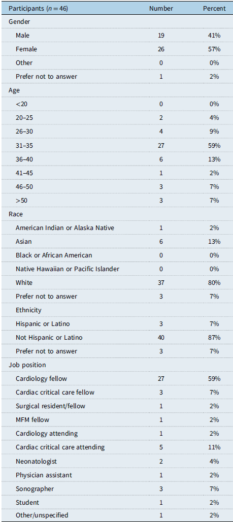
A total of 46 learners completed the survey, 19 (60% response rate) during the 2022 conference, and 27 (68–77% response rate) during the 2023 conference. A majority of survey participants were fellows (70%). There were more female (57%) than male (41%) participants. Demographic data of respondents are presented in Table 1.
Demographic information of participants

Perceived effectiveness of virtual cardiac models
Sixty per cent of respondents rated virtual cardiac models as more effective than textbook diagrams (n = 45) [95% confidence interval: 45–73%], as shown in Figure 2. The virtual cardiac models were rated as equally or more effective than didactic teaching (78% (n = 46) [64–88%]) and specimen videos (78% (n = 46) [64–88%]). Just over half of participants (54% (n = 46) [40–68%]) found the virtual models to be less effective than hands-on cadaveric specimen inspection.
Learners’ perception of the efficacy of virtual cardiac models in comparison to traditional teaching modalities when virtual models were viewed via laptop screen.

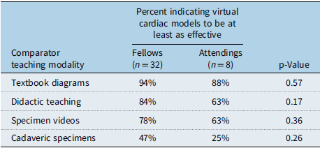
When responses were stratified by training level, as shown in Table 2, a higher percentage of fellows than attendings perceived the models to be at least as effective as each of the comparator teaching modalities. However, this trend was not statistically significant.
Percentage of survey respondents at each training level who perceive virtual cardiac models to be at least as effective as comparator teaching modalities

Perceived effectiveness of virtual reality headset
A majority of learners found the viewing of virtual cardiac models via virtual reality headset to be equally or more effective than textbook diagrams (81% (n = 43) [95% confidence interval: 67–90%]), didactic teaching (66% (n = 44) [51–78%]), and specimen videos (61% (n = 44) [51–78%]), Figure 3. Learners reported the virtual reality headset to be less effective (61% (n = 44) [47–74%]) than hands-on cadaveric specimen review. Most found viewing the virtual cardiac models via headset to be equally or more effective than viewing them via laptop screen (82% (n = 44) [68–90%]).
Learners’ perception of the efficacy of virtual cardiac models in comparison to traditional teaching modalities when virtual models were viewed via virtual reality headset.

Learner attitudes towards the virtual cardiac models
A majority of participants found the models to be an engaging (87%) and enjoyable (85%) way to learn cardiac morphology, as shown in Figure 4. Most participants also reported that after using the virtual models they were better able to retain a concrete mental image of cardiac defects (81%), and they had a deeper understanding of cardiac morphology (79%). A majority of participants reported they would recommend the virtual cardiac models to other learners (87%), and most reported they would use them for self-directed learning at home if they had access (85%). A slight majority (58%) of participants reported that viewing the virtual models via virtual reality headset enhanced their learning compared to viewing on a two-dimensional screen, as shown in Figure 5. However, participant preference for viewing the models via an online platform versus via the virtual reality headset varied, with similar numbers of participants reporting a preference for each mode of viewing, and many reporting no preference.
Attitudes towards the virtual cardiac models when viewed via laptop screen, rated on a 5-point Likert scale ranging from strongly agree to strongly disagree.

Attitudes towards viewing the virtual cardiac models via virtual reality headset, rated on a 5-point Likert scale ranging from strongly agree to strongly disagree.

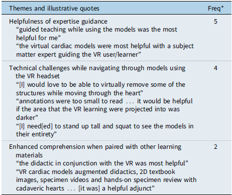
Assessment of the open-ended responses regarding participant experience with the virtual models, Table 3, revealed that learners felt the models to be more effective teaching tools when experts in cardiac morphology were present to guide them through the structures. Additionally, comprehension was enhanced when the virtual models were paired with other teaching modalities. Also, despite the overall positive attitudes towards the virtual reality headset, many participants reported technical challenges when navigating the models via headset that could be improved upon to better facilitate learning.
Survey participant themes regarding their experience with the virtual models (n = 11)

VR, virtual reality.
* Freq: frequencies indicate the number of individuals whose response included a particular theme.
Discussion
This study evaluated learner perceptions of a novel teaching tool in cardiac morphology. Innovative, contemporary educational material is needed to more effectively teach CHD, as current teaching is heterogenous, Reference Rogers, Klein, James and FitzGerald3 learning preferences are changing, Reference Rogers and Cohen9,Reference Windram, Neal and McMahon10 and use of traditional materials, such as heart specimens, is limited by location, time, and resources. Reference Seslar, Shepard and Giroud11
The use of virtual cardiac specimens, created using photogrammetry and viewed on traditional web browsers and in virtual reality, was well received. We found that participants perceived the virtual models to be more effective than textbook diagrams and equally or more effective than didactic teaching and specimen videos. When asked to compare the effectiveness of the virtual models with hands-on specimen review of the cadaveric hearts used to generate the models, responses were mixed, though a slight majority felt reviewing the cardiac specimens in person to be more effective than virtually. This is perhaps unsurprising, as the virtual models are unable to fully replace the kinetic experience of holding and manipulating a heart in one’s own hands. Reference Preece, Williams, Lam and Weller12 However, our data suggest that in circumstances where cadaveric specimens are not accessible, virtual cardiac models are an excellent learning tool.
In addition to accessibility, there are many other advantages that virtual models have over cadaveric specimens. One significant advantage is that of specimen preservation. A bulk of the specimens from large cardiac collections are already over 30–40 years old. Regardless of preservation method, specimens experience wear and tear, having a finite lifespan as a teaching tool. It is estimated that, in some collections, more than half of the specimens have already been rendered useless by time and frequent handling by teachers and trainees. Reference Seslar, Shepard and Giroud11 Our virtual models have the ability to preserve and share examples of cardiac pathology, including rare congenital defects, for many generations of learners to come.
Another benefit of the virtual cardiac models is that they encourage the kind of education promoted by adult learning theory – learning that is active, self-directed, and can be applied to the topic at hand. Reference Knowles13 Learners have control over the pace at which they navigate the models and can access them on any device anytime for a just-in-time refresher. Reference Barnett and Barnett14 Most learners in our study indicated that they would use these virtual models at home for their own self-directed learning and thought of them as an engaging educational tool. Furthermore, these virtual models may be increasingly appealing to newer generations of learners, who often prefer learning modalities that incorporate contemporary technologies. Reference Eckleberry-Hunt and Tucciarone15,Reference Hopkins, Hampton and Abbott16 In our study, a higher percentage of fellows than attendings reported the virtual models to be more efficacious than each traditional teaching modality. While this data was not statistically significant, our study likely lacked the power to fully assess this trend given our small sample size. These models also have the potential to be used as supplemental material to modernise existing curricula or as the basis for the creation of new e-learning curricula.
Limitations
There are several limitations to this study. First, the population surveyed was attendees of two conferences which may limit generalisability, as those who attended may not be representative of all paediatric cardiology learners. Next, we could not verify previous experiences learners had with comparator education modalities, though they were instructed to select “not applicable” for any modalities they lacked prior experience with. Additionally, there may be variables that could influence participants’ perceived efficacy with the virtual cardiac models, such as comfort with technology and familiarity with navigating virtual reality space, that were not explored in this study. Lastly, while this study focused on perceived effectiveness of the virtual models, it was unable to assess improved knowledge of cardiac morphology or changes to clinical practice.
Despite these limitations, we believe that these virtual models are a useful and accessible teaching tool with the potential to enhance knowledge of cardiac morphology through self-driven learning. Next steps would be to survey a larger audience and to design a study to measure objective outcomes, including knowledge acquired and retained through the use of our virtual cardiac models.
Conclusion
This study demonstrated that learners perceive the novel virtual cardiac models as at least equal to, if not more effective than, most current teaching materials including textbook diagrams, didactic teaching, and videos. The study also showed that learners still value working with cadaveric specimens, but that virtual models may be a valuable alternative for teaching cardiac morphology in settings where access to cadaveric specimens is limited. Overall opinion of the virtual reconstructions was exceedingly positive, and learners are eager to utilise these virtual models both via online platforms and using virtual reality headset devices. Virtual cardiac specimens can be an effective educational tool for cardiac morphology, allowing sharing of resources and expertise not available at all teaching institutions.
Supplementary material
The supplementary material for this article can be found at https://doi.org/10.1017/S1047951125000071.
Acknowledgements
The authors thank Javier Gentile, MD, for facilitating virtual reality viewing of cardiac specimens. The authors also thank the Media Lab at Cincinnati Children’s Hospital for helping develop techniques to scan pathology specimens.
Financial support
This research received no specific grant from any funding agency, commercial, or not-for-profit sectors.
Competing interests
None.