INTRODUCTION
Smaller species of Diopatra Audouin & Milne Edwards, 1833 are known to brood their eggs in the parental tube, where they undergo direct development and remain until they are ready to settle as juveniles and build their own tubes (Paxton, Reference Paxton1993; Pires et al., 2012). Diopatra marocensis Paxton et al., 1995 was originally described from near Sidi Boulbra on the south Moroccan Atlantic coast, and subsequently reported from a number of sites on the Portuguese coast (Rodrigues et al., Reference Rodrigues, Pires, Mendo and Quintino2009; Pires et al., Reference Pires, Quintino, Gentil, Freitas and Rodrigues2012) and as its northernmost distribution from northern Spain (Arias et al., Reference Arias, Anadón and Paxton2010). The reproductive biology of D. marocensis was studied by Fadlaoui & Retière (Reference Fadlaoui and Retière1995), Fadlaoui et al. (Reference Fadlaoui, Lechapt and Retière1995) and Pires et al. (Reference Pires, Quintino, Gentil, Freitas and Rodrigues2012). The former paper investigated the population dynamics, oogenesis, reproductive period and recruitment of the Sidi Boulbra population from Morocco, while Fadlaoui et al. (Reference Fadlaoui, Lechapt and Retière1995) studied its larval development. Pires et al. (Reference Pires, Quintino, Gentil, Freitas and Rodrigues2012) studied the reproductive cycle of a population from Ria de Aveiro, western Portugal. Fadlaoui et al. (Reference Fadlaoui, Lechapt and Retière1995) and Pires et al. (Reference Pires, Quintino, Gentil, Freitas and Rodrigues2012) reported brooding of large eggs (about 600 µm) in the parental tube, with most eggs and larvae of a very similar size, and the juveniles leaving the tube when they consisted of 25–34 chaetigers. The Ria de Aveiro population was found to have coelomic gametes and tubes with eggs and larvae in every sampling month, indicating year round reproduction with a peak period from April to September, and a male:female sex-ratio of 1:2 to 1:4 (Pires et al., 2012). For the Sidi Boulbra population, Fadlaoui & Retière (Reference Fadlaoui and Retière1995) reported also a continuous reproductive period throughout the year. Females were found to brood eggs and larvae in the winter and summer months and to contain immature and mature oocytes in their coelomic fluid in every sampling month. The population was reported to recruit juveniles throughout the whole year with a peak in spring and summer and to present three size-classes in the winter of 1990 and 1992, indicating a life span of at least two years (Fadlaoui & Retière, Reference Fadlaoui and Retière1995).
This paper reports on a one-year study of the reproductive biology of the D. marocensis population at the Villaviciosa estuary, northern Spain, with emphasis on gametogenesis and parental care and development, in order to elucidate the sexual strategy of this population.
MATERIALS AND METHODS
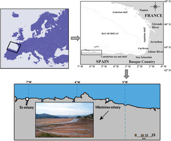
For the study of the reproductive cycle, samples of Diopatra marocensis were collected from the intertidal sandy flats of the Villaviciosa estuary, Asturias, northern Spain (Bay of Biscay), 43°18′–43°32′N 5°29′–5°32′W (Figure 1) at monthly intervals from March 2010 to April 2011. At this site, the D. marocensis density was very low, around one individual/m2, never exceeding two individuals/m2 (Arias et al., Reference Arias, Anadón and Paxton2010). We also re-examined the preserved material of this species (collected during 1976–2009) from the Collection of the Department of Biology of Organisms and Systems (Zoology) of the University of Oviedo (BOS-EUN8; BOS-EUN9; BOS-EUN10; BOS-EUN20). Voucher specimens of D. marocensis from the study site have been deposited at the National Museum of Natural Sciences, Madrid (MNCN 16.01/14333).
Location map of the sampling site: the Villaviciosa estuary.

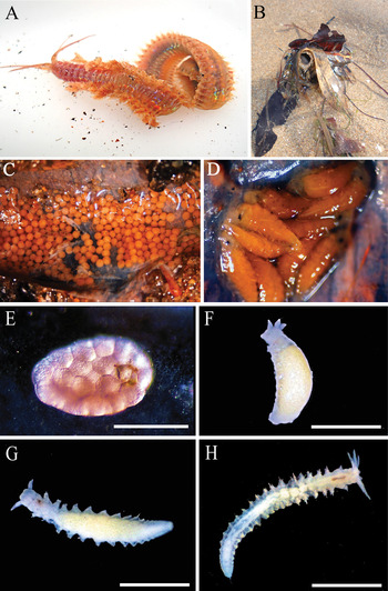
Collected specimens were brought alive to the laboratory anaesthetized in a 7.5% MgCl2 solution isotonic with seawater and extracted from their tubes (Figure 2A). The width of the 10th chaetiger of all specimens was measured with callipers, as size reference. Then, a sample of the coelomic fluid (~1 ml) of about 10–12 worms per month was extracted with a Pasteur pipette after making a short incision in their body wall at about the 30th chaetigerous segment. The extracted coelomic fluid was then mounted fresh on a cavity slide with seawater and examined under a binocular microscope. For each individual, the mean oocyte size from 40 oocytes was calculated using the average value of the longest and shortest oocyte diameter as estimate of oocyte size. All measurements were done monthly on freshly mounted oocytes using a calibrated eyepiece graticule. We also determined the mean number of eggs per brood. The oocyte diameter was used as an index of the stage of maturation. Furthermore, 8–10 tubes (Figure 2B) were sampled monthly during the annual period and fixed in 10% buffered formalin to assess the presence/absence of eggs and larvae. When they were present (Figure 2C, D), they were counted and measured (mean egg diameter; larvae total length and number of segments). The developing individuals are here referred to as larvae as long as they are within the parental tube, and as juveniles after leaving the tube to settle.
Diopatra marocensis: (A) live gravid specimen showing oocytes through the body wall; (B) distal end of tube of D. marocensis projecting from sediment, with attached vegetal material; (C) brooded eggs inside the parental tube; (D) brooded larvae with 5–6 chaetigers inside the parental tube; (E) morula stage of a fertilized egg removed from the coelomic cavity; (F) brooded larva with eight chaetigers; (G) brooded larva with 12 chaetigers; (H) brooded larva with 18 chaetigers. Scale bars: E, 400 µm; F, 2 mm; G, 2 mm; H, 3 mm.

In order to study gametogenesis and the time of spawning, serial histological sections were examined after treating individuals as follows. Specimens were fixed in Bouin's solution for 24 hours, washed thereafter in 70% ethanol for two days prior to dehydration, dehydrated following standard methods, transferred to xylene and embedded in paraffin wax. Serial, 6 µm thin sections were cut with a Leica 2045 microtome and double stained with haematoxylin and eosin. Mature sperm extracted from nephridial chambers of recently fixed animals we prepared for scanning electron microscopy (SEM) in order to study its ultrastructure. The extracted sperm was post-fixed overnight in a cold (T = 4°C) ultrafiltered seawater solution of 2.5% glutaraldehyde (pH = 7.2), rinsed in distilled water and dehydrated in an ascending series of ethanol, critical point dried using acetone as transition liquid and sputter coated with gold. Samples were then analysed using a JEOL 6610 LV scanning electron microscope. To study the sexual strategy, a total of 30 individuals were histologically studied. Since D. marocensis lacks secondary sexual characters, sex determination was accomplished by examining histological or microscopic preparations of wet-mounted coelomic content.
RESULTS
Parental care and development
Animals collected from the estuary ranged in length from 118 to 159 mm and the width of the 10th chaetiger ranged from 2.8 to 4.8 mm; their monthly mean width varied between 3.45 ± 0.6 and 3.95 ± 0.53 mm, with specimens of the largest size-class present in every monthly sample.
Brooding individuals were observed from March to July and were found in all size-classes with the smallest and largest one measuring respectively 2.9 and 4.8 mm in width. They incubated inside their tubes a single brood with synchronously developing embryos covered by a thin mucous lining (Figure 2C, D). Broods collected in March contained large, yellow coloured eggs in advanced cleavage phases, measuring 568–640 µm in size with a mean of 602 µm (N = 30; standard deviation (SD): 5.97) and also a small percentage, around 10%, of small yolky eggs 100–140 µm in size. Oocytes in advanced cleavage phases were also found inside the coelom of brooding females (Figure 2E). The mean fecundity, measured as the total number of eggs per brood, was 206 (range: 189–222; N = 30; SD: 2.08).
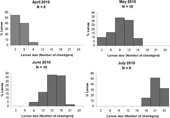
Development was lecithotrophic with larvae completing their development inside the broods. When the larvae attained a length of 2–3 chaetigers they lost their ciliation. Larvae of this size were already observed in April and May, numbering on average 194 larvae per brood (range from 181–202; N = 30; SD: 1.43). The larvae remained inside the brood pouch and were incubated by the parental worm until July. During this period extending from April to July, their mean size increased dramatically from 2–3 to 24 chaetigers (Figure 2F–H; Figure 3). In the same month the size of larvae varied widely among broods but within the same tube, the variation in size was narrow. In June, most larvae were completely developed, presenting an open digestive tube and functional jaws by the time they reached a size of 15–16 chaetigers. In July, inside the tubes only old broods containing larvae with 18–24 chaetigers were found (Figure 2H) and a mean number of 108 juveniles leaving the parental tube with 23 chaetigers were observed (range from 22–24; N = 30; SD: 2.76). Thus, approximately half of the number of deposited eggs developed to viable juveniles.
Size–frequency distribution of larvae of Diopatra marocensis inside the parental tube from Villaviciosa estuary from April 2010 to July 2010. N, number of parental tubes of each sample.

Gametogenesis
OOGENESIS
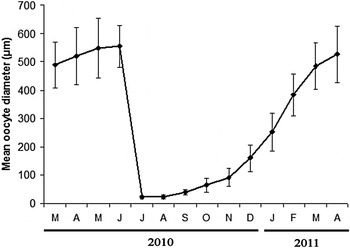
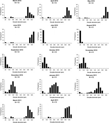
Field observation together with the monthly mean oocyte size variation (Figure 4) and the size–frequency distribution of the oocytes observed in the body cavity of the monthly collected specimens (Figure 5) indicated that in the Villaviciosa estuary the species exhibited a seasonal reproductive activity. During the summer months July and August, the population passed through a resting period in which exclusively inactive oocytes in the smallest size-classes with a mean diameter of 22–23 µm were found free in the coelom (Figure 4). Spawning occurred during a relatively short reproductive season extending from March to June, when ripe eggs in the largest size-classes from 500–620 µm represented more than 70% of the oocytes filling the coelomic cavity (Figure 5) and the mean oocyte diameter of the adults reached up to 485–555 µm (Figure 4). During this period, oocytes in the intermediate size-classes were almost absent in the coelom and only a small fraction of oocytes in the smallest size-classes (less than 10%) were present.
Mean coelomic oocyte diameter of Diopatra marocensis (µm ± standard deviation) per month during the study period.

Monthly size–frequency histograms of coelomic oocyte diameter of Diopatra marocensis from Villaviciosa estuary from March 2010 to April 2011. N, number of individuals of each sample.

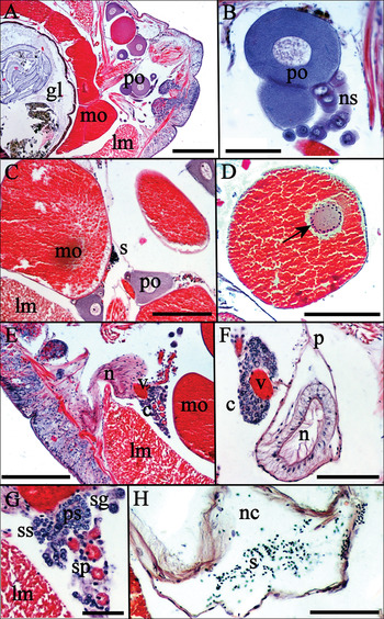
Within the same individual, oogenesis was asynchronous and except for the resting period, oocytes of all size-classes and in different stages of oogenesis could be observed inside the coelomic cavity (Figure 6A). Previtellogenesis and vitellogenesis occurred free in the coelomic cavity, as mentioned before, and eggs were laid in advanced cleavage stages (Figure 2E). The earliest stages of oogenesis were represented by clusters of oogonial cells arising from the coelomic epithelium arranged in raceme-like structures located on either side of the dorsal blood vessel. Some of these oogonial cells formed nests of fertile oogonia and the other neighbour cells were identified as nurse cells, typically associated with onuphid eggs. Then, the oogonia-nurse cell strings were released into the coelom and a single oocyte developed with two nurse cell strings attached (Figure 6B). The previtellogenic oocytes (Figure 6A–C) began cytoplasmic growth in September, attaining sizes ranging from 40–140 µm. Synthesis of yolk granules started in October and continued until the end of June. Vitellogenic eggs (Figure 6A, C) covered a wide range of size-classes from 180–500 µm. Fully ripe eggs fell within the sizes-classes of 540–620 µm. The nucleus in late vitellogenic oocytes is characterized by the presence of many peripheral micronucleoli (~30 nucleoli/nucleus) and the nuclear envelope is surrounded on its outer side by active ooplasm (Figure 6D).
Transverse sections of Diopatra marocensis: (A) gravid adult; (B) previtellogenic oocytes with strings of nurse cells; (C) mature oocytes, previtellogenic oocytes and mature sperm; (D) mature oocyte showing nucleus with micronucleoli; (E) overall view to show the nephridium with associated vessels and sperm cells, and mature oocytes; (F) nephridium and associated blood vessels with different mature stages of sperm cells cluster; (G) cluster of spermatogonial cells, spermatogonia, spermatocytes and spermatids; (H) nephridial chamber containing mature sperm. c, cluster of sperm cells; gl, gut lumen; mo, mature oocytes; po, previtellogenic oocytes; ns, nurse cells strings; v, blood vessel; lm, longitudinal muscle; p, peritoneum; ps, primary spermatocyte; n, nephridium; nc, nephridial chamber; s, sperm; sg, spermatogonia; sp, spermatids; ss, spermatocyte. Scale bars: A, 500 µm; B, 50 µm; C–D, 250 µm; E, 500 µm; F, 100 µm; G, 50 µm; H, 100 µm.

SPERMATOGENESIS
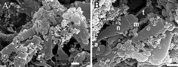
Throughout the whole year, spermatogonial cells appeared associated with the walls of small blood vessels that serve the lateral coelomic cavity and the nephridia. In late winter, part of them formed clusters of small spermatogonia (Figure 6F, G). The spermatogonia formed short chains (Figure 6F, G) and proliferated through mitotic divisions during the winter months of February and March, giving rise to nests of spermatogonia. Shortly before onset of spermatogenesis, peripheral spermatogonia increased slightly in size attaining a diameter ranging from 20–25 µm. They were recognizable by their big eccentric, irregular nuclei surrounded by a thin layer of eosinophilic cytoplasm. Primary spermatocytes, measuring about 10–15 µm in diameter and characterized by their oval or rounded shape and a large, inflated nucleus with clumps of chromatin distributed centrally and peripherally as irregular strands, appeared for the first time in February, indicating onset of spermatogenesis. They could be observed until March forming cysts adjacent to spermatogonia and cysts of secondary spermatocytes (Figure 6G). Secondary spermatocytes measured 7–8 µm in size. Next the spermatids were detached from the cysts and the blood vessels and continued the spermiogenesis free in the coelom close to the nephridia. Mature sperm were aggregated into small groups (Figure 6C) in the coelom and were also found inside modified nephridial chambers (Figure 6H). Mature sperm from the nephridial chambers had an elongated, high conical head topped by a narrow tubular projection, the acrosome (Figure 7A, B). The base of the head was slightly wider than the short ring-shaped mid-piece (Figure 7B). In April, the spermatogenesis ceased and only an exiguous quantity of mature sperm was observed in the coelomic cavity. Sperm production at any stage of the process was relatively low.
Scanning electron micrographs of mature sperm inside the nephridial chamber of Diopatra marocensis: (A) panoramic view of mature sperm masses inside the nephridial chamber; (B) enlarged view of the mature sperm. a, acrosome; m, mid-piece; n, nucleus; t, tail.

Sexual strategy
All gravid specimens were of a pinkish or orange colour, with oocytes visible through the body wall (Figure 2C). No cream-coloured specimens, suggesting ripe males, were encountered during any months. The histological study of gametogenesis of D. marocensis did not detect any specimens that were exclusively male during any month of the year. The only individuals containing spermatocytes were all developing oocytes at the same time, revealing that the population of Villaviciosa consisted of simultaneous hermaphrodites. Reproductive resources were allocated simultaneously to the production of eggs and sperm in the same reproductive segments ranging from chaetigers 22–25 to 60–65. However, the reproductive effort was unevenly distributed among sexes. Most of the reproductive resources were devoted to the production of large, yolk-rich eggs that were filling almost completely the coelomic cavity (Figure 6A, C, D) during the reproductive season, from March to June. The amount of mature sperm found from February to April, by contrast, was exiguous in volumetric terms. Spermatogenesis and oogenesis occurred also in topographically separated regions of the segment and were asynchronous: spermatogenesis took place close to the ventrolateral branches of the blood vessels and started in February, ending in April (Figure 6E, F). The oogenesis was extraovaric and occurred free in the coelom (Figure 6A–D); the early phase of the oogenesis, the previtellogenesis, started in September and vitellogenesis was completed between March and June. Thus, simultaneous hermaphrodites with ripe eggs and sperm were found only during the period extending from March to April. From March to May we observed individuals with modified dorsally extending nephridial chambers that were connected with the coelomic cavity.
DISCUSSION
In the Villaviciosa estuary, the population of Diopatra marocensis has a discontinuous reproductive season with a resting period during the summer months of July and August and a short spawning season from spring to early summer extending from March to June. Furthermore, post-reproductive death was not observed. Individuals in the largest size-classes were observed in all sampling months in a period spanning between two spawning seasons and brooding individuals were found in all size-classes, suggesting that the species is iteroparous. Iteroparity also occurs in the populations of D. marocensis from Morocco (Fadlaoui & Retière, Reference Fadlaoui and Retière1995). However, in Morocco and also in Portugal the reproductive season is continuous (Fadlaoui & Retière, Reference Fadlaoui and Retière1995; Pires et al., 2009). Differences in the mean surface water temperature may account for this.
A review of life history studies in the family Onuphidae (Paxton, Reference Paxton1986) and the genus Diopatra (Paxton, Reference Paxton1993; Pires et al., 2012) showed that members of the family produce relatively large eggs (170–1170 µm) that are not planktotrophic. Some species undergo a short free-swimming lecithotrophic larval stage before settling, but most species deposit their eggs in gelatinous masses or inside the parental tube where they undergo direct development by lecithotrophy. Not much is known about sperm transfer in the family. Hartman (Reference Hartman1967) described brooding in the Antarctic Paradiopatra antarctica (Monro, 1930) (as Paronuphis antarctica) where she observed a tiny specimen firmly appressed to the brooding female that she interpreted as a pygmy male.
The only report of a sperm transfer system is for Kinbergonuphis simoni (Santos et al., 1981), involving spermatophores that the females collect and store in their seminal receptacles (Hsieh & Simon, Reference Hsieh and Simon1990). The sperm of K. simoni have oblong heads, indicating that they are of the ent-aquasperm type as defined by Jamieson & Rouse (Reference Jamieson and Rouse1989). In D. marocensis, the mature sperm with its elongated head and modified ring-shaped mid-piece resembles those of the ent-aquasperm type. Presumably, this sperm may be stored in the modified nephridial chambers which may act as seminal receptacles until the time of fertilization. However, although the worms have internal fertilization, as suggested by the presence of eggs undergoing early cleavage divisions up to the morula stage within the coelom, and by the morphology of the mature sperm, we have no evidence as to how sperm transfer from one individual to the other would be accomplished.
The simultaneous hermaphroditic population of D. marocensis in the Villaviciosa estuary agrees with the general observations on polychaetes made by Heath (Reference Heath1977, Reference Heath1979). This author hypothesized that hermaphroditism is associated with internal fertilization and tube brooding in polychaetes. In fact, in polychaete families with mostly brooding species and sessile tubicolous forms, viz. Sabellidae and Serpulidae, hermaphroditism is widespread (Schroeder & Hermans, Reference Schroeder, Hermans, Giese and Pearse1975; Giangrande, Reference Giangrande1997). Within Onuphidae, hermaphroditism has been previousy reported only in Diopatra sp. (Lieber, Reference Lieber1931) and suggested for Rhamphobrachium ehlersi Monro, 1930 (Paxton, Reference Paxton1986).
According to Ghiselin (Reference Ghiselin, Giese, Pearse and Pearse1987), hermaphroditism should evolve under three conditions: (1) when finding a mate is difficult; (2) in small, genetically isolated populations; and (3) when one sex benefits from being larger or smaller than the other. With regard to how these conditions may apply to the Villaviciosa population, finding a mate may be difficult for D. marocensis, being a sessile tubicolous species existing in low densities (~2 individuals/m2) forming small patchily distributed aggregates. Moreover, D. marocensis is likely to be genetically isolated; the species has a direct development, and thus a low dispersal potential, and the nearest known population in the north of the Iberian Peninsula in the Eo estuary (~150 km distant from Villaviciosa) (Figure 1) is now extinct (personal observation by A.A.).
We hypothesize that in the case of D. marocensis, under the conditions mentioned above, the population in Villaviciosa has evolved simultaneous hermaphroditism with simultaneous production of sperm and oocytes. In populations from Portugal, by contrast, no evidence of simultaneous hermaphroditism has been found. There, only the presence of males and females was reported (Pires et al., 2012). However, sex determination was accomplished only by observing externally the colour of the ripe segments and was not corroborated by histological sections and the existence of hermaphrodites cannot be ruled out. In fact, the sex-ratio reported by Pires et al. (Reference Pires, Quintino, Gentil, Freitas and Rodrigues2012) with females twice or four times as abundant as males deviates from the expected Fisher 1:1 sex-ratio for dioecious species. This together with the fact that the smallest female found was larger than the smallest male, suggests that the population of Portugal may be protandric. The absence of simultaneous hermaphrodites in the population from Portugal may be explained by differences in the population size among both localities. Portuguese D. marocensis populations can reach densities exceeding 10 individuals/m2 (Rodrigues et al., Reference Rodrigues, Pires, Mendo and Quintino2009; Pires et al., Reference Pires, Quintino, Gentil, Freitas and Rodrigues2012), while in Villaviciosa D. marocensis occurs at low densities. It is possible that like in another simultaneous hermaphroditic polychaete species, viz. Ophryotrocha diadema Åkesson, 1976 (Schleicherová et al., Reference Schleicherová, Lorenzi and Sella2006; Lorenzi & Sella, Reference Lorenzi and Sella2008), in D. marocensis the population size influences the individual fitness and its sex allocation. In Villaviciosa, under conditions of low density, D. marocensis individuals may increase their fitness by allocating reproductive resources to the production of both gametes in the same individual. Under conditions of high population density, simultaneous hermaphrodites may be suppressed or reduced and sex allocation may favour the male or female function. In the dorvilleid O. diadema, the number of mates available in a population also influences sex allocation. In promiscuous mating conditions, where the number of reproductive competitors or potential partners increases, the allocation to the male function increases. In monogamous populations, simultaneous hermaphrodites are favoured (Lorenzi & Sella, Reference Lorenzi and Sella2008).
In isolated specimens of D. marocensis from the Villaviciosa estuary, the simultaneous occurrence of mature sperm and oocytes may favour self-fertilization. This seems quite probable, because the quantity of sperm found in gravid individuals was low and generally it is accepted that hermaphroditic polychaetes which are able to self-fertilize have reduced sperm production (Westheide, Reference Westheide1990; Eckelbarger, Reference Eckelbarger, Rouse and Pleijel2006). A lower sperm production indicates reduced male investment, likely due to low sperm competition and limited mating group size (Eckelbarger, Reference Eckelbarger, Rouse and Pleijel2006).
Evidence from the present study indicates that D. marocensis larvae use their own vitellus and other extraembryonic nutritive resources to complete their development. Besides the ripe (presumably fertilized) eggs, the broods of D. marocensis from Villaviciosa estuary contain also yolky eggs that are much smaller in size. These eggs may serve as nurse eggs for the developing larvae that probably engulf them whole by the time the larvae consist of 16–17 chaetigers. At this size, the gut tract begins to open and larvae have functional jaws, are able to manipulate and ingest nurse eggs and may switch from being endolecithotrophic, consuming their own vitellus, to feed on extraembryonic nutritive resources. Adelphophagy during the latter larval stages may also occur. This is supported primarily by the fact that the size of the juveniles leaving the parental tube varies widely within broods, as is common in species that feed on nurse eggs or siblings (Spight, Reference Spight1976; Rivest, Reference Rivest1983). Besides, the number of viable juveniles leaving the parental tube represents about 45% of the number of larvae with 2–3 segments. Adelphophagy occurs commonly in other polychaetes like spionids, especially Polydora Bosc, 1802 (Blake, Reference Blake1969; Woodwick, Reference Woodwick, Reish and Fauchald1977; Mackay & Gibson, Reference Mackay and Gibson1999). Therefore, D. marocensis development may combine the endolecithotrophic strategy at an early stage when the larvae are smaller than 15 chaetigers with the exolecithotrophic one at final stages, when the larvae have more than 16–17 chaetigers. A similar developmental strategy has also been reported in the spionid Boccardia proboscidea Hartman, 1940. This species is capable of producing planktonic larvae that are plantktotrophic only after variable periods of being lecithotrophic (Blake & Kudenov, Reference Blake and Kudenov1978; Gibson, Reference Gibson1997).
Another interesting finding of this study is the presence of peripheral micronucleoli in late vitellogenic oocytes of D. marocensis. Almost a century ago this was reported for Nothria conchylega (Sars, 1835) (as Onuphis conchylega) by Eulenstein (Reference Eulenstein1914), who considered them as remnants of the disintegrating nucleolus. Lieber (Reference Lieber1931) studied the oogenesis of some Diopatra species and remarked on the changes that the nucleoli undergo during oocyte growth. She noted that small parts of the initial two nucleoli broke off and moved toward the nuclear envelope where similar forms closely adhered to its outer side, suggesting a possible exit of nucleoli or eliminating some substances. Dhainaut (Reference Dhainaut1972) described the development of the nucleolus during oogenesis in Nereis pelagica Linnaeus, 1761, where in late vitellogenesis, near the time of sexual maturity the central nucleolar mass becomes rearranged into a number of micronucleoli that migrate towards the nuclear envelope, as we have observed for D. marocensis. The band of active ooplasm observed outside the nuclear envelope may indicate migration of materials in a similar way to the ‘nuage’ of the previtellogenic stage where nucleocytoplasmic granules migrate through the nuclear pores into the perinuclear ooplasm in a number of polychaetes (Eckelbarger, Reference Eckelbarger, Rouse and Pleijel2006).
ACKNOWLEDGEMENT
The first author is supported by a Severo Ochoa fellowship from the FICYT Foundation (Principado de Asturias, Spain).