Pulmonary artery aneurysms are a rare event characterised by aneurysmatic dilatation of the pulmonary vascular bed. Its incidence is 1 in 14,000 individuals, and it is often asymptomatic. It is usually diagnosed incidentally and often has a good prognosis. However, patients who reach the surgical margin experience various symptoms. The symptoms that prompt hospital admission of these patients include cough, dyspnoea, chest pain, and haemoptysis. Life-threatening complications of this pathology include pulmonary artery dissection, massive haemoptysis, and acute coronary syndrome as a result of coronary artery compression.Reference Gupta, Agrawal, Iakovou, Cohen, Shah and Talwar1
The most common causes of pulmonary arterial aneurysm are CHD, connective tissue diseases (e.g., Marfan’s syndrome), vasculitis, infection, neoplasm, and often iatrogenic traumas.Reference Nguyen, Silva, Seely, Chong, Lee and Müller2 A diagnosis of pulmonary artery aneurysm is appropriate when a main pulmonary artery diameter exceeding 43.4 mm in men and 40.4 mm in women or a pulmonary artery 1.5 times the normal diameter is detected.Reference Barney, Brown, Das and Kapoor3
Main pulmonary artery and/or right ventricular systolic pressure value can be used as high or low pressure in pulmonary artery aneurysm classifications.Reference Reisenauer, Said, Schaff, Connolly, Maleszewski and Dearani4,Reference Galiè, Torbicki and Barst5 Coexistence of pulmonary hypertension and right ventricular failure is very important in terms of clinical management.Reference Tuncer, Tuncer, Tas, Erdem and Polat6 In this study, we will share the results of our preferred surgical methods, experience, and approaches used to treat patients with pulmonary artery aneurysm.
Materials and methods
In our study, we retrospectively analysed 33 patients who were operated on in our cardiac surgery clinic from 2012 to 2020. All patients operated on for pulmonary artery aneurysm were included in the study. The patients’ age, gender, weight, diagnosis, past operations, and applied operation were obtained from the hospital’s archives and data processing system. This is a retrospective, observational, single-centre study. No emergency pulmonary artery aneurysm repairs were recorded during the period of our review; all of the cases we reviewed were identified as elective. Aneurysms and pseudoaneurysms in patients who were patched for right ventricular outflow tract reconstruction and corrective surgery with conduit were excluded.
Echocardiogram, CT triggers, and biochemistry tests were performed for all patients prior to surgery. Two methods were used in the surgical interventions: patients with aneurysms without thrombus underwent primary plication to reduce the outer sac without opening the aneurysm sac and aneurysm resection followed by a synthetic or biological graft interposition was performed in all other cases. All patients underwent a median sternotomy as an incision, and all interventions were performed under cardiopulmonary bypass. Arterial cannulation of the patients was aortic, femoral, or axillary, and venous cannulation was unicaval, bicaval, or femoral depending on the case. Routine electrocardiography, pulse oximetry, and intra-arterial blood pressure was monitored under anaesthesia. Bispectral index (BIS™: Covidien, MN, USA) was used to measure the depth of anaesthesia, and near-infrared spectroscopy (NIRS, INVOS 5100: Somanetics Corporation, Troy, MI, USA) was used to evaluate cerebral oxygenation.
The demographic and physical details of the patients included in the study were as follows: 17 (51.5%) were female and 16 (48.5%) were male; they ranged in age from 23 to 61 years (mean 30.66 ± 12.72 years); their heights ranged from 155 to 184 cm (mean 165.18 ± 15.71 cm); and their weights ranged from 57 to 117 kg (mean 88.91 ± 20.73 kg).
Mechanical ventilation settings were adjusted so that tidal volume was 8–10 ml kg−1, positive end-expiratory pressure was 3 cm H2O, and FiO2 was 50%; the respiratory rate was also adjusted so that each patient’s PaCO2 value was between 30 and 40. A pulmonary artery catheter was not placed due to the risk of dissection of the pulmonary artery aneurysm. Continuous cardiac imaging was obtained using a transesophageal echocardiography probe. The patients were placed in moderate hypothermia (31–26°C) during the surgery, and del Nido, blood, and custodiol were used as cardioplegia.
Daily radiological chest X-rays and echocardiographic evaluations were performed in the post-operative ICU follow-up of all patients. To decide which surgical intervention was appropriate for each patient, pulmonary artery diameter (>5 mm), pulmonary symptoms (i.e., presence and degree of pulmonary hypertension), and arterial and right ventricular pressures (>35 mmHg) were evaluated, in line with the decisions taken by the Joint Council of Cardiology and Cardiac Surgery. Surgical treatment planning and timing for each patient were determined according to his or her underlying pathology.
In the post-operative period, patients with pulmonary hypertension (PHT > 30 mmHg) were started on nitric oxide at a dose of 10–30 ppm; when necessary, this was increased by 5 ppm to a maximum of 80 ppm until an adequate response was obtained. Inhaled oral treatment and sildenafil (100–200 mg/day) was started. Thirty days pre- and post-surgery were used as the chronological markers of early mortality.
NumberCruncher Statistical System 2007 (Kaysville, UT, USA) software was used for the statistical analysis. Descriptive statistical methods (mean, standard deviation, median, frequency, ratio, minimum, and maximum) were used to evaluate the study data. Skewness, the Kolmogorov–Smirnov test, and histogram graphs were used as the criteria-of-fit for normal distribution in numerical data. The χ2 and Fischer tests were applied to the categorical data. Mann–Whitney U test was used to analyse numerical data that did not fit the normal distribution on categorical data, and the t-test was used for the normal distributed data. Spearman’s correlation was used for correlation analysis in numerical data. Logistic regression analysis was used to investigate the effect of all parameters on mortality.
This retrospective study was conducted in accordance with the ethical rules defined in the Declaration of Helsinki. The patients’ informed consent was obtained prior to their respective surgeries.
Results
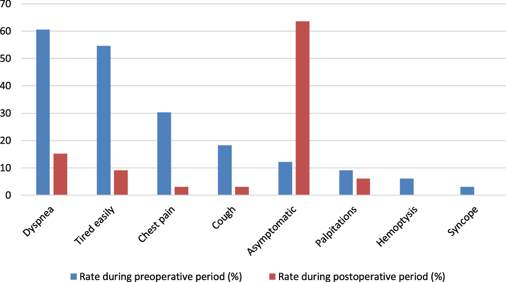
The patients’ preoperative anamnesis revealed that 20 (60.6%) had experienced dyspnoea, 18 (54.54%) tired easily, 10 (30.30%) had experienced episodes of chest pain, 6 (18.18%) had experienced chronic coughing; 3 (9.09%) had experienced palpitations, 2 (6.06%) had haemoptysis, and 1 (3.03%) had syncope. Only four patients (12.12%) were asymptomatic; some patients experienced multiple complaints simultaneously. In the post-operative period, five patients (15.15%) experienced dyspnoea, three (9.09%) reported fatigue, one (3.03%) reported chest pain, one (3.03%) experienced prolonged cough, and two (6.06%) experienced palpitations and other complaints. Haemoptysis and syncope were absent in the post-operative period, and 21 patients (63.63%) were asymptomatic (see Fig 1).
The percentage rates of the patients’ pre-operative and post-operative symptoms.

Cross-clamp times ranged from 0 to 202 minutes (mean 98.06 ± 41.73); total cardiopulmonary bypass times lasted 70–415 minutes (mean 148.18 ± 70.17 minutes); intubation times lasted 3–1800 hours (mean 81.64 ± 311.77 hours); ICU stays lasted 1–75 days (mean 6.27 ± 12.96 days); and hospital stays lasted 1–12 days (mean 4.45 ± 2.95 days). Vasoactive inotropic Score (VIS) scores ranged from 0 to 35 (mean 15.75). Echocardiographic evaluations revealed main pulmonary artery diameters ranging from 51 to 88 mm (mean 62.53 ± 10.46 mm).
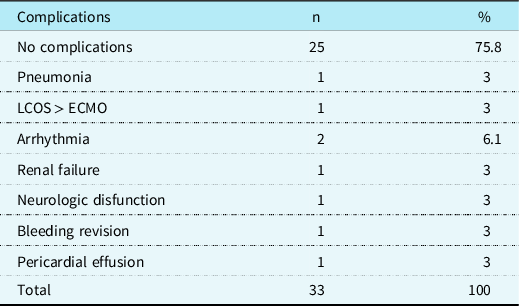
Twenty-five patients experienced no complications. A total of eight patients developed complications. When the complications were evaluated in terms of mortality, the mortality rate in the group without complications was 0% and there was a statistically significant difference in mortality between the two groups (p = 0.001). One patient developed post-operative pneumonia; one patient underwent extracorporeal membrane oxygenation due to low cardiac output syndrome; two patients developed arrythmia; one patient experienced renal failure;one patient developed neurological complications; one patient underwent bleeding revision; and one patient experienced pericardial effusion-related tamponade. The post-operative complications are listed in Table 1.
Post-operative complications and frequency in patients.

LCOS: low cardiac output syndrome.
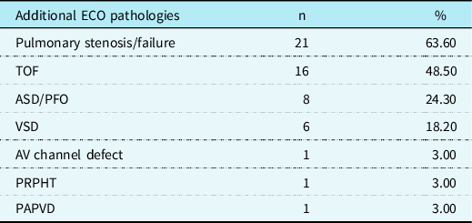
Additional pathologies with pulmonary artery aneurysms in the patients included in the study are presented in Table 2. Twenty-one (63.6%) patients experienced pulmonary valve pathology; 16 (48.5%) had a history of corrected Tetralogy of Fallot (TOF); and the remaining patients experienced Atrial Septal Defect/Ventricular septal defect (ASD/VSD), atrioventricular canal defect, primary pulmonary hypertension, and partial pulmonary venous return anomaly.
Additional pathologies with pulmonary artery aneurysm.

The following surgical procedures were performed to repair aneurysms: 10 patients (30.3%) received a graft interposition and 23 patients (69.7%) underwent pulmonary artery plication. There was no significant difference in mortality between these two groups of patients (p > 0.05). A total of 4 (12.1%) patients died: 3 from the plication group and 1 from the graft group. There was a significant difference in mortality between patients with complications and those without complications (p < 0.05).
Discussion
Increased pulmonary flow and pulmonary artery pressure increase the risk of pulmonary artery aneurysms rupturing. Surgical approaches are preferred in treating pulmonary artery aneurysms that exceed a certain diameter and pressure. However, the risks and long-term results of surgical treatment are not well defined and the results reported in the literature have varied greatly.Reference Graham and Shehata7 In this study, we have shared our own observations and experiences in treating a series of 33 patients who underwent surgery for pulmonary aneurysms.
Although pulmonary artery aneurysm patients can remain asymptomatic, as the diameter of an aneurysm increases, symptoms reported in the literature include haemoptysis, dyspnoea, chest pain, palpitations, and syncope. Cough as a result of bronchial compression by a large pulmonary artery aneurysm causes progressive dyspnoea, cyanosis, and/or pneumonia.Reference Park, Chamarthy, Lamus, Saboo, Sutphin and Kalva8 In our series, dyspnoea and fatigue were the most common symptoms, while syncope was the rarest symptom.
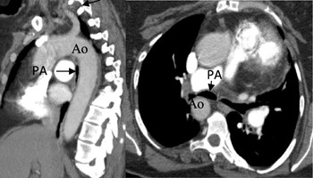
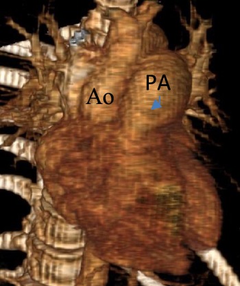
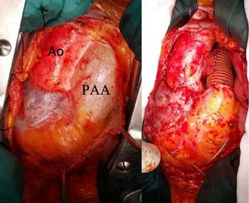
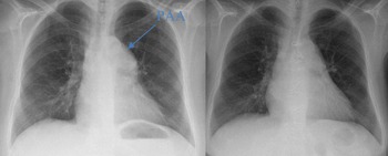
In pulmonary artery aneurysm patients, signs of right ventricular or right atrial hypertrophy on Electrocardiogram (ECG) are usually prominent. Systolic heart sounds during auscultation may accompany diastolic murmur. Hilar enlargement, the appearance of a lung nodule or pulmonary mass on X-ray, and transthoracic or transesophageal echocardiography are important tools and indications for evaluating heart function and pulmonary blood flow can be used to monitor the function of valves and shunts. Bronchoscopy can show compression of the bronchus. Angiography, evaluation of pulmonary vasculature and right-sided haemodynamic pressure, contrast-enhanced computed tomography diagnosis and size, number, location, and extent of pulmonary artery aneurysm, and magnetic resonance imaging can reveal arterial wall thickening, provide information about blood flow, and characterise aortic and pulmonary haemodynamics without exposing patients or health-care providers to radiation.Reference Kreibich, Siepe, Kroll, Höhn, Grohmann and Beyersdorf9 We routinely perform ECGs, chest X-rays, echocardiograms, and CT with contrast and decide whether an interventional angiography is appropriate for each patient individually (see Figs 2–5).
Tracheal stenosis secondary to pulmonary aneurysm in a patient with a pulmonary aneurysm is shown with an arrow. Ao: aorta; PA: pulmonary artery.

3D recon image of a patient with a pulmonary aneurysm in the pre-operative period. Ao: aorta; PA, pulmonary artery.

Pre- and post-operative images of the heart of a patient whose pulmonary aneurysm was repaired with a Dacron graft. Ao: aorta; PAA: pulmonary artery aneurysm.

Pre- and post-operative X-ray images of the lungs of a patient who underwent plication to correct a pulmonary aneurysm. The patient, whose post-operative lung load findings decreased, experienced increased aeration following the surgery. PAA: pulmonary artery aneurysm.

There is no definitive information in the literature on the best timing for operating on pulmonary aneurysms. Management strategies for low- and high-pressure aneurysms should be different.Reference Fakler, Mebus and Kaemmerer10 In a study of 38 patients, Reisenauer et al. detected advanced medial necrosis in their specimens in the pathological examination of pulmonary aneurysms exceeding 8 cm and reported that these patients should be operated on.Reference Reisenauer, Said, Schaff, Connolly, Maleszewski and Dearani4 They suggested that in the treatment of pulmonary artery aneurysms between 5 and 8 cm, decisions should be made according to each patient’s pressure and aetiology.Reference Reisenauer, Said, Schaff, Connolly, Maleszewski and Dearani4 In our clinical practice, we make decisions by considering right ventricular systolic pressure value, pulmonary artery pressure, pulmonary artery diameter, and aetiology while evaluating patients. Early surgery is preferred for all patients, regardless of whether they are symptomatic or asymptomatic, due to the increased risk of rupture caused by medial necrosis in the pulmonary artery wall with a pulmonary artery diameter greater than 8 cm. We decided on surgery for symptomatic patients with an MPA diameter of more than 5 cm. Based on our experience in this case series, although the diameter indication is important, the presence of serious symptoms also influenced our decisions. In our case series, we operated on patients with a pulmonary artery diameter within the surgical margin if severe symptoms such as trachea or bronchial compression were present.
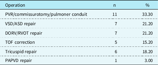
Surgical options for pulmonary artery aneurysms include primary aneurysm repair (plication) and pulmonary artery replacement with allogeneic or synthetic grafts. Any associated abnormalities detected are corrected simultaneously during the surgery. Surgical results are satisfactory and the short-term prognosis is good.Reference Fakler, Mebus and Kaemmerer10 Ten patients received allogeneic or synthetic grafts and the remaining 23 patients in our series received primary plication. The operations performed are listed in Table 3.
Operations performed.

Results from previous case reports have also shown improvements in lung function and Pulmonary Hypertension (PH) after surgery. Data from past case reports and series also indicate that treating pulmonary hypertension with vasodilators does not affect the growth rate of pulmonary artery aneurysms. Lung transplantation may be an option in severe parenchymal or progressive pulmonary vascular disease.Reference Ahmed, Ramani, Ghoreishi, Safdar and Griffith11,Reference Hou, Ma and Liu12 All of our pulmonary hypertension patients are treated with nitric oxide inhalers and sildenafil in the post-operative period, as we believe that it has a positive effect on patients experiencing pulmonary hypertension attacks triggered by cardiopulmonary bypass in the early post-operative period.
Veldtman et al. suggested that the timing of surgery in low-pressure pulmonary aneurysms should be based on changes in right ventricular size and function due to pulmonary stenosis or insufficiency, rather than aneurysm size.Reference Veldtman, Dearani and Warnes13 In cases of severe life-threatening haemoptysis or compression, the indication for surgery is indisputable.Reference Veldtman, Dearani and Warnes13 In our patient series, MPA diameters ranged from 51 to 88 mm (mean 62.53 mm). Surgical treatment was preferred in patients with accompanying compression or other symptoms.
There are several haemodynamic factors that can contribute to pulmonary artery aneurysm, one of which is increased pulmonary blood flow in wide left-to-right shunts. Although congenital lesions are more frequently associated with pulmonary artery aneurysm, the largest pulmonary artery diameters were found to be associated with patients who had tetralogy of Fallot, especially those with an abscessed pulmonary valve. This is thought to be most likely due to pulmonary regurgitation. The turbulence of the blood flow created by pulmonary regurgitation can lead to significant dilatation of the proximal pulmonary artery due to the very low resistance and high compliance of the pulmonary artery. Furthermore, the presence of pulmoner regurgitation may exacerbate haemodynamic stress as the stroke volume increases and could cause large root aneurysms.Reference Gallego, Rodríguez-Puras and Serrano Gotarredona14,Reference Rahman, Cook, Zheng and Rehman15 In our patient series, 21 patients (63.6%) had pulmonary valve pathology, 16 (48.5%) had a history of corrected TOF, and the remaining patients had ASD/VSD, atrioventricular canal defect, primary pulmonary hypertension, or partial pulmonary venous return anomaly. We believe that pulmonary valve insufficiency contributes to the development of pulmonary aneurysms.
Pulmonary aneurysms usually disrupt the normal anatomy of the mediastinum and may compress surrounding tissues. The difficulty of access and the fragile nature of the pulmonary artery due to its posterior location create difficulties during surgical interventions. A multidisciplinary, experienced, and careful approach is required to achieve successful results when treating this rare patient group.Reference Haj-Yahia, Sbaih and Bali16 In our series, four patients died, comprising a mortality rate of 12.1%.
According to Laplace’s law, wall tension is directly proportional to vessel pressure and vessel radius and inversely proportional to wall thickness. Shin et al presented distinctive plication techniques to reduce wall tension in pulmonary aneurysms: they argued that avoiding the use of foreign materials such as grafts reduces bleeding contributes to the reduction of pulmonary pressure.Reference Shin and Suh17 In our own series, we preferred the primary plication method for 21 patients. We also operated on the pulmonary valve in 11 patients with pulmonary valve insufficiency. We believe that correcting the pathologies of the pulmonary valve in patients who are to undergo surgery for pulmonary artery aneurysms will reduce the likelihood of recurrent aneurysms developing and accelerating the improvement of right ventricular function in post-surgical follow-ups.
Conclusion
Pulmonary artery aneurysms are an unexplored area of cardiovascular surgery, and are thus considered a gray area. Pulmonary artery aneurysm surgery is a safe and life-saving treatment that prevents fatal complications such as pulmonary artery aneurysms, but surgical timing and treatment strategies have yet to be addressed in the guidelines clearly. We believe that satisfactory results can be obtained by experienced surgical teams performing plication, reduction pulmonary arterioplasty, or graft replacement to correct pulmonary artery aneurysms. The indications for planning and timing of surgery must be carefully considered, taking into account pulmonary artery pressure, right ventricular systolic pressure value, and pulmonary artery aneurysm diameter in symptomatic patients with aneurysmatic dilatation over 5 cm or asymptomatic patients with aneurysmatic dilatation over 8 cm.
Acknowledgements
No funding was obtained for this study.
Financial support
This research received no specific grant from any funding agency, commercial, or not-for-profit sectors.
Conflicts of interest
None.
Patient consent statement
Consent was not obtained and waived.
Data availability statement
Data sharing is not applicable to this article as no new data were created or analysed in this study.
Institutional review board approval or waiver
The study protocol was approved by hospital management. This retrospective study was conducted in accordance with the principles of the Declaration of Helsinki.