Introduction
Surface modification plays a relevant role in the optimization of surface properties, especially for alloys used in biomedical applications. Indeed, the traditional alloys used in the biomedical industry usually assure satisfying “bulk” mechanical properties for specific implants. However, surface properties are generally not suitable for an optimal electrochemical and biochemical interaction with the surrounding environment. Living tissues, biologic systems and organs are in fact affected by the chemical exchange and mechanical interactions of devices inserted in the body. Inflammation, blood coagulation, and restenosis can be triggered when an undesired interaction with the device takes place.[Reference Qi, Maitz and Huang1]
Co–20Cr–15W–10Ni, commercially known as L605, is a high-performance alloy used for the fabrication of commercial stents, and other medical implant devices.[Reference O'Brien and Carroll2] L605 structure and chemical composition[Reference Shingledecker, Glanton, Martin, Sparks and Swindeman3] are accountable for high-mechanical properties that are needed in cardiovascular applications.[Reference Poncin and Proft4] In spite of this, the alloy is composed of elements (Co, Cr, and Ni) that could present a certain degree of local or systemic toxicity.[Reference Hildebrand and Champy5] In fact, these elements could be responsible for the formation of debris, of compounds and complexes that are noxious for the biologic environment[Reference Haynes, Crotti and Haywood6]; furthermore, even the released ions in the bloodstream could be responsible for metal accumulation in other body parts. Hence, a control of the surface properties, for example by the formation of a cytocompatible surface barrier reducing element selective leaching from the substrate, could constitute a valid method to improve the clinical outcome of metallic cardiovascular devices.[Reference Jaganathan, Supriyanto, Murugesan, Balaji and Asokan7]
The current work describes the surface modification obtained by an oxygen plasma immersion ion implantation procedure applied to L605. The electropolished (EP) samples were subjected to surface modification in a plasma implanter, to study the effect of the antenna power (between 300 and 150 W), and of the treatment durations of 60, 120 and 240 min. The alloy bulk and surface features, before and after the surface treatment, were assessed by means of several techniques, such as scanning electron microscopy (SEM), electron dispersive spectroscopy (EDS), x-ray diffraction (XRD), atomic force microscopy (AFM), sessile drop contact angle (CA), and transmission electron microscopy (TEM).
Surface chemistry was assessed by x-ray photoelectron spectroscopy (XPS), while cytocompatibility was evaluated by direct contact human umbilical vein endothelial cell (HUVEC) tests.
Materials and methods
Material preparation
A 0.4 mm-thick L605 sheet (Rolled Alloys® Laval, QC, Canada), with a nominal composition as given in Table I, was cut into 10 × 10 mm2 pieces. Each sample was hand-cleaned with an industrial detergent (Citranox, Sigma Aldrich), then subjected to a cleaning process consisting of three subsequent steps: (1) a 10 min ultrasonic bath in 20 mL of pure acetone, (2) a 10 min ultrasonic bath in 20 mL of deionized water, and finally (3) a 10 min ultrasonic bath in 20 mL of pure methanol.
After cleaning, the specimens were EP with an electrolyte comprised of 67% v/v H3PO4 (85% purity), 20% v/v industrial grade H2SO4 (95–98% purity), 3% v/v industrial grade HF (50%), 10% v/v deionized water (10%), and ten drops of glycerol. All the reagents were purchased from Laboratoire MAT, Montréal, QC, Canada. A 10 × 10 mm2 piece of L605 sheet was used as a cathode. The distance between the electrodes was 6 cm. The current between the electrodes was 2.5 A, corresponding to a voltage of ~8–10 V and to a self-stabilizing temperature of 45–50 °C. The duration of the procedure was 6 min. After electropolishing, the samples were rinsed 5 min in an ultrasonically stirred (USS) deionized water bath, then for 5 min in an USS 0.5 M NaOH basic solution, again rinsed 5 min in an USS deionized water bath, and finally dried with particle-free compressed air.
Nominal composition of L605, the alloy used as a substrate for the following surface modification.

Plasma implantation
Oxygen implantations were performed with a PBII-300 system (Plasmionique, Varennes, QC, Canada) consisting of a radio frequency inductively coupled plasma reactor connected to a pulsed high voltage source. The base pressure was P base ~2 × 10⁻⁷ Torr; pure oxygen (99.995% pure, Linde Gas Canada) was used as an implantation gas, in the range 4.8–5.0 × 10⁻³ Torr, with a feeding oxygen flow of 10 sccm. The water-cooled antenna was operated at a frequency of 13.56 MHz. The antenna power was in the range of 150–300 W; the distance between the antenna and the substrate holder was ~35 cm. The external antenna was separated from the deposition chamber by a quartz window. The substrates were implanted with a pulsed bias of U bias = −10 kV. The substrate bias was applied by means of short pulses of 50 µs (sharp edges of ~100 ns), with a repetition rate of 100 Hz. Samples were divided into two groups, that is A and B. Samples belonging to group A were deposited with an antenna power P = 300 W and they were exposed to the surface modification treatment for three different durations, respectively, 60 min (A1), 120 min (A2), and 240 min (A3). Group B samples had their surface modified with an exposition time fixed to 240 min, but the antenna power was, respectively, 150 W (B1), 225 W (B2), and 300 W (B3). A3 and B3 have the same deposition parameters; the double name was maintained for the sake of clarity, especially when the sample features of the same group were compared. Table II summarizes the specific implantation conditions for each group of implanted samples.
Sample denomination and specific deposition conditions for each sample group.

Characterization
XPS (PHI 5600-ci spectrometer, Physical Electronics, USA) measurements were carried out with an incident angle of 45° and a residual pressure of 3 × 10⁻⁹ Torr. A survey spectrum covering the range of 0–1400 eV was first recorded using an Al Kα x-ray source (1488.6 eV) at 200 W. High-resolution spectra of O1s, C1s, Co2p, and Cr2p regions were recorded with a magnesium Kα x-ray source (1253.6 eV) at 150 W. No charge neutralization was required. Three samples per condition were investigated respectively by survey and high resolution.
AFM investigations were performed in tapping mode with a Dimension™ 3100 (Digital Instrument, CA, USA) using a T300R silicon tip (VISTA Probes, Nanoscience Instruments Inc., Phoenix, AZ, USA). R q and R a were calculated as indicators of surface roughness,[Reference Gadelmawla, Koura, Maksoud, Elewa and Soliman8] for a reference area of 1 µm × 1 µm. Five specimens for each condition were analyzed.
A JEOL JSM-840A SEM (tungsten filament, acceleration voltage of 15 kV, probe current 1 × 10⁻¹⁰–1 × 10⁻⁹ mA for imaging, and 1 × 10⁻⁸ mA for microanalysis) was used for surface analysis. The microscope was equipped with a Bruker electron dispersive analysis system (EDS).
The relative hydrophobicity of the material surfaces was assessed according to ASTM-D7490: the CA of three samples was measured at three different areas of each sample by a drop shape analysis system VCA optima XE (AST Products, Billerica, MA, USA). An amount of 5.0 µL of deionized water was used as a test fluid; droplet stabilization was carried out for around 20 s.
A Philips® CM 200 microscope operating at 200 kV and equipped with a LaB6 filament was used for TEM analysis. Samples (lamellae) for electron microscopy observations were prepared by the focused ion beam (FIB) technique in a ZEISS NEON Cross Beam 40EsB microscope. Before milling, a layer of Pt was deposited at the place of cutting in order to protect the thin sample against heavy Ga ions during the preparation. Final milling was performed with the 4 keV Ga+ beam.
Potentiodynamic tests were performed in a 0.152 M NaCl solution, using a conventional three electrode cell (Princeton Applied Research Model K47), with two graphite rods and saturated calomel as counter and reference electrodes, respectively. The prepared L605 specimens were used as working electrodes. The tests were carried out according to the ASTM G59-97 standard. The experiment was performed after the open circuit potential settled for 3600 s. A scan rate of 0.166 mV/s, with an applied potential range of 1 V, was used. The experiment was carried out in an aerated environment at 37 ± 1 °C. The solution was mechanically stirred (80 rotations/min). For each condition a minimum of five specimens were tested. The corrosion rates were calculated on the basis of the abovementioned standard.
Viability cell tests
HUVECs were isolated from an umbilical cordon provided by the Saint François d'Assise Hospital. The detailed protocol for the isolation and characterization of cells was described elsewhere.[Reference Montaño-Machado, Noël, Chevallier, Turgeon, Houssiau, Pauthe, Pireaux and Mantovani9]
A resazurin test was performed in order to assess the metabolic activity of the HUVECs. Plasma-modified samples were immersed in ethanol for 5 min and rinsed with phosphate-buffered saline (PBS) right after. A total of 50,000 HUVECs (approximately 30,000 cells/cm2) was added into 400 µL of culture medium. After 1, 3 and 7 days of culture at 37 °C, culture medium was carefully removed and samples were rinsed with PBS. Then, 400 µL of M199 medium containing 10% of resazurin were added. After 3 h of contact with resazurin solution, 150 µL of the medium were collected and placed in a 96-well plate. Absorbance was read at 600 nm using a spectrophotometer ELISA reader (BioRad mod. 450, Mississauga, Ontario, Canada). The remaining medium on samples was removed and replaced by 400 µL of culture medium. Cells seeded directly on the plate of polystyrene were used as a positive control. The absorbance measurements were normalized to a negative control which was the solution of culture medium and resazurin with no cells. The results presented are the mean of three samples. Significant differences were determined by running a one-way analysis of variance which was followed by the Tukey's post-hoc method. A P-value of <0.05 was considered significant (*).
Results and discussion
Figures 1(a)–1(c) show SEM topography images of the base alloy for different conditions, respectively, in the as-received conditions [Fig. 1(a)], after electropolishing [Fig. 1(b)], and after implantation [Fig. 1(c)]. Figure 1(a) shows rolling marks, irregular scratches, pitting, and particles, which can be attributed to the plastic deformation undergone by the alloy.[Reference Favre, Koizumi, Chiba, Fabregue and Maire10] The electropolishing process removed all the previously mentioned defects; the obtained flat surface exhibited the presence of polygonal grains, twins, and a significative number of precipitates, consistent with the presence of carbides [Fig. 1(b)].[Reference Kilner, Pilliar, Weatherly and Allibert11, Reference Poncin, Gruez, Missillier, Comte-Graz, Proft, Venugopalan and Wu12] After plasma implantation, for example for B1 condition, the sample surface appears again flat and grains are not visible any more [Fig. 1(c)]. Figure 1(d) shows three EDS spectra, one for a typical EP sample [that is the one corresponding to Fig. 1(b)], one for an implanted B1 sample [that is the one corresponding to Fig. 1(c)], and another one for the particle in the inlet in Fig. 1(b), pointed by the arrow. The spectra corresponding to the EP and the implanted conditions showed mainly the presence of Co, Cr, W, and Ni peaks. Their spectra are compatible with those of a commercial L605.[Reference Poncin, Gruez, Missillier, Comte-Graz, Proft, Venugopalan and Wu12] The spectrum related to the particle in Fig. 1(b) showed the same main peaks as for the other patterns, but with a different intensity: in detail, the W Mα and the Cr Kα peaks were more relevant than those of the EP alloy. This means that the chemical composition of the precipitate in question is compatible with that one of a W–Cr-based carbides, as already evidenced by Poncin and coworkers.[Reference Poncin, Gruez, Missillier, Comte-Graz, Proft, Venugopalan and Wu12]
SEM surface images of (a) the as-received L605 alloy, (b) after electropolishing (carbides are shown in the inlet), and (c) after implantation (a carbide is shown in the inlet); (d) EDS patterns of panels (b) and (c); (e) bright field TEM image of an implanted sample (A3/B3): the presence of nanocrystals is evidenced by the arrows; and (f) corresponding diffraction pattern showing the sample microstructure.

In order to investigate the inner structure of the oxidized layers, TEM analysis was performed on cross-sectioned samples. Figure 1(e) shows a TEM bright field image of the A2 sample. The implanted layer is clearly visible between the Pt film used for FIB preparation and the Co alloy matrix. The layer thickness was uniform and about 20 nm thick. It is composed of amorphous material inside which small nanoparticles are present. In particular, the dark arrows in the same figure identify two of these nanoparticles where some atomic planes are visible as alternating black/white parallel strings. To identify the crystallographic structure of the nanoparticles, selected area electron diffraction (SAED) measurements have been performed. The SAED pattern of the sample region imaged in Fig. 1(e) is shown in Fig. 1(f). The most intense diffraction spots are due to the face-centered cubic cobalt alloy in the  $[2\overline {11} ]$ zone axis.[Reference Yamanaka, Mori, Kuramoto and Chiba13] There are also some rings having a diffused intensity; the associated interplanar distances are compatible with the lattice planes of the Pt used for FIB preparation. Moreover, some small diffraction spots, outside the rings, are also visible and their associated lattice distances (0.284, 0.242, and 0.203 nm) are compatible with the presence of the Co3O4 oxide (d(200),
 $d(21\bar{1})$, and d(220)). Summarizing, TEM analysis puts in evidence the formation of an amorphous and nanocrystalline layer of mainly Co oxide.
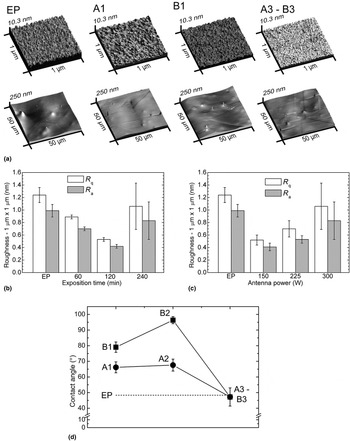
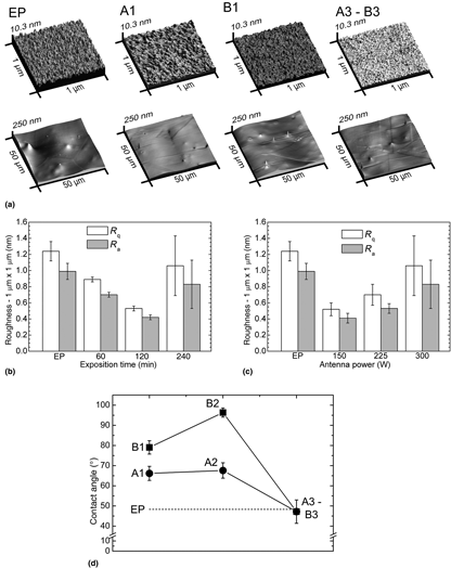
Typical three-dimensional surface topographies of the EP, A1, A3/B3, and B1 samples are shown in the AFM images of Fig. 2(a). The surface morphology is shown for two different area sizes, that is for a 50 µm × 50 µm area and for a 1 µm × 1 µm one. Large scale AFM images highlighted the presence of the same elements already found during SEM investigations, that are twins, grain boundaries, and carbides; the surface wavy shape and the occasional presence of holes can be both attributed to the electropolishing process, as precipitates could be removed from the matrix without being affected by the process.[Reference Bettini, Eriksson, Boström, Leygraf and Pana14]
(a) AFM three-dimensional representations of EP, A1, B1, and A3/B3 sample surfaces for 1 × 1 µm2 and 50 × 50 μm2; R q and R a roughness values for (b) A series samples, and (c) B series samples; (d) CA values for A and B series samples.

The implantation process modified slightly sample roughness [Figs. 2(b) and 2(c)]; the EP and the A3/B3 conditions were the roughest ones (R q = 1.2 nm). For group A samples, a 4-h exposition time to plasma increased the roughness, as it is evident for A1 and A2 conditions (R q = 0.9 nm and R q = 0.6 nm, respectively) compared with the A3/B3 one. Also for B sample group, for an increasing applied antenna power, samples showed an increasing roughness trend: for B1, R q was 0.5 nm, while for B2 R q = 0.7, and for B3, R q = 1.2 nm.
Static CA values [Fig. 2(d)] were the same for EP and A3/B3 samples (~45°); the highest value was found for the B2 condition, that is ~95°; the values for the other conditions were in the range of 65°–80°.
It must be stressed that it is difficult to define some general trends in the final features of the modified zones in our samples, as a function of the experimental conditions (treatment time and RF power), because they are the result of different phenomena. Kaeppelin et al.[Reference Kaeppelin, Carrère, Torregrosa and Mathieu15] studied how the applied power had an influence on the plasma parameters: by increasing the applied power, in the case of N2, n e (electron density, 1/cm3) increased linearly in the range of 100–300 W. The measure of n e can be considered equivalent to the measure of the ion density n i (1/cm3), that is n e ≈ n i: in fact all the ionic species present in the considered plasma, that is N+ and N2+, are monocharged. Nevertheless, the [N+]/[N2+] ratio changes as a function of the applied power. For a pure O plasma with a similar pressure range of this work, O2+ was the dominant species, for an applied power of 300 W.[Reference Wang and Olthoff16] Wang and Olthoff[Reference Wang and Olthoff16] reported a higher amount of O+ ions for other plasma conditions. For example, for ~7.5 mTorr, the O2+/O+ ratio was 1.6, and it was increasing with increasing pressure: for example, it reached a value of ~3 for ~30 mTorr and ~4 for ~45 mTorr; the total amount of ions decreases with pressure.
The flattening of the surface, corresponding to a low roughness for A1 and B2 conditions, is related to an increased adatom mobility, which is a function of the incoming energy, and to a limited sputtering effect. In fact, the preferential sputtering of Cr oxide leaves the surface enriched in Co oxides, and the recrystallization of the surface, for higher fluences, is responsible for an increase in roughness. The CA measures followed the exposed roughness trend.[Reference Nastasi and Mayer17] The synergy of these effects was responsible for the final chemical composition and the morphology of the investigated samples; the energetic particles impinging on the surface remodel the surface, by increasing the thermal flux and providing higher adatom mobility.[Reference Ensinger18] Depending on which effect among the abovementioned was prevailing, a different roughness and surface energy were obtained.[Reference Nastasi and Mayer17]
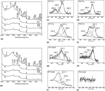
Typical XPS survey spectra for EP, A, and B group samples are shown in Figs. 3(a) and 3(b). Traces of Si were found on implanted samples, due to O etching of the glass window during the implantation process. Sputtering phenomena due to mainly O+ and O2+[Reference Wang and Olthoff16] are caused by the electrostatic coupling from the coil to the plasma; this is known to cause not only the sputtering of dielectrics in proximity of the antenna (quartz window), but also of other materials near the coil.[Reference Suzuki, Konishi, Nakamura and Sugai19] The EP surface showed the presence of elements due to the alloy, such as mainly Co (~7 at.%) and Cr (~11 at.%), and small amounts of Ni and W (2–3 at.%). C (~30 at.%) and O (~45 at%.) were present because of the interaction of the surface with the atmosphere; traces of Na, P, F, and N were also found. The presence of these elements can be attributed to the electropolishing process, in which an HF and H3PO4-based electrolyte was used. The presence of Na could be attributed to the cleaning process after electropolishing, in which a 1 M NaOH basic solution was used. The presence of N could be attributed to N2 adsorption from the atmosphere.
XPS surveys for (a) A and (b) B series samples. High resolution spectra, respectively, for C1s, O1s, Co2p3, and Cr2p3, in the EP (c, e, g, i) and for a typical implanted condition (d, f, h, j).

After implantation, mainly Co was found on the surface (10–15 at.%); C decreased to ~20 at.%, while O increased to 45–55 at.%. The natural oxide formed on the EP samples was a mixture of Cr and Co oxides; the amount of these elements is different from that in the bulk alloy, with a Co/Cr ratio of ~2.5. C [Figs. 3(c) and 3(d)] is present in a very small amount in L605, so that the percentage found on EP and implanted samples could be mainly attributed to the presence of metallic carbides (~282.5 eV), C–C and C–H2 (284.8 eV), –C–OH (~286.4 eV), and –O–C=O (~288.5 eV).[Reference Miller, Biesinger and McIntyre20]
Also for O1s, the same peaks for the EP and the implanted samples were found, as shown in Figs. 3(e) and 3(f); peaks at ~530.0, ~531.2, and ~532.2 eV could be attributed, respectively, to the O present in metal oxides, in hydroxides, and in adsorbed water. For the EP condition, the most relevant peak was the one related to the presence of hydroxides (~65%), hence its low CA value; the peak related to the presence of oxides was the most evident after implantation (~45 at.%).[Reference Biesinger, Payne, Grosvenor, Lau, Gerson and Smart21]
Co2p3 peaks [Figs. 3(g) and 3(h)] presented different features when the EP and the implanted samples were compared. In particular, Co2p3 was composed of contributions located at ~777.5 eV (metallic Co) and ~781.2 eV (CoO or Co(OH)2), ~784.7 and ~788.7 eV are satellites of the previous one. After implantation, the most relevant contribution was at ~780.0 eV, compatible with the presence of Co3O4, confirming the diffraction measurements; all the other peaks are satellites of the main one.
Cr [Figs. 3(i) and 3(j)] was present only in the EP sample, and it disappeared after implantation; the binding energy (BE) of the Cr2p3 region was resolved in three contributions, that is one (~574.3 eV) attributed to metallic Cr, a second one (~576.0 eV) to Cr2O3, and the third one (~574.3 eV) to Cr(OH)3.
An enrichment in Cr at the surface for L605 alloys is reported in an investigation for thermal treatments, both in air and under a low pressure atmosphere[Reference Swami22]; the preferential oxidation of Cr compared with Co is a well-documented behavior in the literature, for different conditions of surface treatment.[Reference Stott, Wood and Hobb23] The presence of metallic Co and Cr on EP samples could be attributed to the reduced thickness of the surface layer formed after electropolishing, and the non-thermodynamic equilibrium nature of the process. The absence of Cr on the surface of implanted samples could be attributed to the preferential sputtering of Cr2O3: in fact, the average density of Co3O4 is higher than the Cr2O3, and the sputtering yield is inversely proportional to the compound density.[Reference Behrisch24]
Figures 4(a) and 4(b) show, respectively, the potentiodynamic diagrams for A and B sample series, compared with the EP condition. The EP sample OCP was decreasing for all the treated conditions, suggesting a dissolution of the surface oxide, enriched in Co, until a thermodynamical stable enriched Cr layer was obtained; the OCP potential for all the implanted materials was however higher than for the EP condition, whose oxide thickness was lowest than for the other conditions. The oxygen ion implantation is responsible for a change in the alloy corrosion properties. For all the implanted samples, E corr was always slightly higher than that of the EP condition. Corrosion current densities showed lower values for the treated samples (from 0.47 to 1.09 µA/cm2) compared with the EP one, 1.98 µA/cm2. Hence, the corrosion rates of all oxygen implanted samples calculated on the base of the current density were lower than that of EP samples. EP samples showed the highest corrosion rate (0.0182 mm/year). Among the investigated oxygen implanted samples, the lowest corrosion rate was recorded for the A2 sample (0.0043 mm/year), which is the one with the lowest R q.[Reference Lutz, Díaz, García, Blawert and Mändl25]
Potentiodynamic tests for (a) A series samples and (b) B series samples; and (c) cell test results.

Figure 4(c) shows the results of the viability tests performed with HUVECs. This cell type was chosen because of its suitability for cardiovascular applications. Indeed, it has been generally accepted in the literature that a stent fast endothelialization decreases the risk of long-term clinical complications. After 24 h, no significant difference was found among EP, A1, B2, A3/B3, and the positive control, showing high-cell viability. However, B1 and A2 showed less promising results with significantly lower metabolic activity detected. At day 3, only EP showed cell viability as high as the positive control, while the rest of the conditions showed significantly lower viability. Finally, in the third measurement at day 7, A3/B3 showed as good cell viability as the positive control and EP. The rest of the samples, namely A1, A2, B1, and B2, showed a significantly lower cell viability. The decrease in cell viability observed at day 3 could be related to a process of adaptation of cells when in contact with the metallic surfaces. Indeed, it is possible that cells decelerate their metabolism at day 3 in order to survive. After 7 days of culture, cells could recover their normal metabolic activity and proliferation rate, once the surface changed because of the interaction not only with cells, but also with the proteins present in the culture medium. At this moment, a positive effect of A3/B3 condition on the HUVECs was confirmed, being this sample the best treatment for cell viability. This finding can be mainly explained by the hydrophobic character of the surfaces. Indeed, the EP and A3/B3 samples showed a similar CA (~45°), while the rest of the samples presented higher values. As expected, a more hydrophilic surface could be providing a more viable environment for the HUVECs. Moreover, even small differences on chemical composition can have an impact on cell viability, as it has been previously reported for the presence of oxygen and cobalt.[Reference Suriyaprabha, Rajendran, Karunakaran and Kannan26–Reference Simonsen, Harbak and Bennekou28]
Conclusions
An oxygen plasma immersion ion implantation process was applied to an L605 alloy, used for the fabrication of cardiovascular stents. The application of an implantation process is responsible for the creation of a surface oxidized layer enriched in Co oxide, compared with the composition of an EP sample, where Cr oxide is also present. The duration of the treatment and the antenna power has an effect on the physical and electrochemical properties of the material, modifying surface roughness and CA but mainly decreasing the corrosion rate in a significant way. Moreover, the longest treatment (240 min) at the largest antenna power (300 W) resulted in a higher endothelial cell viability after 7 days of incubation.
Acknowledgments
This work was partially funded by NSERC-Canada, FRQ-NT-Quebec, and CFI-Canada. VMM was awarded a doctoral scholarship from Conacyt—National Council of Science and Technology, Mexico. LMA was awarded an undergraduate scholarship from CNPq—CAPES Foundation and Ministry of Education of Brazil. CP and DM were recipients of the Linkage Grant from Quebec/Italy sub-commission of the Quebec Ministry of Intl Relations.