
I. INTRODUCTION
Sterilization of biomaterials aims at eliminating microorganisms, including bacteria, spores, and fungi. Reference Park, Navarrete, Baier, Meyer, Tannenbaum, Boyan and Schwartz1 Manufacturers of medical devices must ensure compliance of their products with quality requirements and specifications established in regard to regulations for microbial contamination. Reference Vetten, Yah, Singh and Gulumian2 Different sterilization methods can be used, such as: steam and dry autoclave, ethylene oxide, radiations (ultraviolet and gamma), low temperature chemical sterilization (formaldehyde and peracetic acid), among others. Reference Liu, Zhou, Wu, Chen and Zheng3,Reference Qiu, Sun and Connor4 Each method has advantages and disadvantages and can even modify the surface of the materials as well as promote chemical, physical, morphological, and biological changes. Reference Savaris, Dos Santos and Brandalise5 In the case of implants, these properties directly affect the interactions and biocompatibility with the host tissue. Reference Park, Navarrete, Baier, Meyer, Tannenbaum, Boyan and Schwartz1,Reference Liu, Zhou, Wu, Chen and Zheng3
Sterilization is considered the last stage on the modification of the surface of implants. Therefore, depending on the biomedical device to be used and on its properties, the method must be carefully selected. Reference Qiu, Sun and Connor4,Reference Wilson and Nayak6
Among the biomaterials, titanium and its alloys are widely used due to their mechanical properties, corrosion resistance, and biocompatibility. Reference Chen, Wang and Yuan7 However, in vivo problems related to the corrosion of implanted titanium devices were found. Reference Yu, Addison and Davenport8 In their studies, high titanium levels were detected in humans or animals, and in the case of alloys, such as Ti6Al4V, they found that dissolution of V and Al may lead to Alzheimer’s disease, peripheral neuropathy, and osteomalacia, and that the presence of V at high levels alters the kinetics of the enzymatic activity associated with the inflammatory response. Reference Vasilescu, Drob, Raducanu, Moreno, Popa and Rosca9,Reference Somsanith, Narayanan, Kim, Park, Bae and Lee10 Another important factor in regard to titanium implants and their alloys is that their surfaces constitute an adequate environment for bacterial adhesion and proliferation, favoring biofilm formation. Reference Vasilescu, Drob, Raducanu, Moreno, Popa and Rosca9 Although titanium and its alloys are considered cytocompatible biomaterials, they do not exert a chemical bond with the bones, and, therefore, alternative methods are still being investigated to improve the osseointegration process. Reference Zhao, Milanova, Warmoeskerken and Dutschk11–Reference Xie and Luan13
In this context, one way to modify the metallic implants surface biocompatibility, conserning the osseointegration process, is by the use of functional coatings. In the literature, some methods are reported: plasma spray, anodization, and sol–gel. Reference Ke, Robertson, Dernell, Bandyopadhyay and Bose14–Reference Choudhury and Agrawal16 The versatility of the sol–gel method stands out as it facilitates the coating of complex-shaped surfaces, it has a low cost, it is simple and uses low temperatures, as well as enables the control of the composition and the resulting film thickness. Reference Choudhury and Agrawal16,Reference Juan-Díaz, Martínez-Ibáñez, Lara-Sáez, da Silva, Izquierdo, Gurruchaga, Goñi and Suay17
Some researches have used organic–inorganic hybrid coatings using silicon-based precursors (silanes) for application in biomaterials. Reference Dubruel, Vanderleyden, Bergadà, Paepe, Chen, Kuypers, Luyten, Schrooten, Hoorebeke and Schacht18–Reference Ballarre, Seltzer, Mendoza, Orellano, Mai, García and Ceré20 These coatings consist of the combination of inorganic Si(OR)4 and organic R′(CH2) n Si(OR)3 silanes, where R is an hydrolyzable alkoxy group and R′ is an organofunctional group. These coatings are formed due to the occurrence of hydrolysis and condensation reactions of the precursors, with the formation of a siloxane bond network (Si–O–Si), uniformly and chemically bound to the metallic substrate. Reference Wang and Bierwagen21
In biomaterials, the literature reports that the use of silane hybrid coatings applied to different metal substrates has been widely studied to improve the corrosion performance of metallic prostheses. Reference Zomorodian, Brusciotti, Fernandes, Moura, Fernandes and Montemor22–Reference Zheng, Nie, Zheng, Cheng, Wei and Valiev24 These coatings act as a physical barrier to the action of the electrolyte, avoiding the migration of cytotoxic ions from the metal substrate to the body environment, thus affecting the biocompatibility of the implant. Reference Asri, Harun, Hassan, Ghani and Buyong15,Reference Ballarre, Seltzer, Mendoza, Orellano, Mai, García and Ceré20,Reference Liu, Yue, Romeo, Weber, Scheuermann, Moulton and Wallace25
The influence of the sterilization method on the properties of the metallic substrates and silane hybrid coatings has been studied little. Some authors have reported the effect of the sterilization processes on the properties of the Ti substrates and TiO2 nanotubes. Reference Park, Navarrete, Baier, Meyer, Tannenbaum, Boyan and Schwartz1,Reference Junkar, Kulkarni and Drašler26–Reference Oh, Rammer, Moon, Bae and Jins28 Furthermore, other authors reported the effect of the sterilization processes on hybrid films applied to metallic substrates. Reference Walke, Paszenda, Pustelny, Ziemniak and Basiaga29,Reference Almeida, Joana, Helena, Fernanda and Alvado30 Walke et al. Reference Walke, Paszenda, Pustelny, Ziemniak and Basiaga29 repoted that ethylene oxide sterilization on silane coatings obtained from TEOS and TMOS precursors applied to stainless steel (316L) causes the modification of superficial properties and compromise the barrier effect of the coating after sterilization. Almeida et al. Reference Almeida, Joana, Helena, Fernanda and Alvado30 emphasized that sterilization by gamma irradiation of a silane film obtained from the precursors TEOS and PDMS caused surface modifications and increased the thermal stability of the coating.
Considering that the sterilization processes can modify the surface properties of metallic substrates and hybrid coatings applied to metallic substrates, the present work aims at evaluating the influence of three sterilization processes, namely, steam autoclave, hydrogen peroxide, and ethylene oxide. Morphological, protective, and cytocompatibility properties of a hybrid coating are evaluated. The hybrid coating was obtained from a sol consisting of precursors alkoxysilane Tetraethoxysilane (TEOS) and organoalkoxysilane Methyltriethoxysilane (MTES) applied to Ti6Al4V alloy samples.
II. MATERIALS AND METHODS
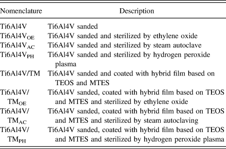
Table I describes the stages performed to obtain and characterize the hybrid coating and the Ti6Al4V alloy samples.
Nomenclature used for the samples during the development of this study.

Table II presents the elemental chemical composition of the alloy used in this study, analyzed by the X-ray Fluorescence technique (Niton® XL3t GOLDD equipment, Thermo Fisher Scientific, Tewksbury, Massachusetts). The percentages obtained for each element present in the alloy samples are in accordance with those established by the standard ASTM F136-13, which refers to the use of the Ti6Al4V alloy in surgical implants.
Chemical composition of the Ti6Al4V alloy analized by X-ray Fluorescence.

A. Surface preparation
Commercial Ti6Al4V alloy samples (Grade 5, supplied by Ti Brasil, São Paulo, Brazil) with dimensions of 15 × 15 × 0.85 mm thickness were used as metal substrates. These were sandblasted with silicon carbide (SiC) up to #1200 grammage, cleaned in an alkaline detergent at 60 °C for 10 min, immersed in an ultrasonic bath with acetone and ethyl alcohol (Anidrol, 99.5%) for 10 min, and dried with a cold jet.
B. Hybrid coating preparation
The silane precursors Tetraethoxysilane (TEOS), Methyltrimethoxysilane (MTES) of Sigma-Aldrich (St. Louis, Missouri; 98%), ethyl alcohol, nitric acid (Neon, Suzano, Brazil; 66.2%), and acetic acid (Neon, 98%) were used for the synthesis of the hybrid coating.
The TEOS/MTES mixture was performed at a 40:60 molar ratio, maintaining the following ratios: TEOS/MTES:H2O (1:2); H2O:C2H6O (1:1); H2O:C2H4O2 (7:1). Reference García, Ceré and Durán31–Reference Ballarre, Manjubala, Schreiner, Orellano, Peter and Ceré33 The hydrolysis of the organic–inorganic precursors was conducted in an acid medium by the addition of nitric acid, with stirring for 24 h at a temperature of 40 °C in the first 3 h.
The deposition of the hybrid coating was performed by using the dip-coating technique, at a withdrawal speed of 40 cm/min and at an immersion time of 5 min. Reference Rodríguez-Cano, Cintas, Fernández-Calderón, Pacha-olivenza, Crespo, González-Martín and Babiano34 After the sol application, the samples were exposed to air for 30 min at room temperature to promote drying and condensation of the film on the substrate. Subsequently, they were taken to the oven for curing at 450 °C for 30 min using a heating rate of 10 °C/min.
C. Coating hygienization
The coating was cleaned by immersing the samples for 10 min in an enzymatic detergent (ENZI-TEC 5) diluted in 2 mL/L. The samples were then rinsed in water and dried with filtered compressed air.
After cleaning, the samples were individually disposed in sterile surgical grade packagings. This process was carried out in the company Esterilizare in Caxias do Sul, RS, Brazil, which is certified by the Brazilian Health Regulatory Agency (ANVISA).
D. Coating sterilization
The coating was sterilized by using three different methods: steam autoclave, ethylene oxide, and hydrogen peroxide plasma.
Saturated steam sterilization was carried out in the company Esterilizare, according to their standard procedure, in a Baumer autoclave (050500001 series, HI-VAC Plus Autoclave Horinzontal model) with a capacity of 0.56 m3. The operational conditions were 134 °C at a pressure of 0.70 atm for 7 min of exposure.
Sterilization by ethylene oxide was carried out in the company Esterilizare, according to their standard procedure. The procedure was conducted at 55 °C at a pressure of 0.65 atm for 180 min of exposure. The gas used in the sterilization process was 90% ethylene oxide and 10% carbon dioxide, supplied by Chemogas (Grimbergen, Belgium; Lot 280715).
Sterilization by the hydrogen peroxide plasma was carried out at the sterilization unit of Pompéia Hospital, in the mucicipality of Caxias do Sul, Brazil. The process was conducted by using a Sterrad NX sterilizer by Johnson and Johnson Company (Irvine, California). The sterilization time was 28 min, at a temperature range from 45 to 55 °C.
These three sterilization methods were selected for this study because they are the usual processes adopted for biomaterials. The parameters used for each type of sterilization were those normally used in establishments certified by ANVISA, without any modification of the processes.
E. Characterization tests
1. Surface properties
The solution after hydrolysis and the hybrid coating were characterized by Fourier Transform Infrared Spectroscopy (FTIR/ATR) in an IS10 Thermo Nicolet device (Thermo Fisher Scientific, Madison, Wisconsin), at a wave number ranging from 4000 to 400 cm−1.
The morphological characterization was carried out using a Field Emission Scanning Electron Microscopy (SEM/FEG), Tescan, model Mira3 (TESCAN, Brno, Czech Republic). The samples were mapped by using energy dispersion spectroscopy (EDS) in Oxford Instruments Model X-Maxd equipment (Oxford Instruments, Abingdon-on-Thames, United Kingdom).
For the adhesion of the coatings to the substrate, Crosshatch and Tape Pull Test analyses were conducted according to ASTM D-3359-09. This standard indicates the manual realization of vertical and horizontal grooves in the deposited coating, with the aid of a cutting device. Thereafter, an adhesive tape is applied and pressed. Finally, the adhesive tape is removed with a slope close to 180°, and the coating detachment areas are analyzed. The degree of adhesion is attributed according to the amount of material adhering to the substrate.
The topography and the nanometric roughness were evaluated by Atomic Force Microscopy (AFM) by using a SHIMADZU SPM-9500J3 microscope (Shimadzu, Kyoto, Japan). The profile analysis was performed in a Mitutoyo Rough Meter, model Surftest SJ-301 (Mitutoyo, Kanagawa, Japan), according to the ISO 4287-1997 standard. The roughness of the surface was monitored by Ra measurements (defined as the arithmetic mean of the deviations from the roughness profile to the midline within the measurement path). Reference Smith35
Contact angle measurements were performed by using the sessile drop method from a device developed by the Research Laboratory Corrosion (LAPEC) at UFRGS (Porto Alegre, Brazil). In this assay, a drop of the simulated body fluid solution (SBF) was deposited on the surface by means of a microsyringe. The drop was observed through a low-magnification lens, and the contact angle was measured through a goniometer.
The thermal properties of the hydrolyzed sol and of the coating after the curing step were analyzed by thermogravimetry (TG), TGA-50, Shimadzu (Kyoto, Japan), with a heating rate of 10 °C/min and temperatures ranging between 10 and 800 °C.
2. Electrochemical properties
The electrochemical behavior of the coating before and after the sterilization processes was evaluated by monitoring the open circuit potential (OCP) and potentiodynamic polarization curves. A three-eletrocde cell was used to perform the analyses, in which the saturated calomel (ECS) was the reference electrode, and the platinum electrode was interconnected to a Potentiostat/Galvanostat, brand IviumStat of the Ivium Technologies, aided by IviumSoft software (Ivium Technologies, Eindhoven, the Netherlands).
The exposed area of the samples for both analyses was 1 cm2. The measurements were performed in a nonagitated medium, naturally aerated, at an approximate temperature of 37 °C, in which the electrolyte used was in the SBF. Reference Kokubo and Takadama36 The open circuit potential was monitored daily for 672 h. Polarization curves were monitored previously and were performed after 1 and 672 h of immersion. The scanning interval was −200 mV below the open circuit potential and 400 mV above that potential, with a scanning speed of 1 mV/s. Reference Ballarre, López, Rosero, Durán, Aparicio and Ceré37
3. Cell culture
Cytotoxicity and cell viability tests were performed by using human osteosarcoma cells (MG-63). Cytotoxicity was assessed by the indirect MTT method test. This method is based on the reduction of MTT [3-(4,5-dimethylthiazol-2-yl)-2,5-diphenyltetrazolium bromide] by the enzyme mitochondrial dehydrogenase, in formation of formazan crystals. Reference Mosmann38,Reference Alley, Dominic, Anne, Miriam, Maciej, Donald, Betty, Joseph, Robert and Michael39 First, to obtain an extraction solution, DMEM (Dulbecco’s modified Eagle Medium) culture medium supplemented with 10% fetal bovine serum (FBS) and 1% penicillin/streptomycin (P/S) was put in contact with Ti6Al4V and Ti6Al4V samples coated with protective film (3 cm2/mL) for 24 h at 37 °C in 5% CO2. Then MG-63 cells were seeded at a density of 5 × 10⁴ cells/mL in 100 μL of DMEM culture medium supplemented with 10% fetal bovine serum (FBS) and 1% penicillin/streptomycin (P/S). After 24 h, the cells were treated with the extraction solution obtained by immersing the samples for 1, 2, and 7 days. DMEM medium (10% FBS and 1% P/S) was used for the negative control and 5% DMSO (dimethylsulfoxide) at 37 °C in 5% CO2 was used for positive control. The medium was removed and 1 mg/mL of MTT in FBS and P/S immune medium was added to the wells. The plates were incubated at 37 °C for 2 h in a humidified atmosphere with 5% CO2. Subsequently, the MTT solution was withdrawn and the formazan crystals were dissolved in 100 μL DMSO. The spectrophotometric reading was performed on a microplate reader (Spectra Max190, Molecular Devices, Sunnyvale, California) at 570 nm and the results were expressed as a viability percentage. The absorbance of the negative control represented 100% viability and the values of the treated cells were calculated as a percentage of the control. Alterations in cell viability analyzed by the indirect method were observed and documented after 1, 2, and 7 days of exposure to the treatment of the sample. The negative control group was treated with DMEM medium (10% FBS and 1% P/S).
Statistical significance was assessed by using the t-test and one-way variance analysis (ANOVA) with the multiple mean comparison test (Tukey) to evaluate statistical differences in the normal distribution cases. Significance was accepted for P lower than 0.05 by using the Statistical Package for Social Sciences (SPSS, version 19.0) for Windows.
To verify the morphological changes in MG-63 cells, the Giemsa staining protocol was performed Reference Nersesyan, Kundi, Atefie, Schulte-hermann and Knasmuller40 for which the cells were seeded in 96 well plates at a density of 5 × 10⁴ cells/mL with 100 μL culture medium DMEM supplemented with 10% fetal bovine serum (FBS) and 1% penicillin/streptomycin (P/S). After 24 h, the cells were treated with the extraction solution, quoted above, and incubated for 1, 2, and 7 days. For the negative control, the DMEM medium (10% FBS and 1% P/S) was used, and for the positive control 5% DMSO was used, incubated at 37 °C in 5% CO2 at the same time as above. The medium was removed, the cells were fixed with methanol for 5 min, and Giemsa’s solution was added for 10 min on the same plate. Subsequently, the plate was visualized in microscopy at 200× magnification.
The cell viability of the substrates was analyzed by performing the direct method with the staining technique by using acridine orange and ethidium bromide in 24 well plates. Samples (1 cm2) with and without coating were arranged in the different wells. MG-63 cells at a density of 3 × 10⁴ cells/plate were then seeded in 1000 mL of DMEM culture medium supplemented with 10% fetal bovine serum and 1% penincillin/streptomycin (P/S), followed by incubation at 37 °C in 5% CO2, for 2 and 7 days. The culture medium was then washed with PBS (Phosphate-buffered saline), the acridine orange dye staining technique (100 μg/mL), and ethidium bromide (100 μg/mL). Cells were counted in apoptosis by fluorescence microscopy at 100× magnification.
The cell morphology and the topographic distribution of the MG-63 cells on the Ti6Al4V and Ti6Al4V samples coated with hybrid film, were evaluated by scanning electron microscopy. Cells were initially seeded in 24 plates at a density of 5 × 10⁴ m/L cells for the 2 and 7 day analyses. In both strains, 1000 μL of the DMEM culture medium supplemented with 10% FBS and 1% penicillin/streptomycin (P/S) on the samples was used for 2 and 7 days. Cells were then fixed with a 3% glutaraldehyde solution in PBS (v/v) for 15 min and with ethanol 30, 50, 70, 90, and 100% (v/v) for 10 min at each concentration. Afterward, the samples were kept in a desiccator until the SEM/FEG analysis was performed.
III. RESULTS AND DISCUSSION
A. Hybrid coating characterization
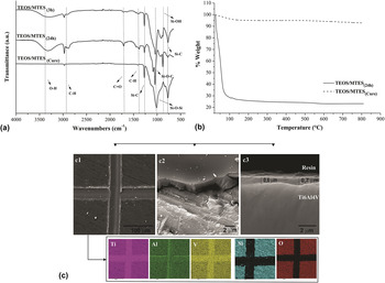
Figure 1(a) shows the FTIR spectra of the solution for 3 and 24 h of hydrolysis and after condensation and curing, respectively.
(a) FTIR spectra of the solution after 3 (TEOS/MTES3h), 24 h (TEOS/MTES24h) hydrolysis and hybrid coating after curing (TEOS/MTEScure), (b) TG curves of the hydrolyzed solution for 24 h and the cured hybrid coating and, (c1) micrographs obtained in SEM/FEG top view and EDS mapping after adhesion assay, (c2) 30° slope in the region of the displacement after adhesion test, and (c3) cross section of the hybrid coating.

The wide absorption between 3200 and 3700 cm−1 is a characteristic of the axial deformation of –OH, arising from silanol groups (Si–OH) and the presence of solvents (water and alcohol), together with the band reference to the C=O bond present in the catalyst structure, acetic acid (1716 cm−1). Bands at about 2900 cm−1 are associated with the C–H bonds, symmetrical and asymmetrical stretches (CH2 and CH3), on the aliphatic chains of the alkoxysilane (TEOS) and organoalkoxysilane (MTES). The time of 3 h of hydrolysis for this sol has been applied in other researches. Reference García, Ceré and Durán31–Reference Ballarre, Manjubala, Schreiner, Orellano, Peter and Ceré33 However, in the FTIR analysis, after 24 h of hydrolysis, the Si–OH band is more intense than that after 3 h, evidencing a higher occurrence of this reaction and, for this reason, the time of 24 h of hydrolysis was applied in the present study. However, it is important to note that the Si–O–C bands, at 1044 cm−1, attributed to the symmetrical stretching due to the movement of the oxygen atoms belonging to the group (Si–O–C) still indicated the incomplete hydrolysis of TEOS and/or MTES. Reference Baldin, Kunst, Beltrami, Lemos, Quevedo, Ferreira, Santos, Sarmento and Malfatti41
The infrared spectrum obtained after the curing step carried out for 30 min at 450 °C confirmed the occurrence of the condensation reaction between the silanols groups, resulting in the formation of a reticulated grid. This is due to the observation of the band referring to the symmetrical and asymmetrical vibration modes of the Si–O–Si bond in 1189 cm−1, the main structural bond of the hybrid material. Finally, the permanence of the bands related to Si–C binding, corresponding to Si–CH3, Reference Juan-Díaz, Martínez-Ibáñez, Lara-Sáez, da Silva, Izquierdo, Gurruchaga, Goñi and Suay17 should be emphasized.
Figure 1(b) shows the TG curves of the hydrolyzed solution after 24 h of hydrolysis and the coating after condensation and curing.
For the hydrolyzed solution, a characteristic mass loss event was observed, in the temperature range between 25 and 120 °C, attributed to the evaporation of solvents, such as ethanol, water, and acetic acid, present in the coating composition. This loss was responsible for the degradation of approximately 70% of the mass of the material.
In the temperature range of 120–800 °C, no significant events of mass loss or gain were observed. However, Wang et al. Reference Wang, Wang, Chen and Gu42 reported that the presence of the methyl group in TEOS/MTES coatings contributes to the higher hydrophobicity of the formed film, thus improving the protective behavior of the film. Innocenzi, Abdirashid and Guglielmi Reference Innocenzi, Abdirashid and Guglielm43 used the curing temperature of 450 °C and justified their choice based on the fact that the degradation and oxidation of the methyl groups present in the organosilane MTES occur in the range of 450–560 °C. The chosen temperature was the threshold to obtain a coating with the highest degree of sintering of the inorganic grid, presenting a compact grid, without compromising the thermomechanical and structural properties and maintaining the functionality of the CH3 group. This mass loss event previously reported was not detected in the present study and can be justified due to the low mass percentage (2%) of the methyl groups compared to the totality of the analyzed composition.
The thermal stability of the hybrid coating after curing (performed during 30 min at 450 °C) was evidenced by the constancy of the mass as a function of temperature increase during the TG analysis, Fig. 1(b). This result may be related to obtaining a structure with a dense and crosslinked siloxane bond grid. Reference Kunst, Beltrami, Cardoso, Santana, Sarmento, Müller and Malfatti44
Figure 1(c) shows the micrographs obtained in the SEM/FEG top view and EDS performed after the adhesion test; the intense signal of the Si and O elements (main constituents of the silane film) was observed in the region where the film adhered to the Ti6Al4V substrate. A high degree of adhesion of the coating to the substrate was also observed since the detachment and removal of the coating occurred in small regions beyond those demarcated according to ASTM D-3359. The thickness of the hybrid coating deposited on the metal substrate was on the order of 0.6 μm. This result is consistent with that obtained by Omar et al. and Ballare et al., Reference Omar, Ballare and Ceré19,Reference Ballarre, Seltzer, Mendoza, Orellano, Mai, García and Ceré20 which used the same silicon precursors on the AISI 316L substrate.
B. Surface properties characterization after sterilization
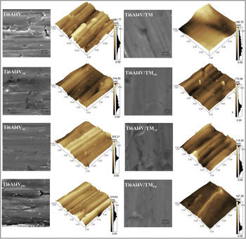
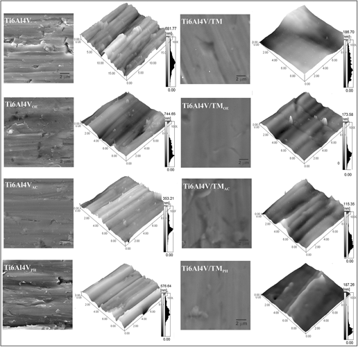
Figure 2 presents the micrographs obtained in the SEM/FEG with the top view of the Ti6Al4V substrate and the protective coating and the profiles of nanometric roughnesses before and after the different methods of sterilization.
Micrographs obtained in SEM/FEG and topography obtained in AFM of the Ti6Al4V and Ti6Al4V/TM before and after sterilization by the three proposed methods.

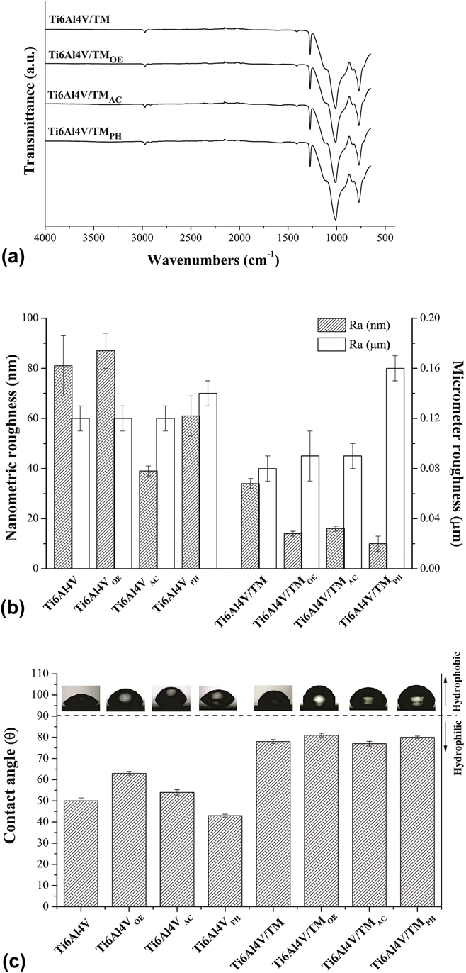
Figure 3(a) shows the FTIR spectra of the hybrid coating before and after the different methods of sterilization. Figure 3(b) presents the average nanometric and micrometric roughnesses for Ti6Al4V and Ti6Al4V/TM sterilized by the three proposed methods, and Fig. 3(c) presents the values obtained for the contact angles.
(a) FTIR spectra of the Ti6Al4V/TM before and after sterilization by the three proposed methods, (b) nanometric and micrometric mean roughness, and (c) contact angle measurements of the Ti6Al4V and Ti6Al4V/TM before and after sterilization by the three proposed methods.

From the SEM/FEG images (Fig. 2), an irregular surface related to the sanding process was observed for the Ti6Al4V substrate. Visually, the hydrogen peroxide sterilization method (Ti6Al4VPH) did not alter the morphology of the Ti6Al4V alloy, but the increase of the micrometric surface roughness of this sample was observed. According to the literature, sterilization by hydrogen peroxide plasma is effective for the removal of surface contaminants and oxides and can cause an increase in surface roughness. Reference Hirano, Kozuka and Asano27,Reference Oh, Rammer, Moon, Bae and Jins28,Reference Gan and Berndt45 Moreover, the increased wettability evidenced by the decrease of the contact angle to 43° in relation to the nonsterilized sample, which presented a contact angle of 50°, is in agreement with the results reported in the literature. Reference Hirano, Kozuka and Asano27,Reference Oh, Rammer, Moon, Bae and Jins28,Reference Gan and Berndt45
From the SEM/FEG images of the Ti6Al4VAC sample, a more regular surface morphology was observed due to the decrease of protrusions and depressions when compared to the Ti6Al4V substrate before sterilization. This can be evidenced by a significant reduction of the nanoscale roughness (Ra) from 81 to 39 nm, after sample sterilization. These morphological modifications can be related to the increase in the oxide layer thickness or contaminant deposition caused by the autoclave sterilization process on the Ti6Al4V substrate. Reference Wilson and Nayak6,Reference Müller, Benkhai, Matthes, Finke, Friedrichs, Geist, Langel and Kramer46 The use of pressure and temperature during this sterilization process can modify the surface morphology of the oxide layer. Reference Park, Navarrete, Baier, Meyer, Tannenbaum, Boyan and Schwartz1 The possible occurrence of these changes in Ti6Al4VAC can be confirmed by the increase of the contact angle from 50 to 54° after Ti6Al4V sterilization, evidencing the reduction of wettabilty of this surface (Ti6Al4VAC) compared to the nonsterilized sample.
Pegueroles et al. Reference Pegueroles, Gil, Planell and Aparicio47 detected a higher presence of carbonaceous species in titanium samples sterilized in ethylene oxide by autoclaving; Liu et al. Reference Liu, Zhou, Wu, Chen and Zheng3 also obtained a reduction in the hydrophilicity of Mg and MgCa surfaces sterilized by ethylene oxide and autoclave and related this change to the presence of inorganic contaminants. Both studies may be related to the hydrophobicity result obtained in this study for Ti6Al4VOE in relation to the nonsterilized sample (from 50 to 63°).
The deposition of the hybrid silane coating (Ti6Al4V/TM) promoted a decrease in the nanometric (from Ra = 81 to Ra = 24 nm) and micrometric (from Ra = 0.12 to Ra = 0.08 µm) roughness compared to the uncoated Ti6Al4V substrate. Figure 2 shows, by observing the SEM/FEG analysis and the roughness profiles, that the coating (Ti6Al4V/TM) is free of discontinuities and presents a uniform morphology compared to the uncoated Ti6Al4V. In addition, we found that deposition of the coating on the metal substrate has resulted in the reduction of its wettability, which can explain the different chemical compositions of the coatings. The results obtained in this work are in agreement with the approach proposed by Wenzel cited by Pequeroles et al., Reference Pegueroles, Gil, Planell and Aparicio47 in which the increasing surface roughness promotes a decrease in contact angle values due to the increased surface areas. Wang et al. Reference Wang, Hsieh and Lee48 observed this relation for the alteration of both the nanoscale and micrometric roughness in titanium substrates. In addition, such modifications may influence the biological response of MG-63 cells. Reference Rodríguez-Cano, Cintas, Fernández-Calderón, Pacha-olivenza, Crespo, González-Martín and Babiano34
By analyzing the bands presented in the FTIR spectra in Fig. 3(a), it is clear that the sterilization processes did not cause structural chemical changes in the hybrid coating, since there is no functional groups formation, nor even the alteration of the bands already present. Galante et al. Reference Galante, Ghisleni, Paradiso, Alves, Pinto, Colaço and Serro49 also did not observe structural changes in silicone hydrogel samples sterilized by the same methods evaluated in this work. However, by the analysis of Fig. 3(b), the influence of the sterilization processes on the morphology of the same is evident.
All the sterilization methods promoted the reduction of the mean nanoscale roughness for Ti6Al4V/TM. This leveling can be associated to the different sterilization processes.
For the Ti6Al4V/TMOE e Ti6Al4V/TMPH samples, although the sterilization methods used use low temperatures, the observed nanometric leveling may have occurred as a consequence of the oxidative nature of the gases used. Reference Vetten, Yah, Singh and Gulumian2,Reference Lee, Kim, Kim, Lee, KIm, Lee and Park50 Similarly, even if no changes were observed in the FTIR spectra, another possible explanation for the reduction of the Ti6Al4V/TMOE nanometric roughness would be the retention of residues from this sterilization process on the sample surface. Reference Lee, Kim, Kim, Lee, KIm, Lee and Park50 These residues have toxic characteristics that can negatively affect cell growth on biomaterials. Reference Park, Navarrete, Baier, Meyer, Tannenbaum, Boyan and Schwartz1,Reference Lee, Kim, Kim, Lee, KIm, Lee and Park50–Reference Holyak, Wang, Liu and Bunch52
Walke et al. Reference Walke, Paszenda, Pustelny, Ziemniak and Basiaga29 also obtained such results by sterilizing surfaces coated with silane films with ethylene oxide. França et al. Reference França, Doris, Samani, Tien, Mateescu, Yahia and Sacher53 reported that the chemical method of sterilization by ethylene oxide promoted an oxidation reaction of the chitosan-containing surface. Lee et al. Reference Lee, Kim, Kim, Lee, KIm, Lee and Park50 observed a reduction of approximately 50% of the porosity of the material in the sterilization of poly(lactic-co-glycolic acid) (PLGA) polymer sterilized by plasma of hydrogen peroxide, which also contributed to its leveling. Finally, Yavuz et al. Reference Yavuz, Oliaei and Celikta54 identified that sterilization of poly(methyl methacrylate) (PMMA) by ethylene oxide promoted more reduction of surface roughness than the autoclave method.
The autoclave sterilization method, in addition to promoting the nanometric reduction of Ti6Al4V/TMAC, presented a more discontinuous coating with a greater number of surface defects compared to the others, as shown in Fig. 2. Although the thermal stability of the coating was detected in the TG curve, Fig. 1(b), the use of high temperatures combined with pressure may have caused the thickening of the SiO2 layer and the appearance of surface defects. Reference Fleith, Ponche, Bareille, Amédée and Nardin51 Such modification may have acted as adsorption sites of –OH or caused a variation of CH3 groups due to sensitization to the pressure, thus increasing the wettability of this sample. Reference Hirano, Kozuka and Asano27,Reference Oh, Rammer, Moon, Bae and Jins28,Reference Fleith, Ponche, Bareille, Amédée and Nardin51
Likewise, all the sterilization methods increased the micrometric roughness of the hybrid coating. The most significant was obtained by the sterilization method of hydrogen peroxide plasma.
The sample Ti6Al4V/TMPH presented an increase of roughness (Ra) of 0.08–0.16 μm, which also may be associated to the mode of action of this process, in which the ionized gas bombards the surface of the material promoting the removal of a surface layer. The layer thickness removed as well as the modifications caused in the material are dependent on the type and purity of the gas used, the applied pressure, and the position of the sample in the equipment during the sterilization process. Reference Park, Navarrete, Baier, Meyer, Tannenbaum, Boyan and Schwartz1,Reference Gan and Berndt45
C. Protective properties characterization after sterilization
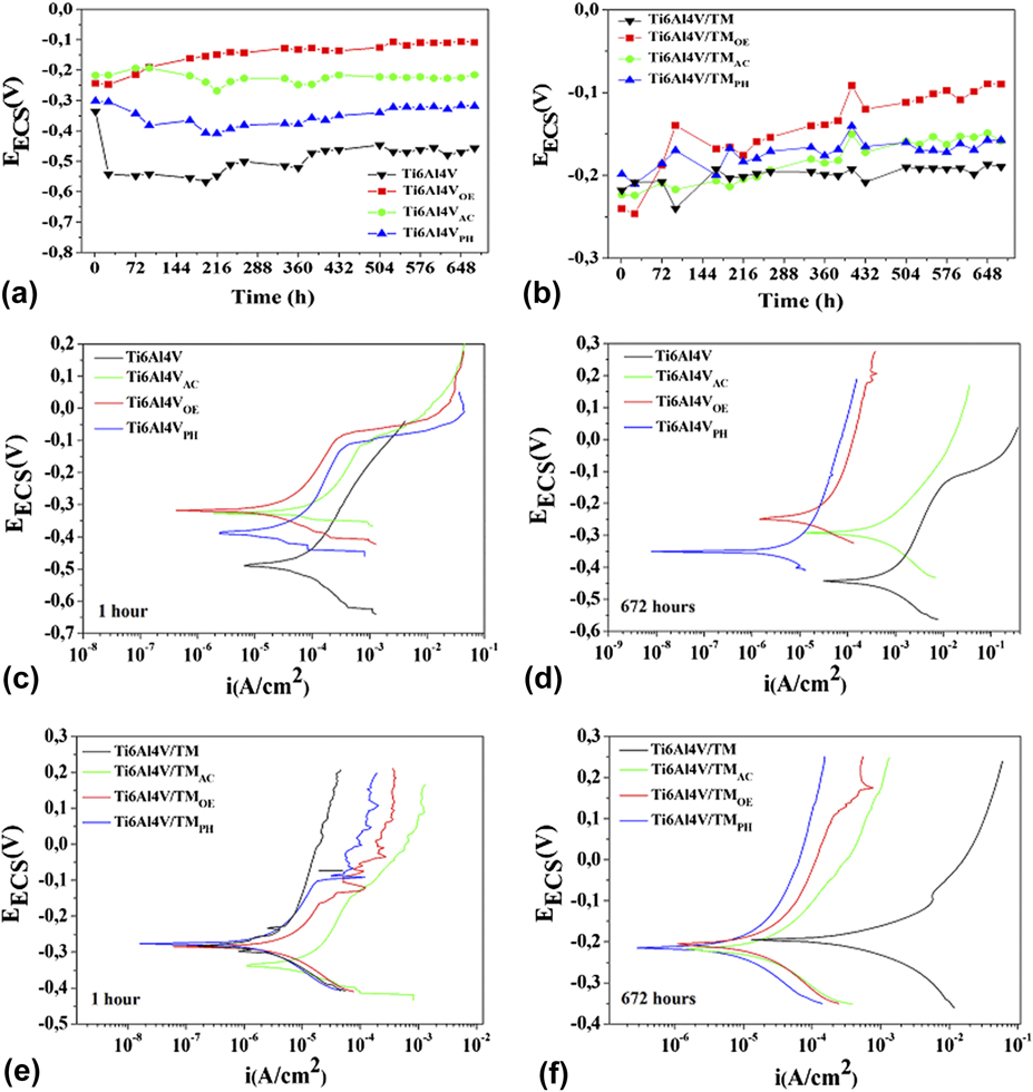
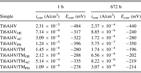
Figures 4(a) and 4(b) show the open circuit potential monitoring measured during 672 h of immersion, and Figs. 4(c)–4(f) show the polartization curves in the SBF solution (1 and 672 h of immersion) for Ti6Al4V and Ti6Al4V TM sterilized by the three proposed methods. Table III presents the corrosion potential values (E corr) and current densities (i corr) obtained by extrapolating the Tafel lines from the polarization curves, Figs. 4(c)–4(f).
Monitoring the open circuit potential (OCP) daily for 672 h (a) Ti6Al4V and (b) Ti6Al4V/TM sterilized by the three proposed methods and polarization curves (c, d) Ti6Al4V and (e, f) Ti6Al4V/TM, sterilized by the three proposed methods, after 1 and 672 h of immersion, both in SBF at 37 ± 3 °C.

Corrosion potential values (E corr) and current densities (i corr) for Ti6Al4V e Ti6Al4V/TM sterilized by the three proposed methods, after 1 and 672 h of immersion.

The effect of sterilization is clearly observed during the OCP monitoring for both systems. Samples of sterilized Ti6Al4V presented a displacement of the potential values in a more positive sense in relation to Ti6Al4V. Furthermore, the values associated with the increase presented by Ti6Al4VOE were constant, which can be attributed to the decrease of the reactivity of the substrate with the immersion time, usually associated to the increase of the oxide layer thickness resulting from the reaction of the Ti6Al4V alloy with the electrolyte. Reference Liu, Yue, Romeo, Weber, Scheuermann, Moulton and Wallace25 In regard to the hybrid coating, sterilized samples showed more noble potentials as a function of time than nonsterilized samples. Such behavior was associated to the compromise of the barrier effect of the coatings after sterilization, which allowed the permeability of the electrolyte through them, causing superficial modifications in the metallic substrate that contributed to the protective properties.
The presence of the hybrid coating acting as a physical barrier between Ti6Al4V and the electrolyte Reference Ribeiro, Hammer, Vaz and Rocha55 can be observed during the first hours of immersion due to the more positive values of Ti6Al4V/TM potentials in relation to Ti6Al4V. However, upon sterilization of Ti6Al4V by ethylene oxide and hydrogen peroxide, surface modifications showed similar potential values to those presented by the sterilized coatings.
By analyzing the values of the corrosion potentials and the current densities presented in Table III, together with the interpretation of the profiles of the curves, it was possible to verify the influence of the sterilization processes. After 1 h of immersion, more positive potential corrosion values were obtained for Ti6Al4VOE and Ti6Al4VAC followed by Ti6Al4VPH and Ti6Al4V, as per Fig. 4(c). This increase, added to the displacement of anodic and cathodic curves to lower values of current density of the sterilized samples in relation to nonsterilized, according to López et al., Reference López, Fauré, Cabrera and García56 was due to the formation of a passive layer on the substrates. As a consequence, the occurrence of electrochemical reactions acting on the deterioration of the Ti6Al4V alloy reduced.
After 672 h of immersion, all Ti6Al4V samples shifted the potential corrosion values to more positive numbers. According to Szewczenko et al., Reference Szewczenko, Grygiel, Walke, Nowinska, Granieczny, Kaczmarek and Marciniak57 this shift is related to the existence of a porous oxide layer on Ti6Al4V, which, immersed in a SBF solution, tends to partially degrade, allowing the permeability of the electrolyte and the formation of an internal oxide layer when in contact with Ti6Al4V, Fig. 4(d). Jamrozik et al. Reference Kiel-Jamrozik, Szewczenko, Basiaga and Nowińska58 also mentioned this behavior in the study of the autoclaved Ti6Al4V alloy and observed that pitting corrosion was simultaneously suppressed by the recreation of an oxide layer on the surface of the material. Paszenda et al. Reference Paszenda, Walke and Jadacka59 observed a higher electrochemical stability of autoclave-sterilized titanium oxides than that of nonsterilized ones and associated this with the use of steam under pressure during the execution of the method.
In addition, after 672 h, the positive effect caused by the sterilization methods on the Ti6Al4V alloy was also observed due to the displacement of the curves to higher values of current density presented by Ti6Al4V in relation to the sterilized samples. Also, as a function of time, the superiority of the anodic behavior of Ti6Al4VPH, Ti6Al4VOE, and Ti6Al4VAC compared to Ti6Al4V was also observed. However, the Ti6Al4VPH sample presented the lowest current density (i corr) in relation to the others.
The Ti6Al4V/TM sample, after 1 h of immersion [Fig. 4(e)], showed potential corrosion values similar to Ti6Al4V/TM, Ti6Al4V/TMOE, and Ti6Al4V/TMPH. Furthermore, it presented an inferior behavior than that of Ti6Al4V/TMAC, due to the displacement to lower values. In addition, cathodic and anodic curves shifted to higher current density values. Both behaviors confirm the inferiority of the barrier behavior presented by the autoclave coating, related to the greater morphological alteration observed previously, as well as with the greater hydrophilicity it presented in relation to the others.
The cathodic curves of the Ti6Al4V/TM, Ti6Al4V/TMOE, and Ti6Al4V/TMPH were also similar, but with different anodic behavior, with the displacement to higher current density values. Therefore, after the first hour of immersion, the negative effect of the sterilization processes on the protective properties of the hybrid coating was verified. However, after 672 h of immersion, as shown in Fig. 4(f), the nonsterilized coating presented a lower barrier behavior due to the displacement of its curves to higher current density values. This behavior is a different behavior from the sterilized samples, for which current density values remained almost unchanged, close to 10−6. Walke et al. Reference Walke, Paszenda, Pustelny, Ziemniak and Basiaga29 justified this difference by the formation of a protective layer above the SiO2 coating sterilized and immersed in a simulated body solution, improving its resistive properties.
Although the barrier effect is noticed to be given the more positive potential corrosion values of the coatings in relation to the metallic substrates as a function of the immersion time, an approximation of the current density values was observed, mainly for the sample Ti6Al4V/TMAC. Such an approximation was associated with the permeability of the electrolyte through the films, and according to Basiaga et al., Reference Basiaga, Walke, Paszenda and Kajzer60 it is related to the contact of the electrolyte with the metallic substrate interface, which promotes the formation of titanium oxide passive or the formation of corrosion products with protective properties. Reference García, Ceré and Durán61
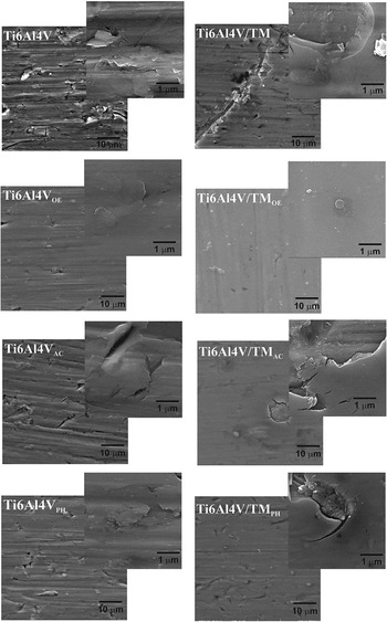
Figure 5 shows the SEM/FEG micrographs after polarization tests of immersed samples for 672 h in SBF.
Micrographs obtained in SEM/FEG after the polarization test of immersed samples for 672 h in SBF at 37 ± 3 °C of the Ti6Al4V and Ti6Al4V/TM before and after sterilization by the three proposed methods.

The images obtained by SEM/FEG for the Ti6Al4V samples show similar surfaces for Ti6Al4VPH and Ti6Al4V. Moreover, the leveling presented by the sample Ti6Al4VOE after the time of immersion in a body fluid can be observed, in addition to the permanence and the greater intensity of the surface discontinuities presented by Ti6Al4VAC.
For the Ti6Al4V/TMOE sample, a regular appearance coating, free from significant surface defects, was presented, except from clusters located in some regions of the surface. For Ti6Al4V/TMPH, the presence of imperfections, with small corrosion points and cracks distributed along the surface, was visualized, similar to the Ti6Al4V/TM sample. The Ti6Al4V/TMAC sample presented imperfections due to the its rupture in several points in a greater magnitude than those previously reported.
The morphologies presented by the coatings after 672 h are in agreement with the phenomena previously discussed.
D. Cell culture
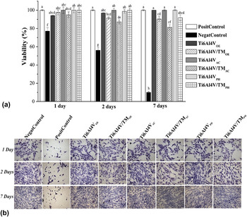
Figure 6(a) shows a comparison of the cytotoxic activity of the extract obtained by the immersion of Ti6Al4V and Ti6Al4V/TM substrates sterilized by the proposed methods, negative and positive control in MG-63 cells after 1, 2, and 7 days.
(a) Cytotoxic effect of extract of Ti6Al4V e Ti6Al4V/TM with the three proposed sterilizations methods. MG-63 cells were treated for 1, 2, and 7 days with the obtained extract. Positive control (cells treated with 5% DMSO). Results were obtained from three independent experiments. Each bar represents the average percentage of viability ± SD relative to negative control (fluid body), which was normalized to 100%. *Letters correspond to statistically significant differences using ANOVA-Tukey test (P ≤ 0.05). (b) Cell morphology of MG-63 treated for 1, 2, and 7 days visualized under light microscopy of inverted light with increase of 200×.

At different treatment times, all Ti6Al4V substrates presented cell viability values above 75%, indicating a reduced cytotoxic activity, a fact that was not observed for the positive control (DMSO 5%), which presented reduced viability with values around 10% on the seventh day of analysis. Among all evaluated samples, the ones with the lowest percentages of cell viability were Ti6Al4V/TMOE after 1 day and Ti6Al4V/TMAC at 2 and 7 days. They still presented indices higher than 70%, in accordance with the values recommended by ISO 10993-5.
The influence of the sterilization methods on the protective coating can be noticed after 7 days of exposure, in which a reduction in cell viability was obtained, that was accentuated for Ti6Al4V/TMAC. However, all the samples of sterilized Ti6Al4V/TM remained viable above 75%, again indicating a beneficial coating behavior at the concentrations and parameters tested.
The morphology of MG-63 cells was evaluated by the analysis of the images obtained with Giemsa staining in light microscopy, Fig. 6(b). The cellular alterations are compatible with the results presented by MTT.
The images show that the negative control cells were surrounded by abundant cytoplasm, well defined and intact, which is opposite to the positive control, in which a substantial increase in cellular cytotoxicity and cellular morphological alterations were expected. After 1 and 2 days of exposure with the different extracts, an increase in cell density was observed. After 7 days, the cell density showed to be more intense for all the conditions evaluated, with little evidence of condensation of the cellular structures. Thus, the results obtained for the substrates tested in this study corroborated with the absence of cytotoxic activity previously discussed.
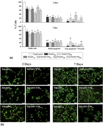
Figure 7(a) shows the percentage of cell viability events, initial apoptosis, and late apoptosis and/or necrosis of MG-63 cells when in direct contact with Ti6Al4V and Ti6Al4V/TM sterilized by the three methods and stained by acridine orange and ethidium bromide.
(a) Percentage of cells stained with acridine orange and ethidium bromide after performing a direct contact assay according to ISO 10993-5-2009-2, with modifications. *Letters correspond to the statistically significant diferences using the ANOVA-Tukey test (P ≤ 0.05). (b) MG-63 cells stained with acridine orange and ethidium bromide viewed under a fluorescence microscope at 200× magnitude (green colored cells represent viable cells and red colored cells represent apoptosis and/or necrosis).

Numerous researches highlight the importance of surface parameters in cellular response. In addition, MG-63 osteoblast cells respond differentially to these parameters and to the composition of the material. Reference Györgyey, Ungvári and Kecskeméti62 In this study, it was possible to verify the influence of the composition of the material, given that the cell viability profile was lower for the samples with the hybrid coating than that of the metal substrates.
The roughness of the metallic surface is known to have a considerable in vitro effect on parameters such as cell adhesion, proliferation, and differentiation. Reference Likibi, Jiang and Li63,Reference Wirth, Grosgogeat, Jaffrezic-Renault and Ponsonnet64 Specifically, nanometric surface roughness is directly related to protein and extracellular matrix sizes and therefore plays an important role in osteoblast differentiation and tissue regeneration. Reference Gittens, McLachlan, Olivares-Navarrete, Cai, Berner, Tannenbaum, Schwartz, Sandhage and Boyan65 In the literature, it is reported that surfaces with micrometric roughness favor cell adhesion and promote, in vivo, the increase of bone-implant contact area influencing the osseointegration process. Reference Likibi, Jiang and Li63,Reference Moon, Kim, Kim and Jang66 However, Moon et al. Reference Moon, Kim, Kim and Jang66 obtained similar percentages of cell viality when evaluating osteoblastic cells on nano and microstructured Ti6Al4V topographies.
In addition, Park et al. and Oh et al. Reference Park, Navarrete, Baier, Meyer, Tannenbaum, Boyan and Schwartz1,Reference Oh, Rammer, Moon, Bae and Jins28 observed that the cellular response is influenced by surface parameters and that these are still dependent on the different sterilization processes used. For Liu et al., Reference Liu, Zhou, Wu, Chen and Zheng3 wettability is one of the most important parameters that affects the cellular response of the materials used for implants. This interferes directly in protein adsorption and cell adhesion. It is possible that the cells prefer hydrophilic surfaces, but proteins tend to preferentially absorb on hydrophobic surfaces.
After 2 days of incubation, the highest amount of viable cells was obtained for the Ti6Al4V and Ti6Al4V/TM samples sterilized by hydrogen peroxide, followed by ethylene oxide and autoclave. Most probably, the proteins adsorb in titanium with the most hydrophobic moiety facing the metal surface, which may justify the higher viability presented by Ti6Al4VPH and Ti6Al4V/TMPH. Reference Pegueroles, Gil, Planell and Aparicio47
As previously mentioned, some authors refer that the use of micrometric rough surfaces favors cellular behavior, Reference Wirth, Grosgogeat, Jaffrezic-Renault and Ponsonnet64 justifying again the greater viability presented by samples sterilized by hydrogen peroxide.
Zhao et al. Reference Zhao, Mei and Wang67 observed that the degrading effects caused by the autoclave sterilization of titanium nanotubes are responsible for the lower adhesion and cell proliferation, causing a reduction in bioactivity. According to Pegueroles et al., Reference Pegueroles, Gil, Planell and Aparicio47 the increase in oxide layer thickness and the presence of contaminants after a sterilization process, changes similar to those previously observed in this study, affect the biological response of biomaterials, justifying the observed rates for Ti6Al4V/TMAC after 2 days of incubation.
Conversely, according to Martin et al., Reference Martin, Schwartz, Hummert, Schraub, Simpson, Lankford, Dean, Cochran and Boyan68 at longer exposure periods, MG-63 cells proliferated better on smooth surfaces, corroborating with the increase in percentage of cell viability obtained for the samples sterilized by autoclave from 2 to 7 days of incubation. The results obtained in cell viability are in agreement with those obtained by the MTT method.
The initial levels of apoptosis in the second day for all coated samples were lower than those for the metallic substrate regardless of sterilization methods. Similarly, it was also observed that the levels of late apoptosis and/or necrosis were lower for all the samples but with the higher indices presented by the coated samples. This may be related to the low average roughness values and the more hydrophobic behaviors presented by the coatings compared to the metal substrates, both of which may have contributed to the impairment of cell adhesion, inducing the cells to the death process.
After 7 days of incubation, the initial apoptosis profile became heterogeneous and the Ti6Al4V/TMAC sample presented the highest percentage, around 29%. In addition, the higher level of apoptosis and/or necrosis presented by Ti6Al4V/TMOE (25%), which may have happened due to the retention of residues from the process Reference Park, Navarrete, Baier, Meyer, Tannenbaum, Boyan and Schwartz1,Reference Holyak, Wang, Liu and Bunch52 that were released over time, motivating increases in cell rates of late apoptosis and necrosis as a function of time. Despite the percentages presented for apoptosis, they are considered low compared to those obtained for cell viability.
Figure 7(b) shows the fluorescence microscopy images of MG-63 cells stained with acridine orange and ethidium bromide in the direct contact assay. By applying the fluorescence analysis technique, a direct measurement of the cellular morphological alterations could be performed. For the cell interaction, this method allows viable cells to be differentiated from those in the process of apoptosis or necrosis. Reference Sharma, Bano and Ghosh69 The images show that the deleterious effect of the cells was not verified, only the increase of cell viability from 2 to 7 days of incubation. These results are in accordance with the ones showed above.
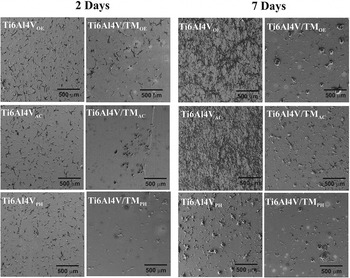
Figure 8 depicts the micrographs obtained by scanning microscopy (SEM/FEG) of the cells on the substrates evaluated after 2 and 7 days of incubation.
Morphology of MG-63 cells on the surfaces of Ti6Al4V and Ti6Al4V/TM sterilized by the three proposed methods after 2 and 7 days of incubation.

In SEM/FEG different cellular morphologies were observed for Ti6Al4V and TiAl4V/TM, but no significant differences were found based on the sterilization process used. This is because, for all Ti6Al4V samples, the MG-63 cells presented a fusiform morphology, being such a behavior accentuated as a function of time. Among the sterilization methods, the cells observed on Ti6Al4VPH appear to present more morphological changes.
According to Junkar et al., Reference Junkar, Kulkarni and Drašler26 the surface parameters, besides modifying the physiology, alter the cellular morphology. According to the authors, the oxygen peroxide sterilization of titanium sheets promoted an increase in the thickness of the oxide, which consequently provided a greater interaction of the same with Hbo-type cells. This observation is consistent with the results found for Ti6Al4VAC and Ti6Al4VOE due to the fact that the presence of elongated and more distributed cells was verified in the analyzed surfaces.
Although the percentage of cell viability was high, the cells on the protective coatings sterilized by the three proposed methods presented an attenuated spherical morphology, regardless of the incubation time. This result indicates low interaction with the surface. Reference Junkar, Kulkarni and Drašler26 Such morphology was also observed in MG-63 cells on Mg and MgCa alloys sterilized by similar methods. Ti6Al4V/TMOE and Ti6Al4V/TMPH showed cell agglomerates that became dispersed as a function of time. Reference Liu, Zhou, Wu, Chen and Zheng3 On the other hand, for Ti6Al4V/TMAC, a small membrane elongation was observed, which is a very similar behavior to Ti6Al4VAC. This elongation is related to the lower barrier behavior in the sample, which causes the formation of oxides from the metal substrate.
IV. CONCLUSION
The combination of the alkoxysilane TEOS with the organoalcoxysilane MTES applied by dip coating resulted in an adherent hybrid coating with a homogeneous coverage on the Ti6Al4V substrate.
The proposed sterilization methods promoted changes in the morphological and physicochemical properties of the Ti6Al4V substrate, as well as of the hybrid silane film applied to Ti6Al4V.
Regarding the electrochemical properties, the TEOS/MTES hybrid coating presented a protective character to the metallic substrate of the Ti6Al4V alloy. However, for the hybrid coating, the protective behavior was compromised regardless of the sterilization method used. Furthermore, the effect of the sterilization processes on the anodic behavior of the substrate was notable. Ti6Al4V presented a decrease on the current densities compared to the nonsterilized sample.
The Ti6Al4V and Ti6Al4V/TM substrates did not present cytotoxicity to the MG-63 cells. Nevertheless, they behaved differently due to the superficial modifications caused by each of the sterilization methods. The hydrogen peroxide plasma sterilization showed a higher percentage of viable cells. However, it caused more morphological alterations than what was verified for the Ti6Al4V substrates. The hybrid coatings presented lower percentages of viable cells and higher rates of late apoptosis and necrosis. After 7 days of incubation, among the hybrid coatings, Ti6Al4V/TMOE presented the lowest percentage of viability and the highest cell death rate, respectively.
Supplementary Material
To view supplementary material for this article, please visit https://doi.org/10.1557/jmr.2017.429.
ACKNOWLEDGMENTS
The authors would like to express their gratitude to CNPq (Project PVE 401211/2014-2), CAPES, UFRGS, UCS, Esterilizare RS/Brazil and hospital Pompéia RS/Brazil for the investment and support in this research.